VSports手机版 - Skip to main page content
U.S. flag

An official website of the United States government

Dot gov

The . gov means it’s official. Federal government websites often end in . gov or . mil VSports app下载. Before sharing sensitive information, make sure you’re on a federal government site. .

Https

The site is secure V体育官网. The https:// ensures that you are connecting to the official website and that any information you provide is encrypted and transmitted securely. .

. 2015 Jul 10;11(7):e1004224.
doi: 10.1371/journal.pcbi.1004224. eCollection 2015 Jul.

"VSports app下载" Geometry of the Gene Expression Space of Individual Cells

Affiliations

Geometry of the Gene Expression Space of Individual Cells

Yael Korem et al. PLoS Comput Biol. .

Abstract

There is a revolution in the ability to analyze gene expression of single cells in a tissue. To understand this data we must comprehend how cells are distributed in a high-dimensional gene expression space. One open question is whether cell types form discrete clusters or whether gene expression forms a continuum of states. If such a continuum exists, what is its geometry. Recent theory on evolutionary trade-offs suggests that cells that need to perform multiple tasks are arranged in a polygon or polyhedron (line, triangle, tetrahedron and so on, generally called polytopes) in gene expression space, whose vertices are the expression profiles optimal for each task. Here, we analyze single-cell data from human and mouse tissues profiled using a variety of single-cell technologies VSports手机版. We fit the data to shapes with different numbers of vertices, compute their statistical significance, and infer their tasks. We find cases in which single cells fill out a continuum of expression states within a polyhedron. This occurs in intestinal progenitor cells, which fill out a tetrahedron in gene expression space. The four vertices of this tetrahedron are each enriched with genes for a specific task related to stemness and early differentiation. A polyhedral continuum of states is also found in spleen dendritic cells, known to perform multiple immune tasks: cells fill out a tetrahedron whose vertices correspond to key tasks related to maturation, pathogen sensing and communication with lymphocytes. A mixture of continuum-like distributions and discrete clusters is found in other cell types, including bone marrow and differentiated intestinal crypt cells. This approach can be used to understand the geometry and biological tasks of a wide range of single-cell datasets. The present results suggest that the concept of cell type may be expanded. In addition to discreet clusters in gene-expression space, we suggest a new possibility: a continuum of states within a polyhedron, in which the vertices represent specialists at key tasks. .


V体育官网入口 - Conflict of interest statement

The authors have declared that no competing interests exist.

Figures

Fig 1
Fig 1. Overview of Pareto archetype analysis of single-cell datasets to discover polytopes in gene expression space and infer tasks.
Datasets from different human and mouse tissues analyzed by different groups with different technologies were analyzed by Pareto archetype analysis. Best fit polytopes and their significance were found. Tasks were inferred from the genes maximally enriched in the cells closest to each vertex of the polytope.
Fig 2
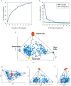
Fig 2. Human colon crypt cells fall in a tetrahedron in gene expression space.
(a) For k = 2–11 we found the k-polytope that best fit the data using PCHA algorithm, considering all 76 dimensions. Explained variance of best fit polytopes with k = 2–11 vertices begins to saturate at k = 4 or k = 5 vertices. (b) Comparison between the variance explained by the first k principal components of the data to the variance explained by the k principal components of shuffled data suggests that effective data dimensionality is three or four. Blue line: variance explained by PCA of intestinal data. Green line: variance explained by PCA of shuffled data. Points represent mean values. Error bars, representing 5%-95% variation intervals, are smaller than line width. Points for which the real data EV is higher than the randomized data EV are marked with *. (c) Data displayed in first 3 PCs axes resembles a tetrahedron, and its projections on principal planes (d)-(f) resemble triangles. Archetypes and their variation upon data resampling (bootstrapping) are shown as colored ellipses (see S1B Text). Thin lines—tetrahedron edges.
Fig 3
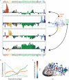
Fig 3. Expression profiles of the four colon crypt archetypes are each enriched for markers of specific cell types.
(a) The expression profiles of the four archetypes, with enriched genes colored. Enriched genes were determined by leave-1-out enrichment analysis, binning the cells according to distance from each archetype and seeking when average expression in the bin closest to the archetype is maximal, as described in Methods: 1D Gene enrichment at archetypes (See full enriched genes list in S2 Table). Light blue—enterocyte archetype, yellow—Nodal archetype, green—stem cells archetype, red—goblet cell archetype. Genes that are not enriched, or enriched in more than one archetype, are in dark blue. Zero level represents the average expression of each gene in the dataset. (b) Leave-1-out enrichment plot: expression of a gene (SLC26A3—an enterocyte marker) as a function of distance from archetype in equal mass bins of cells (Methods: 1D Gene enrichment at archetypes), line color indicates archetype. This gene is maximally enriched only at the enterocyte archetype (blue line). For enrichment plots for additional genes see S5 Fig. (c) A two dimensional enrichment plot of SLC26A3, in which its expression is plotted on the plane of the first 2PCs of the data, indicating expression is maximal in the cells closest to the enterocyte archetype. Contours are expression density estimated using a Gaussian kernel (Methods: 2D Gene enrichment at archetypes). Archetype positions and PCs were calculated without the tested gene.
Fig 4
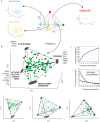
Fig 4. Progenitor crypt cells fall in a tetrahedron.
(a) Enterocytes, goblet cells and nodal cells analyzed separately do not form significant polytopes. Cells are color coded by type in the tetrahedron of Fig 3, and each cell class is plotted in its own first 3PC. (b) Progenitor cells analyzed separately fall uniformly in a tetrahedron. The best fit tetrahedron is shown (PCHA delta = 0.5). Arrow represents direction of development according to Axin2 levels, see S7b Fig. Also shown are projections on the principal planes, which resemble triangles or quadrangles. Archetypes and their variation upon data resampling (bootstrapping) are shown as gray ellipses. (c) Explained variance as a function of polytope order k or number of PCs D both suggest a tetrahedron (k = 4, D = 3).
Fig 5
Fig 5. Mouse and human colon lower crypt cells fall on similar triangles and show a similar distribution within the triangle.
(a) Mouse colon lower crypt cells dataset by [37], plotted on its first 2PCs plane. Inset: PCA explained variance analysis suggest k = 3 vertices and D = 2 dimensions, namely a triangle. (b) Human lower crypt dataset plotted on its first 2PC plane. Inset: explained PCA explained variance analysis. The 24 genes common to the two datasets were used. Arrow indicates projection of the nodal cell archetype on the triangle.
Fig 6
Fig 6. Different tissues analyzed by different single-cell technologies show polytopes and tasks.
(a) Human bone marrow cells analyzed by single-cell mass cytometry [13,25] in which proteins are detected using mass-tagged antibodies is well described by a 4D simplex (a polytope with 5 vertices). The simplex is shown projected on the first 3PCs, for other projections see S12c Fig. The archetypes correspond to cell types as indicated. Cell density peaks near each archetype. (b) Mouse spleen LPS stimulated dendritic cells analyzed by single-cell RNA-Seq [3] are well described by a tetrahedron. Archetypes are labeled with functions inferred from genes maximally enriched in cells near each archetype. For more details see S1G and S1H Text.

References

    1. Blainey PC, Quake SR. Dissecting genomic diversity, one cell at a time. Nat Methods. 2014;11: 19–21. - PMC - PubMed
    1. Dalerba P, Kalisky T, Sahoo D, Rajendran PS, Rothenberg ME, Leyrat AA, et al. Single-cell dissection of transcriptional heterogeneity in human colon tumors. Nat Biotechnol. 2011;29: 1120–1127. 10.1038/nbt.2038 - DOI - PMC - PubMed
    1. Jaitin DA, Kenigsberg E, Keren-Shaul H, Elefant N, Paul F, Zaretsky I, et al. Massively Parallel Single-Cell RNA-Seq for Marker-Free Decomposition of Tissues into Cell Types. Science. 2014;343: 776–779. 10.1126/science.1247651 - DOI (V体育ios版) - PMC - PubMed
    1. Landry ZC, Giovanonni SJ, Quake SR, Blainey PC. Optofluidic cell selection from complex microbial communities for single-genome analysis. Methods Enzymol. 2013;531: 61–90. 10.1016/B978-0-12-407863-5.00004-6 - DOI - PMC - PubMed
    1. Sanchez-Freire V, Ebert AD, Kalisky T, Quake SR, Wu JC. Microfluidic single-cell real-time PCR for comparative analysis of gene expression patterns. Nat Protoc. 2012;7: 829–838. 10.1038/nprot.2012.021 - DOI - PMC - PubMed

"V体育2025版" Publication types

"V体育2025版" Substances

LinkOut - more resources