Skip to main page content
U.S. flag

An official website of the United States government

Dot gov

The . gov means it’s official. Federal government websites often end in . gov or VSports app下载. mil. Before sharing sensitive information, make sure you’re on a federal government site. .

Https

The site is secure. The https:// ensures that you are connecting to the official website and that any information you provide is encrypted and transmitted securely V体育官网. .

Review
. 2015 Jul 20;28(7):1391-7.
doi: 10.1021/acs.chemrestox.5b00208. Epub 2015 Jul 2.

"VSports手机版" Adverse Outcome Pathways and Drug-Induced Liver Injury Testing

Affiliations
Review

V体育2025版 - Adverse Outcome Pathways and Drug-Induced Liver Injury Testing

"V体育ios版" Mathieu Vinken. Chem Res Toxicol. .

Abstract

Drug-induced liver injury is a prominent reason for premarketing and postmarketing drug withdrawal and can be manifested in a number of ways, such as cholestasis, steatosis, and fibrosis. The mechanisms driving these toxicological processes have been well characterized and have been emdedded in adverse outcome pathway frameworks in recent years. This review evaluates these constructs and simultaneously illustrates their use in the preclinical testing of drug-induced liver injury. VSports手机版.


V体育2025版 - Figures

Figure 1
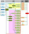
Figure 1. AOP for drug-induced liver fibrosis
The MIE (blue) is considered protein alkylation and covalent protein binding in the liver. This serves as a trigger to provoke hepatocyte injury, including apoptosis, which in turn activates Kupffer cells. As a result, transforming growth factor beta 1 (TGF-β1) expression is induced, which is a key factor for stellate cell activation. The latter goes hand in hand with the occurrence of inflammation and oxidative stress. The different KEs at the cellular level (green) are interconnected in several ways. The overall end result is accumulation of collagen and changes in the extracellular matrix composition in the liver (orange), which becomes clinically manifested as the adverse outcome, namely liver fibrosis (red) (reproduced with permission from).
Figure 2
Figure 2. AOP for drug-induced liver steatosis
Activation of the liver X receptor (LXR), which is the MIE (blue), induces a number of transcriptional changes, including activation of the expression of carbohydrate response element binding protein (ChREBP), sterol response element binding protein 1c (SREBP-1c), fatty acid synthase (FAS) and stearoyl-coenzyme A desaturase 1 (SCD1). As a result, de novo synthesis of fatty acids is enhanced in the liver. At the same time, fatty acid translocase (CD36) production is upregulated, which mediates increased hepatic influx of fatty acids from peripheral tissues. All together, these steps drive the accumulation of triglycerides, which is considered a KE (dark green). At the organelle level, this evokes cytoplasm displacement, distortion of the nucleus and mitochondrial disruption. This ultimately burgeons into the appearance of fatty liver cells (orange) and further into the clinical diagnosis of liver steatosis (red) (reproduced with permission from).
Figure 3
Figure 3. AOP for drug-induced cholestasis
The response matrix between the MIE (dark blue) and adverse outcome (red), the inhibition of the bile salt export pump (BSEP) and cholestasis, respectively, spans over the cellular and organ levels. Identified KEs (dark green) include the accumulation of bile, the induction of oxidative stress and inflammation, and the activation of the nuclear receptors pregnane X receptor (PXR), farnesoid X receptor (FXR) and constitutive androstane receptor (CAR). Together with a number of other steps, these KEs drive both a deteriorative cellular response (yellow), which underlies directly caused cholestatic injury, and an adaptive cellular response (purple), which is aimed at counteracting the primary cholestatic insults. Direct inducing and inhibiting effects are indicated with green and red arrows, respectively. Secondary inducing and inhibiting effects of oxidative stress and/or inflammation are indicated with blue and orange arrows, respectively (5′-NT, 5′-nucleotidase; ALP, alkaline phosphatase; ALT, alanine aminotransferase; AST, aspartate aminotransferase; CYP2B10/3A4/7A1, cytochrome P450 2B10/3A4/7A1; GGT, gamma-glutamyl transpeptidase; MPP, mitochondrial permeability pore; MRP2/3, multidrug resistance-associated protein 2/3; NTCP, sodium/taurocholate cotransporter; OATP1B1, organic anion transporter 1B1; OSTα/β organic solute transporter α/β; SHP, small heterodimeric partner; SULT2A1, dehydroepiandrosterone sulfotransferase; UGT2B4, uridine 5′-diphosphate-glucuronosyltransferase 2B4) (reproduced with permission from).

References

    1. Przybylak KR, Cronin MT. In silico models for drug-induced liver injury: current status. Expert Opin. Drug Metab. Toxicol. 2012;8:201–217. - PubMed
    1. Faa G, Ekstrom J, Castagnola M, Gibo Y, Ottonello G, Fanos V. A developmental approach to drug-induced liver injury in newborns and children. Curr. Med. Chem. 2012;19:4581–4594. - PubMed
    1. Holt MP, Ju C. Mechanisms of drug-induced liver injury. AAPS J. 2006;8:E48–E54. - PMC - PubMed
    1. Verma S, Kaplowitz N. Diagnosis, management and prevention of drug-induced liver injury. Gut. 2009;58:1555–1564. - PubMed
    1. Andrade RJ, Agundez JA, Lucena MI, Martinez C, Cueto R, Garcia-Martin E. Pharmacogenomics in drug induced liver injury. Curr. Drug Metab. 2009;10:956–970. - PubMed

"V体育ios版" MeSH terms