VSports app下载 - Skip to main page content
U.S. flag

An official website of the United States government

Dot gov

The . gov means it’s official. Federal government websites often end in . gov or . mil VSports app下载. Before sharing sensitive information, make sure you’re on a federal government site. .

Https

The site is secure. The https:// ensures that you are connecting to the official website and that any information you provide is encrypted and transmitted securely V体育官网. .

. 2015 May 15;7(1):45.
doi: 10.1186/s13073-015-0168-9. eCollection 2015.

Widespread intron retention diversifies most cancer transcriptomes

Affiliations

Widespread intron retention diversifies most cancer transcriptomes

Heidi Dvinge et al. Genome Med. .

Abstract

Background: Somatic mutations affecting components of the RNA splicing machinery occur with high frequencies across many tumor types. These mutations give rise to distinct alterations in normal splice site and exon recognition, such as unusual 3' splice site preferences, that likely contribute to tumorigenesis. VSports手机版.

Methods: We analyzed genome-wide patterns of RNA splicing across 805 matched tumor and normal control samples from 16 distinct cancer types to identify signals of abnormal cancer-associated splicing. V体育安卓版.

Results: We found that abnormal RNA splicing, typified by widespread intron retention, is common across cancers even in the absence of mutations directly affecting the RNA splicing machinery V体育ios版. Almost all liquid and solid cancer types exhibited frequent retention of both alternative and constitutive introns relative to control normal tissues. The sole exception was breast cancer, where intron retention typified adjacent normal rather than cancer tissue. Different introns were preferentially retained in specific cancer types, although a small subset of introns enriched for genes encoding RNA splicing and export factors exhibited frequent retention across diverse cancers. The extent of intron retention correlated with the presence of IDH1 and IDH2 mutations in acute myeloid leukemia and across molecular subtypes in breast cancer. Many introns that were preferentially retained in primary cancers were present at high levels in the cytoplasmic mRNA pools of cancer cell lines. .

Conclusions: Our data indicate that abnormal RNA splicing is a common characteristic of cancers even in the absence of mutational insults to the splicing machinery, and suggest that intron-containing mRNAs contribute to the transcriptional diversity of many cancers VSports最新版本. .


Figures
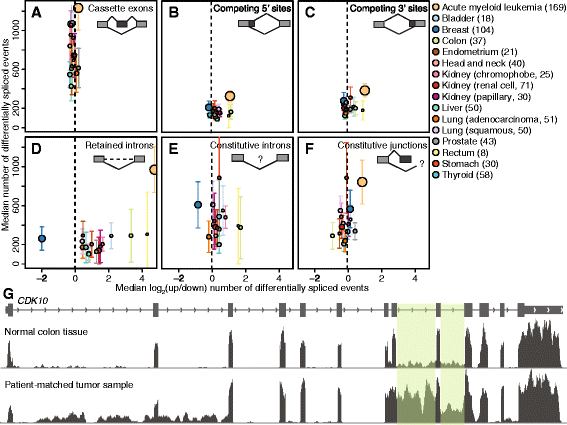
Fig. 1
Fig. 1
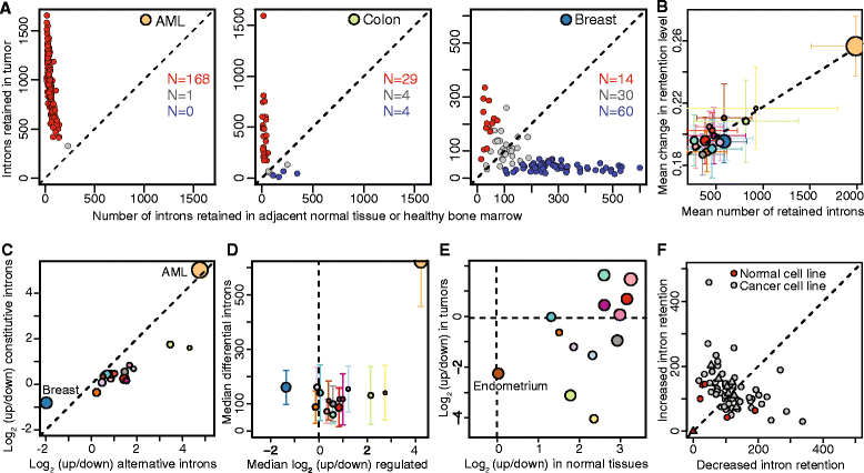
Differential splicing across 16 distinct cancer types. The median numbers of (a) cassette exons, (b, c) alternative 5′ and 3′ splice sites, and (d) retained introns that are differentially spliced between patient-matched cancer and normal samples (y axis) versus log2 of the ratio of the number of events that are up- and downregulated in cancer versus normal controls (x axis). The median is computed across all samples for each cancer type. Upregulation is defined as increased cassette exon inclusion, usage of intron-proximal 5′ or 3′ splice sites, or increased intron retention. Bars indicate the standard deviation across samples for each cancer type, and circle sizes are proportional to the number of samples for each cancer type. See figure for color legend. (e, f) As panels A-D, but for (e) retention of constitutive introns or (f) alternative splicing of constitutive junctions. Upregulation is defined as increased constitutive intron retention or decreased alternative splicing of constitutive junctions. For constitutive introns, AML is outside the figure limits (to the right). (g) RNA-seq read coverage of CDK10 for a patient-matched cancer and normal sample from the colon. Shaded boxes mark introns that are most frequently retained in the cancer sample
Fig. 2
Fig. 2
Intron retention characterizes all analyzed cancer types except for breast. (a) Numbers of introns with increased or decreased retention in cancer versus control normal samples in AML, colon cancer, and breast cancer. Red/blue illustrate cancer samples that exhibit an enrichment >1.5-fold for increased/decreased intron retention relative to control normal samples. (b) The average number of differentially retained introns versus the mean absolute change in retention level, computed for each cancer type. Error bars indicate standard deviation across the individual patient samples. Colors as Fig. 1. (c) Median log2 of the ratio of the number of introns that exhibit increased and decreased retention in cancer versus normal controls, where the median is taken over all samples for each cancer type. The x and y axes are, respectively, restricted to annotated alternative and constitutive introns. Colors as Fig. 1. (d) As Fig. 1d, but computed using only reads crossing the exon-intron boundary. (e) Median log2 of the ratio of the number of introns that exhibit increased and decreased retention in normal breast versus other normal control tissues (x axis), and breast cancer versus other cancer types (y). (f) Number of differentially retained introns in breast cancer cell lines (gray) compared to non-cancerous epithelial breast cells (red). Triangles [23]; circles [24]
Fig. 3
Fig. 3
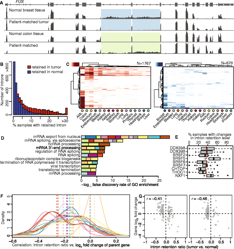
Intron retention preferentially affects genes encoding RNA processing factors. (a) RNA-seq read coverage of FUS for patient-matched tumor and normal samples from breast and colon. Shaded boxes indicate differentially retained introns. (b) Histograms illustrating the numbers of introns exhibiting increased (red) or decreased (blue) retention in cancer relative to normal samples, computed across all cancer types. (c) Hierarchical clustering of all retained introns (rows) and all cancer types (columns). Analysis restricted to introns that exhibit increased (red) or decreased (blue) retention relative to normal controls in >10 % of samples for at least one cancer type. Clustering is based on Euclidean distances computed over intron retention frequencies and Ward’s agglomeration method. (d) The combined -log10 false discovery rate of the most significant Biological Process Gene Ontology (GO) terms enriched among genes containing differentially retained introns in at least 20 % of samples within each cancer type. Colors as in Fig. 1. (e) Percentage of samples within cancer types with differential intron retention for select genes mapped to the ‘mRNA export from nucleus’ GO term (GO: 0006406). Dashed line, average across all genes and cancer types. (f) Distribution of Pearson correlation coefficients between intron retention and gene expression across all samples within each cancer type. Dashed line, median taken over all samples for each cancer type. Colors as in Fig. 1. (g) Scatter plots comparing intron retention to fold-change of the corresponding parent genes for two colon adenocarcinoma samples relative to their patient-matched normal control
Fig. 4
Fig. 4
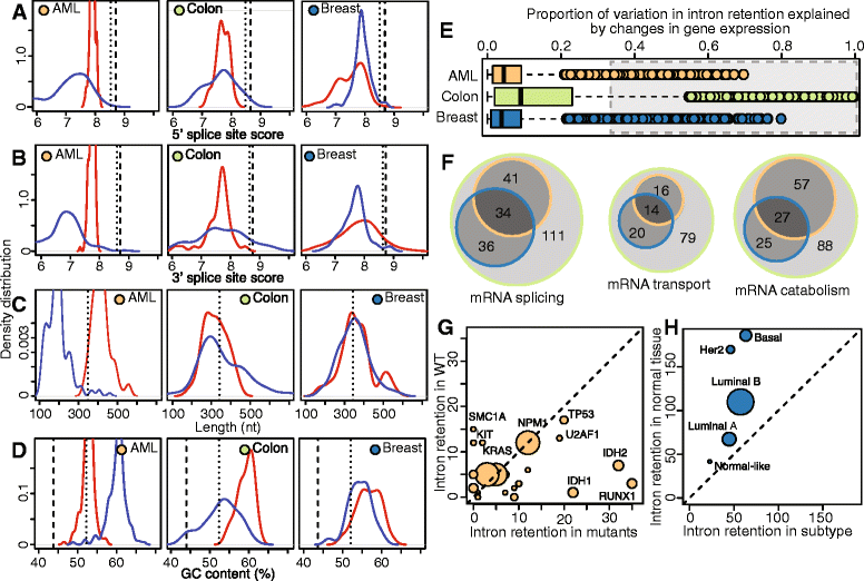
Cis-acting sequence features and trans-acting factors affect the degree of intron retention. (a) 5′ splice site scores, (b) 3′ splice site scores, (c) length, and (d) GC content of introns exhibiting increased (red) or decreased (blue) retention in cancer relative to normal controls. Each distribution illustrates the sample medians per cancer type. Dashed lines, median across all constitutive introns; dotted lines, median across 67,000 constitutive introns <1 kb (resulting in a length distribution similar to alternative introns). The median constitutive intron length of 1.4 kb is outside the range in (c). (e) Proportion of variation in intron retention that can be explained by a General Additive Model (GAM) of differential expression of RNA processing, transport and degradation genes. Each dot is a specific intron/mRNA combination. Gray box, combinations where more than one-third of the intron retention level can be explained by expression of trans-acting factors. (f) Overlap across cancer types for mRNAs within the gray box in (e), divided by Gene Ontology term. (g) Numbers of introns exhibiting increased or decreased retention in AML samples mutated in a specific gene compared to all wild-type AML samples for each gene. Dot size is proportional to mutation frequency. Analysis restricted to genes mutated in >5 samples. Mutations with >12 differentially spliced introns are highlighted. Note that the subgroup analyses illustrated here is statistically distinct from previous analyses, which were computed per-sample between matched tumor-normal pairs. This plot only illustrates introns that are consistently differentially spliced across the subgroup relative to other samples. (h) Numbers of introns exhibiting increased or decreased retention in breast cancer subtypes relative to all control breast samples (subgroup analysis, as in (g)). Analysis is over all 1,080 cancer and 104 normal breast samples. Dot size is proportional number of samples within each subtype
Fig. 5
Fig. 5
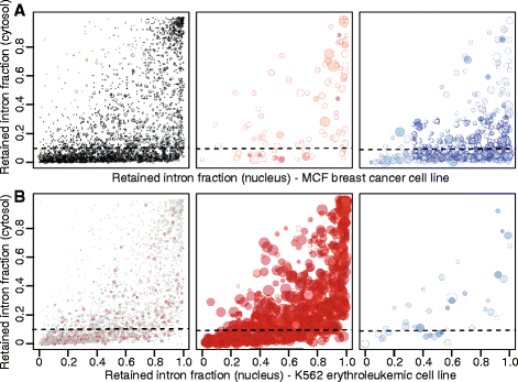
Retained introns are frequently found in the cytoplasm. Intron retention in the nuclear versus cytoplasmic subcellular fractions of MCF-7 (a) and K562 (b) cell lines, categorized by how frequently the introns are differentially spliced in primary breast cancer or AML samples. Left, introns that are differentially spliced in <25 % of samples from the corresponding primary cancer; center, introns that are retained in more cancer samples than normal controls; right, introns that are retained in more normal controls than cancer samples. The left, center, and right panels are mutually exclusive. Each dot represents a single intron. The dot size is proportional to the number of patients the intron is differentially spliced in, and the dot color intensity represents the proportion of patients where it is retained in the tumor (red) versus the control (blue). Introns lying above the dashed line are retained in >10 % of cytosolic transcripts

References

    1. Yoshida K, Sanada M, Shiraishi Y, Nowak D, Nagata Y, Yamamoto R, et al. Frequent pathway mutations of splicing machinery in myelodysplasia. Nature. 2011;478:64–9. doi: 10.1038/nature10496. - DOI - PubMed
    1. Graubert TA, Shen D, Ding L, Okeyo-Owuor T, Lunn CL, Shao J, et al. Recurrent mutations in the U2AF1 splicing factor in myelodysplastic syndromes. Nat Genet. 2012;44:53–7. doi: 10.1038/ng.1031. - DOI - PMC - PubMed
    1. Papaemmanuil E, Cazzola M, Boultwood J, Malcovati L, Vyas P, Bowen D, et al. Somatic SF3B1 mutation in myelodysplasia with ring sideroblasts. New Eng J Med. 2011;365:1384–95. doi: 10.1056/NEJMoa1103283. - DOI - PMC - PubMed
    1. Visconte V, Makishima H, Jankowska A, Szpurka H, Traina F, Jerez A, et al. SF3B1, a splicing factor is frequently mutated in refractory anemia with ring sideroblasts. Leukemia. 2012;26:542–5. doi: 10.1038/leu.2011.232. - DOI - PubMed
    1. Quesada VV, Conde LL, Villamor NN, Ordóñez GRG, Jares PP, Bassaganyas LL, et al. Exome sequencing identifies recurrent mutations of the splicing factor SF3B1 gene in chronic lymphocytic leukemia. Nat Genet. 2012;44:47–52. doi: 10.1038/ng.1032. - DOI - PubMed