<u lang="oyiHZUjN"></u><center lang="yuVmWzsN"></center> "VSports app下载" Skip to main page content
U.S. flag

An official website of the United States government

Dot gov

The . gov means it’s official VSports app下载. Federal government websites often end in . gov or . mil. Before sharing sensitive information, make sure you’re on a federal government site. .

Https

The site is secure. The https:// ensures that you are connecting to the official website and that any information you provide is encrypted and transmitted securely V体育官网. .

. 2015 Jun 16:6:7390.
doi: 10.1038/ncomms8390.

"VSports" Programmed cell death 5 mediates HDAC3 decay to promote genotoxic stress response

Affiliations

"VSports最新版本" Programmed cell death 5 mediates HDAC3 decay to promote genotoxic stress response

V体育安卓版 - Hyo-Kyoung Choi et al. Nat Commun. .

Erratum in

"V体育平台登录" Abstract

The inhibition of p53 activity by histone deacetylase 3 (HDAC3) has been reported, but the precise molecular mechanism is unknown. Here we show that programmed cell death 5 (PDCD5) selectively mediates HDAC3 dissociation from p53, which induces HDAC3 cleavage and ubiquitin-dependent proteasomal degradation. Casein kinase 2 alpha phosphorylates PDCD5 at Ser-119 to enhance its stability and importin 13-mediated nuclear translocation of PDCD5 VSports手机版. Genetic deletion of PDCD5 abrogates etoposide (ET)-induced p53 stabilization and HDAC3 cleavage, indicating an essential role of PDCD5 in p53 activation. Restoration of PDCD5(WT) in PDCD5(-/-) MEFs restores ET-induced HDAC3 cleavage. Reduction of both PDCD5 and p53, but not reduction of either protein alone, significantly enhances in vivo tumorigenicity of AGS gastric cancer cells and correlates with poor prognosis in gastric cancer patients. Our results define a mechanism for p53 activation via PDCD5-dependent HDAC3 decay under genotoxic stress conditions. .


"V体育ios版" Figures
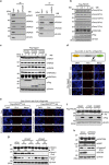
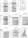
Figure 1
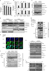
Figure 1. PDCD5 selectively binds to and mediates caspase-3-dependent cleavage of HDAC3 at Asp-391.
(a) PDCD5 is an HDAC3-associating protein among PDCD proteins. Proteins from HCT-116 (p53+/+) whole-cell lysate were immunoprecipitated and subsequently immunoblotted with the indicated antibodies. (b) Overexpression of PDCD5 induces C-terminal cleavage of HDAC3. Cells were transfected with increasing amounts of Flag-PDCD5 plasmid. Whole-cell lysates were immunoblotted with the indicated antibodies. Arrow indicates cleaved HDAC3. (c) Overexpression of PDCD5 selectively triggers the cleavage of HDAC3, but not other class I HDACs. Cells were transfected with increasing amounts of PDCD plasmids. Whole-cell lysates were immunoblotted with the indicated antibodies. Arrow indicates cleaved HDAC3. (d,e) In vivo validation of PDCD5-mediated HDAC3 cleavage at Asp-391. HCT-116 cells were transfected with the indicated plasmids. Permeabilized cells were incubated with antibodies against HA and Flag, and then PLA probes were added. Positive signals were analysed using confocal microscopy. Red dots display uncleaved HDAC3. Representative images of three independent experiments are shown. (f) Inhibition of caspase-3 abrogates PDCD5-induced HDAC3 cleavage. HCT-116 cells were transfected with Flag-PDCD5 plasmid and treated with the indicated caspase inhibitors. (g) Depletion of caspase-3 abrogates ET-induced HDAC3 cleavage. Cells were transfected with siRNAs as indicated and treated with ET (100 μM, 12 h). Whole-cell lysates were immunoblotted with the indicated antibodies. (h) PDCD5 is required for caspase-3-dependent HDAC3 cleavage during ET treatment. Either shcontrol or stable shPDCD5-expressing HCT-116 cells was treated with ET. Whole-cell lysates were analysed by western blotting with the indicated antibodies. Scale bar, 10 μm.
Figure 2
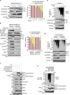
Figure 2. PDCD5 stabilizes p53 by inducing dissociation of the HDAC3–p53 complex and concomitantly triggering cytosolic cleavage of HDAC3.
(a) Overexpression of PDCD5 reduces the activity of HDAC3. HCT-116 cells were transfected with the indicated plasmids. Whole-cell lysates were immunoprecipitated with anti-HDAC3 antibody, and then HDAC3 activity was measured. Error bars, s.d. (n=3). *P<0.05. (b) Depletion of PDCD5 abolishes the ET-induced reduction of HDAC3 activity. Cells were transfected with the indicated siRNAs. Cells were treated with ET (100 μM, 12 h) or STS (1 μM, 12 h) and assayed for HDAC3 activity. Error bars, s.d. (n=3). *P<0.05. (c) Knockdown of PDCD5 abrogates the association of HDAC3 with active caspase-3 in response to ET treatment. Whole-cell lysates were immunoprecipitated with anti-HDAC3 antibody, and subsequently immunoblotted with the indicated antibodies. Arrow indicates cleaved HDAC3. (d) Knockdown of PDCD5 prevents cytoplasmic cleavage of HDAC3 in response to ET treatment. Following cell fractionation, fractions were immunoblotted with the indicated antibodies. (e) Overexpression of PDCD5 enhances ET-induced cytoplasmic translocation of HDAC3. Immunofluorescence analysis was performed as described in the Supplementary Experimental Procedures Section. Representative images of three independent experiments are shown. (f) PDCD5 knockdown diminishes the ET-induced dissociation of HDAC3 from p53. Indicated shRNA-expressing HCT-116 cells were treated with ET. Whole-cell lysates were analysed by western blotting with the indicated antibodies. (g) Overexpression of PDCD5 dissociates HDAC3 from p53. Cells were transfected with PDCD5 plasmids. Immunoprecipitation and immunoblotting analyses were performed with the indicated antibodies. (h,i) PDCD5 increases p53 acetylation and stability via mediating HDAC3 cleavage. Cells were treated with ET (h) or cycloheximide (i) and subsequently immunoblotted with the indicated antibodies. Scale bar, 10 μm.
Figure 3
Figure 3. PDCD5 mediates ubiquitin-dependent proteasomal degradation of HDAC3 via C-terminal cleavage of HDAC3.
(a) MG132 treatment induces accumulation of cleaved HDAC3. Cells were treated with ET (50 μM) and/or MG132 (10 μM, 3 h). Whole-cell lysates were immunoblotted with the indicated antibodies. Arrow indicates cleaved HDAC3 (left panel). Intensities of protein bands obtained from the immunoblotting assay were quantified with ImageJ (right panel) and normalized with respect to that of β-actin. Relative % intensity was calculated by dividing the normalized intensity by the sum of intensities from both cleaved and full-length HDAC3. Error bars, s.d. (n=3). (*P<0.01 versus without ET.) (b) PDCD5 knockdown diminishes the reduction and cleavage of full-length HDAC3. shcontrol or stable shPDCD5-expressing HCT-116 cells were treated with ET and/or MG132. Whole-cell lysates were immunoprecipitated and immunoblotted with the indicated antibodies (left panel). Relative % intensity was calculated as described above (right panel). Error bars, s.d. (n=3). (*P<0.01 versus without ET.) (c) HDAC3 ubiquitination increases in a time-dependent manner in response to ET treatment. Cells were transfected with HA-Ub plasmid and treated with MG1432 and/or ET. Whole-cell lysates were immunoprecipitated with anti-HDAC3 (C) antibody and immunoblotted with the indicated antibodies. (d) PDCD5 knockdown diminishes HDAC3 ubiquitination in response to ET. Whole-cell lysates were immunoprecipitated with anti-HDAC3 (C) antibody and immunoblotted with the indicated antibodies. (e) Mutation of Asp-391 abolishes ET-induced HDAC3 ubiquitination. Cells were transfected with HA-Ub and the indicated Flag-tagged plasmids, and treated with ET and/or MG132. Whole-cell lysates were immunoprecipitated with anti-Flag antibody and immunoblotted with the indicated antibodies. (f) Mutation of Asp-391 potentiates the action of HDAC3 in the inhibition of PDCD5-mediated p53 acetylation. Cells were transfected with the indicated Flag-tagged plasmids. Whole-cell lysates were immunoblotted with the indicated antibodies.
Figure 4
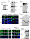
Figure 4. Casein kinase 2α enhances the stability and IPO13-mediated nuclear translocation of PDCD5 during genotoxic stress responses.
(a) CK2α knockdown diminishes the effect of MG132 on PDCD5 stability. HCT-116 (p53+/+) cells were transfected with MG132 or shCK2α and immunoblotted with the indicated antibodies. (b) CK2α knockdown abolishes ET-induced HDAC3 cleavage, PDCD5 induction and PDCD5 phosphorylation. Either shcontrol or stable shCK2α-expressing HCT-116 cells were treated with ET. Whole-cell lysates were immunoblotted with the indicated antibodies. (c) CK2α overexpression inhibits PDCD5 ubiquitination. Cells were transfected with HA-Ub and the indicated Flag-tagged plasmids, and treated with MG132. Whole-cell lysates were immunoprecipitated with anti-Flag antibody and immunoblotted with the indicated antibodies. (d) CK2-mediated phosphorylation is required for ET-induced PDCD5 nuclear translocation. Cells were transfected with the indicated Flag-PDCD5 plasmids and treated with ET. Immunofluorescence analysis was performed as described in the Supplementary Experimental Procedures Section. (e) IPO13 selectively interacts with PDCD5 upon genotoxic stress response. Cells were transfected with HA-PDCD5 and the indicated Flag-tagged plasmids, and treated with ET. Whole-cell lysates were immunoprecipitated and immunoblotted with the indicated antibodies. (f) Knockdown of IPO13 abrogated the nuclear translocation of endogenous PDCD5 in response to ET. HCT-116 cells were transfected with the indicated siRNAs and treated with ET. Immunofluorescence analysis was performed as described in the Supplementary Experimental Procedures Section. (g) IPO13 knockdown abolishes the nuclear translocation of phosphor-PDCD5. HCT-116 cells were transfected with the indicated siRNAs and Flag-PDCD5 plasmid, and treated with ET. Immunofluorescence analysis was performed as described in the Supplementary Experimental Procedures Section. (h) Phospho-mimetic PDCD5S119D mutant further promotes p53 acetylation and stabilization. Cells were transfected with the indicated Flag-PDCD5 plasmids. Whole-cell lysates were immunoblotted with the indicated antibodies. Scale bar, 10 μm.
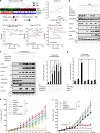
Figure 5
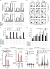
Figure 5. PDCD5 promotes p53-dependent apoptosis via selective inhibition of HDAC3.
(a) Overexpression of HDAC3 suppresses the PDCD5-mediated transcription of p53-target genes. Cells were transfected with indicated siRNAs and/or plasmids and treated with either ET or STS. The levels of indicated genes were analysed by real-time PCR. Error bars, s.d. (n=3). *P<0.05. (b) PDCD5 promotes p53-dependent apoptosis in a caspase-3-dependent manner. Cells were transfected with indicated plasmids and treated with ET and/or Z-DQMD. Annexin V-positive cells were assessed by flow cytometry. A representative figure of three independent experiments is shown. (c) HDAC3, but not other class I HDACs tested, selectively antagonizes PDCD5-enhanced apoptosis. Annexin V-positive cells were assessed by flow cytometry. Error bars, s.d. (n=3). *P<0.05. (d) Knockdown of HDAC3 significantly enhances ET-induced apoptosis. Annexin V-positive cells were assessed by flow cytometry. Error bars, s.d. (n=3). *P<0.05; **P<0.01. (e) PDCD5 is required for ET-induced recruitment of the p53–p300 complex to the promoter region of Bax. Cells were transfected with indicated plasmids and/or shPDCD5, and then treated with ET. ChIP and re-ChIP assays were performed with the indicated antibodies. Precipitated samples were analysed by real-time PCR, and results are presented as the percentage of input. Error bars, s.d. (n=3). *P<0.05, **P<0.01 versus without ET; #P<0.05 versus without ET; ##P<0.05 versus ET+HA-p53WT.
Figure 6
Figure 6. PDCD5 is required for genotoxic stress-induced HDAC3 cleavage and p53 activation.
(a) Depletion of PDCD5 abrogated the ET-induced HDAC3 cleavage and p53 activation. MEFs were infected with Ad-Cre or Ad-GFP and then electroporated with Myc-p53 or treated with ET (50 μM, 8 h). Whole-cell lysates were immunoblotted with indicated antibodies. Arrow indicates cleaved HDAC3. (b) PDCD5 promotes ET-induced p53 activation and HDAC3 cleavage. PDCD5−/− MEFs were electroporated with indicated plasmids, treated with ET, lysed and then analysed by immunoblotting. (c) Restoration of PDCD5 into PDCD5−/− MEFs induces the recruitment of the p53–p300 complex to the promoter region of Bax. ChIP assays were performed with the indicated antibodies. Error bars, s.d. (n=3). *P<0.05, **P<0.01. (d) Both PDCD5 and p53 are mutually required for ET-induced activation of apoptosis. Stable shPDCD5-expressing p53−/− MEFs were electroporated with indicated plasmids and treated with ET. (e) Knockdown of HDAC3 rescues the suppression of p53 caused by depletion of PDCD5. PDCD5−/− MEFs were electroporated with indicated plasmids and/or shHDAC3, and cell lysates were analysed by immunoblotting. (f) Negative effect of cleavage at Asp-391 on the anti-apoptotic function of HDAC3. Cell lysates were analysed by immunoblotting. (g) Ablation of PDCD5 abolishes the genotoxic stress response in vivo. Mice were injected with ET (10 mg kg−1) for the indicated days. Adenovirus expressing GFP or Cre recombinase was injected in mice 6 days before ET injection, as indicated. Tissues from individual livers were harvested and processed for western blotting. Total RNA was isolated from individual livers, and qRT–PCR was performed for the indicated genes. Error bars, s.d. (n=8). *P<0.05 versus without ET; #P<0.05 versus ET (2 days).
Figure 7
Figure 7. Reduction of both PDCD5 and p53 synergistically enhance in vivo tumorigenicity of gastric cancer cells.
(a) Reduction of PDCD5 and p53 significantly correlates with poor survival in stage 2b gastric cancer patients. Kaplan–Meier plots and log-rank test were used to estimate the prognostic differences of categorized patient groups. (b) Depletion of PDCD5 diminishes the effect of HDAC3 knockdown on p53 acetylation and activation. AGS cells were transfected with siHDAC3 and/or shPDCD5 as indicated, and treated with or without ET (75 μM, 8 h). Whole-cell lysates were immunoblotted with indicated antibodies. (c) Restoration of PDCD5 with HDAC3 knockdown potentiates ET-induced p53 activation. Stable shPDCD5-expressing AGS cells were transfected with indicated plasmids and/or siHDAC3, and then treated with ET. DNA damage of cells was determined by the TUNEL assay. Error bars, s.d. (n=3). *P<0.01. (d) Reduction of PDCD5 and p53 synergistically reduces the genotoxic response of AGS cells. Stable shPDCD5-expressing AGS cells were transfected with indicated plasmids and/or siRNA, and then the cells were treated with ET. DNA damage of cells was determined by the TUNEL assay. Error bars, s.d. (n=3). *P<0.01. (e) Reduction of PDCD5 and p53 significantly reduces the chemosensitivity of AGS cells. Stable AGS cells were injected subcutaneously into the right flank of nude mice. Four weeks after injection, mice with comparable-sized tumours (100∼200 mm3) were selected for treatment with ET (10 mg kg−1), with 2-day intervals for 8 weeks. Tumour volumes were measured for 12 weeks. Error bars indicate s.d. (n=6). *P<0.05 versus without ET+shPuro. (f) Knockdown of HDAC3 reversed the impaired chemosensitivity of AGS cells by depletion of PDCD5. Stable shCon or shPDCD5 AGS cells were injected subcutaneously into the right flank of nude mice. Four weeks after injection, mice with comparable-sized tumours (100∼200 mm3) were selected for treatment with etoposide (10 mg kg−1), with 2-day intervals for 8 weeks. Detailed procedure for siHDAC3 treatment is described in the Methods. Tumour volumes were measured for 12 weeks. *P<0.05 versus shCon without ET; **P<0.05 versus shCon with ET; #P<0.05 versus shPDCD5 with ET. Error bars indicate s.d. (n=6).

References

    1. Vazquez A., Bond E. E., Levine A. J. & Bond G. L. The genetics of the p53 pathway, apoptosis and cancer therapy. Nat. Rev. Drug Discov. 7, 979–987 (2008). - V体育2025版 - PubMed
    1. Vousden K. H. p53: death star. Cell 103, 691–694 (2000). - PubMed (VSports最新版本)
    1. Bode A. M. & Dong Z. Post-translational modification of p53 in tumorigenesis. Nat. Rev. Cancer 4, 793–805 (2004). - PubMed
    1. Kruse J. P. & Gu W. Modes of p53 regulation. Cell 137, 609–622 (2009). - "VSports手机版" PMC - PubMed
    1. Oliner J. D. et al.. Oncoprotein MDM2 conceals the activation domain of tumour suppressor p53. Nature 362, 857–860 (1993). - PubMed

Publication types

"V体育平台登录" MeSH terms