Skip to main page content
U.S. flag

An official website of the United States government

Dot gov

The . gov means it’s official VSports app下载. Federal government websites often end in . gov or . mil. Before sharing sensitive information, make sure you’re on a federal government site. .

Https

The site is secure. The https:// ensures that you are connecting to the official website and that any information you provide is encrypted and transmitted securely V体育官网. .

. 2015 Jun 15;194(12):5872-84.
doi: 10.4049/jimmunol.1402154. Epub 2015 Apr 29.

Invariant NKT cells require autophagy to coordinate proliferation and survival signals during differentiation

Affiliations

Invariant NKT cells require autophagy to coordinate proliferation and survival signals during differentiation

Bo Pei et al. J Immunol. .

"VSports" Abstract

Autophagy regulates cell differentiation, proliferation, and survival in multiple cell types, including cells of the immune system. In this study, we examined the effects of a disruption of autophagy on the differentiation of invariant NKT (iNKT) cells. Using mice with a T lymphocyte-specific deletion of Atg5 or Atg7, two members of the macroautophagic pathway, we observed a profound decrease in the iNKT cell population. The deficit is cell-autonomous, and it acts predominantly to reduce the number of mature cells, as well as the function of peripheral iNKT cells. In the absence of autophagy, there is reduced progression of iNKT cells in the thymus through the cell cycle, as well as increased apoptosis of these cells. Importantly, the reduction in Th1-biased iNKT cells is most pronounced, leading to a selective reduction in iNKT cell-derived IFN-γ. Our findings highlight the unique metabolic and genetic requirements for the differentiation of iNKT cells. VSports手机版.


Figures

Figure 1
Figure 1
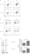
Mice deficient for autophagy genes had reduced iNKT cells. iNKT cells were analyzed by gating on CD1d tetramer+ cells as described in Experimental Procedures. Depicted are the results from cells from the indicated organs from eight-to-nine-week old mice with a CD4-Cre mediated deletion of Atg5 (A,B) or Atg7 (C,D) and controls. Shown are the percentages (A,C) and absolute cell numbers (B,D) compared with Atg5f/f or Atg7f/f mice without the Cre transgene. Representative flow cytometry analyses are from one of at least three independent experiments. *p<0.01, **p<0.005, ***p<0.0001, n≥8.
Figure 2
Figure 2
Deficiency for autophagy genes inhibited iNKT cell maturation. (A,C) Thymus and spleen cells from eight-to-nine-week old mice were analyzed for iNKT cell phenotype. Representative flow cytometry plots from one of four experiments showing gated iNKT cells from Atg5f/f CD4-Cre (A) or Atg7f/f CD4-Cre (C) mice and controls lacking the Cre transgene. Absolute cell numbers of thymic iNKT cells at different stages were shown in (B, Atg5f/f CD4-Cre and D, Atg7f/f CD4-Cre). The analysis of all stages was carried out simultaneously, but because stages 0 and 1 have much fewer cells, for visual clarity their numbers are plotted on a separate graph (left). *p<0.0005, **p<0.0001, n≥10. Error bars are SD.
Figure 3
Figure 3
Cell-intrinsic defects impaired Atg5 deficient iNKT cell development. (A) Bone marrow cells from Atg5f/f CD4-Cre mice or wild type controls (both CD45.2+), together with CD45.1+, wild type bone marrow cells, were co-transferred into recipients. Reconstituted iNKT cells from the indicated organs were analyzed after 11–12 weeks with flow cytometry. Data are representative from one of at least three separate experiments. (B) The ratios of reconstituted iNKT cells (CD45.2+/CD45.1+). *p<0.0001, n>10.
Figure 4
Figure 4
Atg5 or Atg7 deficiency did not cause significant DP thymocyte death and skewed TCR α rearrangements in thymocytes. (A-C) Thymocytes were cultured overnight and cells undergoing apoptosis and dead cells were identified, shown here as flow cytometry analyses (A) and the geometric MFI results indicating cells undergoing apoptosis (B) and dead cells (C). Data in (A) are representative of at least four independent experiments. (B and C) n≥5. Error bars are SD. (D). Equivalent TCR Vα14-Jα18 rearrangements were observed in Atg5 or Atg7 deficient DP thymocytes. cDNA reverse transcribed from total RNA of DP thymocytes depleted of αGalCer-CD1d tetramer+ cells was used as template and titrated at serial dilutions (200 ng, 60 ng and 20 ng) for semi-quantitative PCR. Representative data are from three separate experiments.
Figure 5
Figure 5
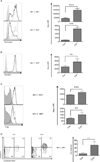
Loss of Atg5 expression led to cell cycle arrest. (A) Atg5 deficient iNKT cells exhibited enhanced incorporation of BrdU. 5 h after injection, thymic iNKT cells were collected and analyzed for BrdU incorporation and Ki-67 expression. (B) Thymic iNKT cells from Atg5f/f CD4-Cre mice accumulated in S-phase. 2 h post injection of BrdU, thymic iNKT cells were stained with 7-AAD and anti-BrdU antibody to evaluate cell cycle progression. (C) Increased expression of p21cip1 in Atg5 deficient iNKT cells. Shown are the representative flow cytometry analysis (left) and geometric MFI (right) of data pooled from at least three independent experiments gating on either NK1.1 or NK1.1+, CD1d tetramer+ thymocytes. Filled histogram: isotype control; solid line: wild type mice; dotted line: Atg5f/f CD4-Cre mice. * p<0.01, n≥4. Error bars are SD.
Figure 6
Figure 6
Atg5 deletion resulted in increased mitochondria, super oxide and cell death. Augmented mitochondrial mass (A) and mitochondrial superoxide production (B) in Atg5 deleted, thymic, NK1.1 and NK1.1+iNKT cells. (C) Higher expression of Fas on Atg5 deficient iNKT cells compared to wild type controls. At least four independent experiments were performed with consistent results, both representative flow cytometry plots (left) and geometric MFI values (right) are shown. Filled histogram: isotype control; solid line: wild type mice; dotted line: Atg5f/f CD4-Cre mice. (D) Dramatically increased apoptosis leading to cell death for NK1.1+ thymic iNKT cells devoid of Atg5. Cells were analyzed after overnight culture and compared with wild type controls. Representative flow cytometry analyses from three separate experiments (left) and geometric MFI (right) of dead cells (Annexin V+, Live/Dead Yellow+) are shown. *p<0.05, **p<0.01, ***p<0.001, ****p<0.0001, n≥4. Error bars are SD.
Figure 7
Figure 7
Atg5 deficient thymic iNKT cells exhibited an increase in active caspase 8 and caspase 3. Atg5 deleted thymocytes were cultured overnight, followed by analysis by flow cytometry of active caspase 8 (A and B) and active caspase 3 (C and D) in thymic iNKT cells (A and C) and TCRβ+ thymocytes (B and D). Shown here are both flow cytometry plots (left) and geometric MFI (right). Data are representative of at least two independent experiments. Filled histogram: isotype control; solid line: wild type cells; dotted line: Atg5 deficient. *p<0.05, n≥3. Error bars are SD.
Figure 8
Figure 8
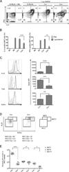
Atg5 deficiency differentially affected iNKT cell functional subsets. (A and B) Impaired iNKT cell activation and polarized cytokine outcome in Atg5f/f CD4-Cre mice after αGalCer immunization. Intracellular cytokine production in splenic iNKT cells was analyzed 2 h after αGalCer injection. Flow cytometry plots (A), geometric MFI of intracellular IL-4 and IFNγ (B) are representative of three independent experiments. (C) Altered expression profile of transcription factors in autophagy deficient thymic iNKT cells. Representative flow cytometry plots from three independent experiments and geometric MFI are indicated. Filled histogram: isotype control; solid line: wild type mice; dotted line: Atg5f/f CD4-Cre mice. (D) iNKT cell subset analysis in autophagy deficient mice compared with wild type controls. Representative flow cytometry plots of three independent experiments are shown. (E) Absolute cell number of thymic iNKT cell functional subsets defined by transcription factor expression levels in Atg5f/f CD4-Cre mice and controls. *p<0.05, **p<0.01, ***p<0.001, ****p<0.0001, n≥4. Error bars are SD.
Figure 9
Figure 9
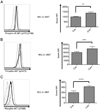
Unbalanced proliferative and apoptotic signaling in Atg5 deficient iNKT cells. Thymic iNKT cells were analyzed ex vivo for activation of mTOR signaling pathway. Phosphorylation of mTORC1 substrate, 4E–BP1 (pT37/pT46) (A), and phosphorylation of mTORC2 substrate, AKT(pS473) (B), were analyzed. (C) Phosphorylation of AKT at T308 was measured to evaluate the PI3K/PDK1 signaling pathway. Both Cre- and Cre+ were pooled from at least four samples. Shown here is the representative flow cytometry analysis of three independent experiments. Filled histogram: isotype control; solid line: wild type mice; dotted line: Atg5f/f CD4-Cre mice. *p<0.05, **p<0.01, ***p<0.005, ****p<0.0005, n≥4. Error bars are SD.

References

    1. Mizushima N, Yoshimori T, Ohsumi Y. The role of Atg proteins in autophagosome formation. Annual review of cell and developmental biology. 2011;27:107–132. - PubMed (V体育官网入口)
    1. Weidberg H, Shvets E, Elazar Z. Biogenesis and cargo selectivity of autophagosomes. Annual review of biochemistry. 2011;80:125–156. - PubMed
    1. Mizushima N, Komatsu M. Autophagy: renovation of cells and tissues. Cell. 2011;147:728–741. - "VSports" PubMed
    1. Virgin HW, Levine B. Autophagy genes in immunity. Nature immunology. 2009;10:461–470. - PMC - PubMed
    1. Levine B, Mizushima N, Virgin HW. Autophagy in immunity and inflammation. Nature. 2011;469:323–335. - PMC (V体育ios版) - PubMed

Publication types

MeSH terms (V体育官网)