Skip to main page content
U.S. flag

An official website of the United States government

Dot gov

The . gov means it’s official. Federal government websites often end in VSports app下载. gov or . mil. Before sharing sensitive information, make sure you’re on a federal government site. .

Https

The site is secure. The https:// ensures that you are connecting to the official website and that any information you provide is encrypted and transmitted securely V体育官网. .

. 2015 May 29;290(22):14120-9.
doi: 10.1074/jbc.M114.633057. Epub 2015 Apr 20.

Rictor Undergoes Glycogen Synthase Kinase 3 (GSK3)-dependent, FBXW7-mediated Ubiquitination and Proteasomal Degradation

Affiliations

Rictor Undergoes Glycogen Synthase Kinase 3 (GSK3)-dependent, FBXW7-mediated Ubiquitination and Proteasomal Degradation

Junghui Koo et al. J Biol Chem. .

Abstract

Rictor, an essential component of mTOR complex 2 (mTORC2), plays a pivotal role in regulating mTOR signaling and other biological functions. Posttranslational regulation of rictor (e. g. via degradation) and its underlying mechanism are largely undefined and thus are the focus of this study. Chemical inhibition of the proteasome increased rictor ubiquitination and levels. Consistently, inhibition of FBXW7 with various genetic means including knockdown, knock-out, and enforced expression of a dominant-negative mutant inhibited rictor ubiquitination and increased rictor levels, whereas enforced expression of FBXW7 decreased rictor stability and levels. Moreover, we detected an interaction between FBXW7 and rictor. Hence, rictor is degraded through an FBXW7-mediated ubiquitination/proteasome mechanism. We show that this process is dependent on glycogen synthase kinase 3 (GSK3): GSK3 was associated with rictor and directly phosphorylated the Thr-1695 site in a putative CDC4 phospho-degron motif of rictor; mutation of this site impaired the interaction between rictor and FBXW7, decreased rictor ubiquitination, and increased rictor stability VSports手机版. Finally, enforced activation of Akt enhanced rictor levels and increased mTORC2 activity as evidenced by increased formation of mTORC2 and elevated phosphorylation of Akt, SGK1, and PKCα. Hence we suggest that PI3K/Akt signaling may positively regulate mTORC2 signaling, likely through suppressing GSK3-dependent rictor degradation. .

Keywords: Akt PKB; FBXW7; GSK3; mTOR complex (mTORC); protein degradation; rictor; signal transduction; ubiquitylation (ubiquitination). V体育安卓版.


Figures

FIGURE 1.
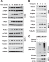
FIGURE 1.
Rictor is regulated by ubiquitination/proteasome-mediated degradation. A and B, both H460 and MCF-7 cells were treated with 10 μm MG132 (A) or 1 μm MLN4924 (B) for the indicated times and then harvested for preparation of WCLs and subsequent WB to detect the given proteins. C, H460 cells were transfected with HA-ubiquitin (HA-Ub) or empty vector for 48 h and then treated with DMSO or 10 μm MG132 for an additional 2 h. The WCLs prepared from these cells were then immunoprecipitated with anti-rictor antibody, and ubiquitinated rictor (Ub-rictor) was detected by WB using anti-HA antibody.
FIGURE 2.
FIGURE 2.
Inhibition of FBXW7 increases rictor levels (A–C) with increased stability (D) and reduced ubiquitination (E). A, WCLs were prepared from the indicated cell lines. B, twenty-four hours after transfection with control or FBXW7 siRNA, H460 cells were further transfected with FLAG-FBXW7β plasmid. After an additional 48 h, the cells were harvested for preparation of WCLs. For HCT116 cells, WCLs were prepared after transfection of control or FBXW7 siRNA for 48 h. C, 293T cells were transfected with different amounts of dnFBXW7 as indicated and then harvested after 24 h for preparation of WCLs. WB was conducted to detect the indicated proteins in the WCLs. D, the indicated cells line were exposed to CHX (10 μg/ml) and then harvested at the different time points as indicated for preparation of WCLs. The indicated proteins were detected with WB and quantified with NIH ImageJ software (Bethesda, MA). Rictor levels were normalized to tubulin and plotted as the relative rictor levels compared with those at time 0 of CHX treatment (right panel). E, 24 h after transfection with control or FBXW7 siRNA, H460 cells were further transfected with HA-ubiquitin or empty vector for an additional 48 h. The cells were then treated with 10 μm MG132 for 2 h and harvested for preparation of WCLs. These lysates were then immunoprecipitated with an anti-rictor antibody, and ubiquitinated rictor (Ub-rictor) was determined by WB using an anti-HA antibody. The asterisk indicates IgG heavy chain.
FIGURE 3.
FIGURE 3.
Enforced expression of FBXW7 decreases rictor levels (A and B) and stability (C). A and B, 293T cells were transfected with empty vector, FLAG-FBXW7α, -β, or -γ plasmids (A) or different amounts of FLAG-FBXW7β as indicated (B). After 24 h, the cells were harvested for preparation of WCLs and subsequent WB to detect the indicted proteins. C, 293T cells were co-transfected with myc-rictor plus FLAG-FBXW7β or empty vector. After 24 h, the cells were treated with CHX (10 μg/ml) and then harvested at the different time points as indicated for preparation of WCLs. The indicated proteins were detected with WB. Protein levels were quantified with NIH ImageJ software (Bethesda, MA) and were normalized to actin. The results were plotted as the relative rictor levels compared with those at time 0 of CHX treatment (bottom panel).
FIGURE 4.
FIGURE 4.
FBXW7 interacts with rictor. 293T cells were transfected with FLAG-FBXW7β plasmid (A, B, and D), empty vector (V), FLAG-dnFBXW7 (dn), or FLAG-FBXW7β (WT) plasmid (C). After 48 h the cells were harvested for preparation of WCLs followed by IP with an anti-FLAG (A and C) or anti-rictor antibody (B) and subsequent WB to detect the indicated proteins. In addition, the WCLs were also treated with λ-phosphatase (1000 units) at 30 °C for 1 h before the indicated IP-WB experiments (D).
FIGURE 5.
FIGURE 5.
Identification of a putative CPD in rictor (A) and demonstration of its function in FBXW7-mediated rictor degradation (B–E). A, a putative CPD motif was identified at positions 1693–1704 of rictor. B and D, 293T cells were co-transfected with FLAG-FBXW7 and myc-rictor or myc-rictor (T1695G) for 24 h (B) or 48 h (D). The cells were then harvested for preparation of WCLs and subsequent WB (B) or IP/WB (D). C, 293T cells were co-transfected with FLAG-FBXW7 and myc-rictor or myc-rictor (T1695G) and 24 h later treated with 10 μg/ml CHX. At the indicated time points, the cells were harvested for preparation of WCLs. The indicated proteins were detected with WB and quantified with NIH Image J. After being normalized to actin, the results were then plotted as the relative protein levels compared with those at time 0 of CHX treatment (bottom panel). E, FBXW7β was co-expressed with myc-rictor or myc-rictor (T1695G) in 293T cells, and after 24 h, cells were transfected with an empty vector (V) or HA-ubiquitin (HA-Ub). After another 24 h, cells were treated with 10 μm MG132 for 2 h and then subjected to preparation of WCLs and subsequent IP/WB to detect ubiquitinated rictor.
FIGURE 6.
FIGURE 6.
Modulation of GSK3 activity alters rictor levels (A–C) and ubiquitination (E and F). A, H460 and MCF-7 cells were treated with the indicated concentrations of SB216763 or CHIR99021 for 6 h. B, H460 or 293T cells were transfected with control (Ctrl) and GSG3α/β siRNA and harvested after 48 h. C, H460 cells were transfected with control or FBXW7 siRNA and after 48 h treated with 50 mm LiCl or 20 μm SB216763 for an additional 6 h. D, 293T cells were transfected with different amounts of GSK3α or GSK3β as indicated. After 24 h the cells were harvested for preparation of WCLs and subsequent WB. E, H460 cells were transfected with vector or HA-ubiquitin (HA-Ub) and after 24 h were treated with 50 mm LiCl for 4 h followed by exposure to 10 μm MG132 for another 2 h. F, H460 cells were transfected with control (Ctrl) or GSK3α siRNA followed after 24 h with transfection of vector or HA-Ub plasmid. After 24 h the cells were treated with 10 μm MG132 for an additional 2 h. After the aforementioned treatments in E and F, the cells were harvested for preparation of WCLs for subsequent IP/WB to detect the indicated proteins or protein ubiquitination.
FIGURE 7.
FIGURE 7.
GSK3 interacts with rictor (A and B) and phosphorylates rictor at Thr1965 (C and D). A and B, 293T cells were transfected with Myc-GSK3β or FLAG-GSK3α, and after 48 h IP was performed for GSK3β with an anti-Myc antibody (A) or GSK3α with an anti-FLAG antibody (B) followed by WB to detect the given proteins. C and D, phosphorylation of purified GST-tagged RF1 and RF1m by GSK3 β (C) or GSK3 α (D) was carried out by mixing 1 μCi of [γ-32P]ATP with kinase buffer and incubating at 37 °C for 30 min. β-Catenin (positive control (PC)), non-substrate (negative control (NC)), and GST-only protein (GST) were used as controls. The protein mixtures were separated by 10% SDS-PAGE gels to detect phosphorylation by direct exposure to x-ray film and GST-tagged protein by WB with an anti-GST antibody.
FIGURE 8.
FIGURE 8.
Enforced activation of Akt increases the levels of rictor and phosphorylation of Akt, SGK1, and PKCα (A) and enhances the assembly of the mTORC2 (B); these data suggest a model for Akt positive regulation of the mTORC2 activity and signaling (C). A and B, The given cell lines were infected with the indicated adenoviruses for 48 h and then harvested for preparation of WCLs and subsequent WB to detect proteins as indicated (A), The WCLs were also used for IP with mTOR antibody followed with WB to detect the indicated proteins (B). C, a working model for Akt positive regulation of the mTORC2. Akt may enhance the assembly and activity of the mTORC2 by inhibiting GSK3-dependent rictor degradation.

"V体育官网" References

    1. Guertin D. A., Stevens D. M., Saitoh M., Kinkel S., Crosby K., Sheen J. H., Mullholland D. J., Magnuson M. A., Wu H., Sabatini D. M. (2009) mTOR complex 2 is required for the development of prostate cancer induced by Pten loss in mice. Cancer Cell 15, 148–159 - VSports app下载 - PMC - PubMed
    1. Sparks C. A., Guertin D. A. (2010) Targeting mTOR: prospects for mTOR complex 2 inhibitors in cancer therapy. Oncogene 29, 3733–3744 - PMC - PubMed
    1. Roulin D., Cerantola Y., Dormond-Meuwly A., Demartines N., Dormond O. (2010) Targeting mTORC2 inhibits colon cancer cell proliferation in vitro and tumor formation in vivo. Mol Cancer 9, 57. - VSports app下载 - PMC - PubMed
    1. Lee K., Nam K. T., Cho S. H., Gudapati P., Hwang Y., Park D. S., Potter R., Chen J., Volanakis E., Boothby M. (2012) Vital roles of mTOR complex 2 in Notch-driven thymocyte differentiation and leukemia. J. Exp. Med. 209, 713–728 - PMC - PubMed
    1. Gao D., Wan L., Inuzuka H., Berg A. H., Tseng A., Zhai B., Shaik S., Bennett E., Tron A. E., Gasser J. A., Lau A., Gygi S. P., Harper J. W., DeCaprio J. A., Toker A., Wei W. (2010) Rictor forms a complex with Cullin-1 to promote SGK1 ubiquitination and destruction. Mol. Cell 39, 797–808 - VSports注册入口 - PMC - PubMed

Publication types

MeSH terms