Skip to main page content
U.S. flag

An official website of the United States government

Dot gov

The . gov means it’s official VSports app下载. Federal government websites often end in . gov or . mil. Before sharing sensitive information, make sure you’re on a federal government site. .

Https

The site is secure. The https:// ensures that you are connecting to the official website and that any information you provide is encrypted and transmitted securely. V体育官网.

. 2015 Feb 19;34(1):18.
doi: 10.1186/s13046-015-0136-7.

Long noncoding RNA CCAT1 promotes hepatocellular carcinoma progression by functioning as let-7 sponge

Affiliations

Long noncoding RNA CCAT1 promotes hepatocellular carcinoma progression by functioning as let-7 sponge

Liang Deng et al. J Exp Clin Cancer Res. .

VSports在线直播 - Abstract

Background: Long noncoding RNAs (lncRNAs) have been identified as having functional roles in cancer biology and are deregulated in many cancers VSports手机版. The present study aimed to determine the expression, roles and functional mechanisms of a long noncoding RNA CCAT1 in the progression of hepatocellular carcinoma (HCC). .

Methods: CCAT1 expression levels in 66 pairs of HCC tissues and pair-matched noncancerous hepatic tissues were tested by real-time PCR. The effects of CCAT1 on HCC cells proliferation and migration were assessed using in vitro cell proliferation and migration assays. A computational screen of microRNAs (miRNAs) target sites in CCAT1 was conducted to search for specific miRNAs binding to CCAT1. The specific binding between CCAT1 and miRNAs was confirmed by RNA immunoprecipitation assay combined with luciferase reporter assay V体育安卓版. .

Results: CCAT1 levels are markedly increased in HCC tissues compared with pair-matched noncancerous hepatic tissues V体育ios版. Up-regulation of CCAT1 is correlated with tumor size, microvascular invasion, AFP and poor prognosis. CCAT1 promotes the proliferation and migration of HCC cells. CCAT1 functions as a molecular sponge for let-7, antagonizes its functions, and leads to the de-repression of its endogenous targets HMGA2 and c-Myc. The effect of CCAT1 on HCC cell proliferation and migration is dependent upon its competitively binding to let-7. .

Conclusions: These data suggest that CCAT1 plays a pivotal role in HCC progression via functioning as let-7 sponge, and implicate the potential application of CCAT1 for the prognosis and treatment of HCC VSports最新版本. .


"V体育官网" Figures
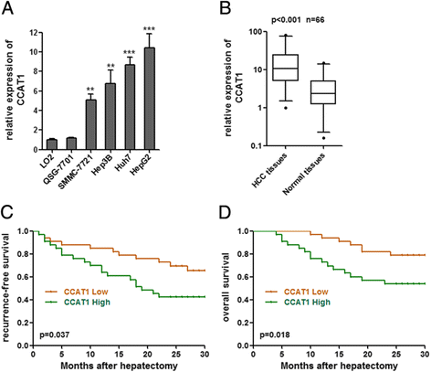
Figure 1
Figure 1
CCAT1 expression in HCC and its association with patients’ prognosis. (A) Expression of CCAT1 in four HCC cell lines and two liver normal cell lines. Data are presented as mean ± standard error based on at least three independent experiments. **p < 0.01, ***p < 0.001. (B) Differences in CCAT1 expression levels between HCC tissues and pair-matched noncancerous hepatic tissues. The expression of CCAT1 was normalized to that of 18S rRNA, which is an abundant and constitutively expressed non-coding RNA. Statistical differences between samples were analyzed using the Wilcoxon signed-rank test (n = 66, p < 0.001). (C, D) Kaplan–Meier analyses of correlations between the CCAT1 expression level and recurrence-free survival (C) or overall survival (D) of 66 HCC patients. The median expression level was used as the cut-off.
Figure 2
Figure 2
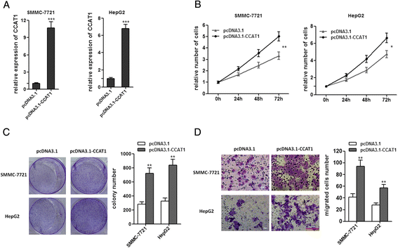
Enforced expression of CCAT1 promotes the proliferation and migration of HCC cells. (A) The expression of CCAT1 in SMMC-7721 and HepG2 cells with CCAT1 stably overexpressed. (B) CCAT1 promotes the proliferation of SMMC-7721 and HepG2 cells. Cell number was determined by the CCK-8 assay, and the relative number of cells to 0 h is presented. (C) The colony number of CCAT1 overexpressed SMMC-7721 and HepG2 cells per well in 6-well plates. (D) CCAT1 promotes cell migration by transwell assays. All values are presented as mean ± standard error based on at least three independent experiments. *p < 0.05, **p < 0.01, ***p < 0.001.
Figure 3
Figure 3
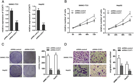
Attenuated expression of CCAT1 inhibits the proliferation and migration of HCC cells. (A) The expression of CCAT1 in SMMC-7721 and HepG2 cells with CCAT1 stably repressed. (B) Repression of CCAT1 decreases the proliferation of SMMC-7721 and HepG2 cells. Cell number was determined by the CCK-8 assay, and the relative number of cells to 0 h is presented. (C) The colony number of CCAT1 repressed SMMC-7721 and HepG2 cells per well in 6-well plates. (D) Repression of CCAT1 decreased cell migration by transwell assays. All values are presented as mean ± standard error based on at least three independent experiments. *p < 0.05, **p < 0.01, ***p < 0.001.
Figure 4
Figure 4
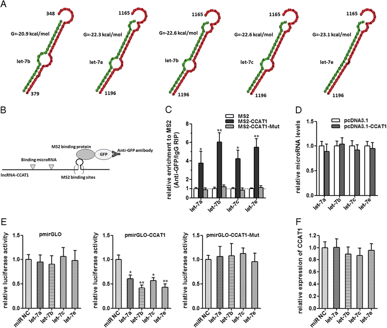
CCAT1 acts as a molecular sponge for let-7. (A) Bioinformatics predicted let-7 binding sites at two distinct positions in CCAT1. Partial sequences of CCAT1 and sequences of four let-7 subtypes are shown. Numbers are in nucleotides relative to the transcriptional start site of CCAT1. (B) Schematic outlining the MS2-RIP strategy to validate endogenous miRNA:CCAT1 binding. (C) MS2-RIP followed by miRNA qRT-PCR to detect miRNAs endogenously associated with CCAT1. (D) Expression levels of let-7 after the transfection of pcDNA3.1-CCAT1 or pcDNA3.1 into SMMC-7721 cells. (E) Luciferase activity in SMMC-7721 cells cotransfected with let-7 and luciferase reporters containing nothing, CCAT1 or mutant CCAT1. Data are presented as the relative ratio of firefly luciferase activity to renilla luciferase activity. (F) CCAT1 expression levels after the transfection of let-7 mimics or the miRNA negative control into SMMC-7721 cells. All values are presented as mean ± standard error based on at least three independent experiments. *p < 0.05, **p < 0.01.
Figure 5
Figure 5
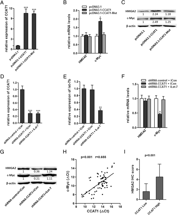
Regulation of endogenous let-7 targets by CCAT1. (A) The expression of CCAT1 in stable SMMC-7721 cell clones. (B) HMGA2 and c-Myc mRNA levels in stable SMMC-7721 cell clones. (C) HMGA2 and c-Myc protein levels in stable SMMC-7721 cell clones. (D, E) The expression of CCAT1(D) or let-7b (E) in indicated HepG2 cell clones after transfection of let-7 inhibitor. (F) HMGA2 and c-Myc mRNA levels in stable HepG2 cell clones. (G) HMGA2 and c-Myc mRNA levels in stable HepG2 cell clones. For A-G, all values are presented as mean ± standard error based on at least three independent experiments. *p < 0.05, **p < 0.01, ***p < 0.001. (H) The correlation between CCAT1 transcript level and c-Myc mRNA level was measured in 66 HCC tissues. The delta-Ct values (normalized to 18S rRNA) were subjected to Pearson correlation analysis. (I) Immunohistochemical scores of CCAT1 in HCC tissues of different CCAT1 expression (Mann–Whitney test, p < 0.001).
Figure 6
Figure 6
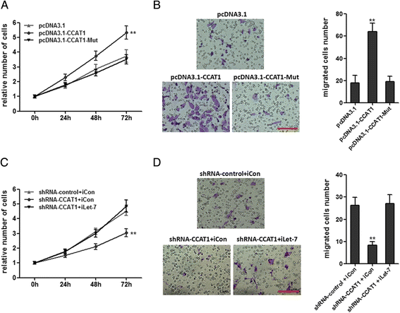
CCAT1 promotes HCC progression by competitively binding let-7. (A) The mutation of let-7 binding sites attenuated the promoting proliferation effect of CCAT1 in SMMC-7721 cells. Cell number was determined by the CCK-8 assay, and the relative number of cells to 0 h is presented. (B) The mutation of let-7 binding sites attenuated the promoting migration effect of CCAT1 in SMMC-7721 cells by transwell assays. (C) Repression of let-7 overcame the inhibitory effects of decreasing CCAT1 on cell proliferation by the CCK-8 assay. (D) Repression of let-7 overcame the inhibitory effects of decreasing CCAT1 on cell migration by transwell assays. All values are presented as mean ± standard error based on at least three independent experiments. **p < 0.01, ***p < 0.001.

References

    1. Jemal A, Bray F, Center MM, Ferlay J, Ward E, Forman D. Global cancer statistics. CA Cancer J Clin. 2011;61:69–90. doi: 10.3322/caac.20107. - DOI - PubMed
    1. Xu X, Fan Z, Kang L, Han J, Jiang C, Zheng X, et al. Hepatitis B virus X protein represses miRNA-148a to enhance tumorigenesis. J Clin Invest. 2013;123:630–45. - PMC - PubMed
    1. Yang JD, Roberts LR. Hepatocellular carcinoma: A global view. Nat Rev Gastroenterol Hepatol. 2010;7:448–58. doi: 10.1038/nrgastro.2010.100. - DOI - PMC - PubMed
    1. Forner A, Llovet JM, Bruix J. Hepatocellular carcinoma. Lancet. 2012;379:1245–55. doi: 10.1016/S0140-6736(11)61347-0. - DOI - PubMed
    1. Fan MQ, Huang CB, Gu Y, Xiao Y, Sheng JX, Zhong L. Decrease expression of microRNA-20a promotes cancer cell proliferation and predicts poor survival of hepatocellular carcinoma. J Exp Clin Cancer Res. 2013;32:21. doi: 10.1186/1756-9966-32-21. - DOI - PMC - PubMed

MeSH terms

Substances