Skip to main page content
U.S. flag

An official website of the United States government

Dot gov

The . gov means it’s official. Federal government websites often end in . gov or . mil. Before sharing sensitive information, make sure you’re on a federal government site VSports app下载. .

Https

The site is secure. The https:// ensures that you are connecting to the official website and that any information you provide is encrypted and transmitted securely V体育官网. .

. 2015 Dec;22(12):1957-69.
doi: 10.1038/cdd.2015.43. Epub 2015 Apr 17.

Mitofusins deficiency elicits mitochondrial metabolic reprogramming to pluripotency

Affiliations

"V体育ios版" Mitofusins deficiency elicits mitochondrial metabolic reprogramming to pluripotency

M J Son et al. Cell Death Differ. 2015 Dec.

Abstract

Cell reprogramming technology has allowed the in vitro control of cell fate transition, thus allowing for the generation of highly desired cell types to recapitulate in vivo developmental processes and architectures. However, the precise molecular mechanisms underlying the reprogramming process remain to be defined. Here, we show that depleting p53 and p21, which are barriers to reprogramming, yields a high reprogramming efficiency. Deletion of these factors results in a distinct mitochondrial background with low expression of oxidative phosphorylation subunits and mitochondrial fusion proteins, including mitofusin 1 and 2 (Mfn1/2). Importantly, Mfn1/2 depletion reciprocally inhibits the p53-p21 pathway and promotes both the conversion of somatic cells to a pluripotent state and the maintenance of pluripotency. Mfn1/2 depletion facilitates the glycolytic metabolic transition through the activation of the Ras-Raf and hypoxia-inducible factor 1α (HIF1α) signaling at an early stage of reprogramming. HIF1α is required for increased glycolysis and reprogramming by Mfn1/2 depletion VSports手机版. Taken together, these results demonstrate that Mfn1/2 constitutes a new barrier to reprogramming, and that Mfn1/2 ablation facilitates the induction of pluripotency through the restructuring of mitochondrial dynamics and bioenergetics. .


VSports最新版本 - Figures
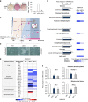
Figure 1
Figure 1
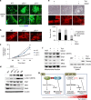
Mitochondrial function is downregulated during early-stage reprogramming of p53- and p21-KO somatic cells. (a) WT, p53−/−, and p21−/− MEFs were reprogrammed via retroviral transduction of the OSKM reprogramming factors. Representative images of AP+ colonies (left). The total numbers of AP+ colonies were determined on day 11 (D11) of reprogramming (right). (b) The stages of sample preparation for the transcriptome and metabolome analyses are depicted. (c) Representative cell morphology on D7 of reprogramming. (d) The expression of genes encoding major enzymes (left) and the relative quantities of each metabolite (right) related to glycolysis were determined through real-time PCR analysis and capillary electrophoresis time-of-flight mass spectrometry, respectively. The fold changes of metabolites in p53−/− and p21−/− reprogramming cultures compared with the WT control at D7 are represented by a color-coded index bar. (e) Transcriptome analysis of mitochondrial function in OSKM-transduced WT, p53−/−, and p21−/− MEFs on D7 of reprogramming. (f) Real-time PCR analysis of the mitochondria-encoded OXPHOS subunits ND1 and Atp6ap1 and the mitochondrial fusion genes Mfn1/2 in WT, p53−/−, and p21−/− D7 reprogramming cultures. The data are presented as the mean±S.E. (n= 3). *P<0.05; **P<0.01 (Student's t-test). Scale bar=50 μm
Figure 2
Figure 2
p53- and p21-KO cells and pluripotent reprogramming intermediates express low levels of mitochondrial fusion proteins. (a) OSKM-transduced WT, p53−/−, and p21−/− MEFs on D7 of reprogramming were stained with Tom20 (mitochondria, green) and DAPI (nuclei, blue) (top). High-magnification images (bottom). (b) Mitochondrial morphology of WT, p53−/−, and p21−/− MEFs stained with Mitotracker (red) and enlarged images (inset in top right corner). (c) Cell proliferation of WT, p53−/−, and p21−/− MEFs were determined by cell number counting. (d) Western blot analysis of Cyclin B1 and mitochondrial fission (Drp)-fusion (Mfn) components. β-actin was used as an internal control. (e) Reprogramming intermediates were sorted based on Thy1 and SSEA1 expression using MACS on D11. Representative images of each subgroup (top), the mitochondrial morphology stained with Mitotracker (red) (middle), and enlarged images (inset in middle right corner) on D14. The percentages of cells with fragmented/intermediate/fused mitochondria were determined in each subgroup (bottom). (f) Western blot analysis of mitochondrial fission (Drp)-fusion (Mfn) components in the mitochondrial fraction and of the pluripotency marker Nanog in the whole-cell lysates of MEFs and each subpopulation. HSP60 and β-actin were used as internal controls. (g) Schematic presentation of the p53- and p21-KO cells. Scale bar=50 μm
Figure 3
Figure 3
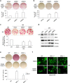
Mfn1/2 ablation promotes the acquisition and maintenance of pluripotency. (a) MEFs and (b) HFFs were transduced with control (shCon), Mfn1 (shMfn1), and Mfn2 (shMfn2) lentiviral shRNAs together with retroviral OSKM reprogramming factors. Representative images of AP+ colonies (each top). The total numbers of AP+ colonies were determined on D11 (a) and D28 (b) of reprogramming, respectively (each bottom). (c) H9 hESCs were transfected with control (siCon), Mfn1 (siMfn1), and Mfn2 (siMfn2) siRNAs and cultured with unconditioned medium (UM; differentiated). Conditioned medium (CM)-cultured hESCs were used as an undifferentiated control. Representative images of AP+ colonies (top) and the relative fold difference in AP+ colonies under feeder-free conditions on D5 (bottom). (d) Western blot analysis of Mfn1/2 and the pluripotency markers Oct3/4 and Nanog in siRNA-transfected hESCs on D5. β-actin was used as an internal control. (e) WT, Mfn1−/−, and Mfn2−/− MEFs were reprogrammed via retroviral transduction of OSKM in the absence (top) and presence (middle) of 50 μM Mdivi1, an inhibitor of mitochondrial division. Representative images of AP+ colonies (top and middle). The total numbers of AP+ colonies were determined on D11 of reprogramming (bottom). (f) The mitochondrial morphology of each reprogramming culture was observed using stably expressed Mito-EYFP (green) on D7. Enlarged images (inset in right corner). The data are presented as the mean±S.E. (n=3). *P<0.05; **P<0.01 (Student's t-test). Scale bar=50 μm
Figure 4
Figure 4
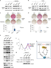
Mfn1/2 knockdown facilitates glycolytic conversion in early-stage reprogramming. (a) Transcriptome analysis of gene sets related to glycolysis in MEFs transduced with OSKM and the indicated shRNAs on D7 of reprogramming. The ratios are indicated by a color-coded index bar. (b) The expression of genes encoding major enzymes (left) and the relative quantity of each metabolite (right) related to glycolysis were determined via real-time PCR analysis and capillary electrophoresis time-of-flight mass spectrometry, respectively. The fold changes of metabolites in Mfn1/2 shRNA-transduced reprogramming cultures compared with the control at D7 are represented by a color-coded index bar. (c) Lactate production was determined in the cell lysates of each group. The data are presented as the mean±S.E. (n=3). *P<0.05; **P<0.01; ***P<0.001 (Student's t-test)
Figure 5
Figure 5
Reciprocal inhibition of p53/p21 and Mfn1/2 activates the Ras-Raf-HIF1α pathway. (a) WT, Mfn1−/−, and Mfn2−/− MEFs were reprogrammed via retroviral transduction of OSKM in the absence and presence of 25 μM Nutlin3a, an MDM2 inhibitor that stabilizes p53. Western blot analysis of p53 and p21 on D11 of reprogramming. (b) WT, p53−/−, and p21−/− MEFs were reprogrammed via retroviral transduction of OSKM with or without retroviral Mfn1 overexpression. Western blot analysis of Mfn1/2 on D11 of reprogramming. (c and d) Representative images of AP+ colonies (top and middle) and the total numbers of AP+ colonies (bottom) were obtained in each indicated group. (e) Western blot analysis of the indicated Ras-Raf signaling proteins in MEFs transduced with OSKM and Mfn1/2 shRNAs on D7 of reprogramming. (f) Cellular ROS levels in MEFs transduced with OSKM and Mfn1/2 shRNAs on D7 of reprogramming. (g) Western blot analysis of HIF1α in Mfn1/2 knockdown reprogramming cultures absence and presence of indicated concentration of N-acetylcysteine. (h) Model showing the activation of Ras-Raf-HIF1α signaling and ROS-mediated HIF1α stabilization in Mfn1/2-depleted cells. β-actin was used as an internal control. The data are presented as the mean±S.E. (n=3). *P<0.05; **P<0.01 (Student's t-test)
Figure 6
Figure 6
Increased glycolysis and reprogramming by Mfn1/2 knockdown is HIF1α-dependent. (a) MEFs were transduced with OSKM and the indicated shRNAs. Western blot analysis was performed on D7 of reprogramming. β-actin was used as an internal control. (b) Lactate production was determined in the cell lysates of each group. (c) Representative images of AP+ colonies (top and middle) and the total numbers of AP+ colonies (bottom) were obtained in each indicated group. The data are presented as the mean±S.E. (n=3). *P<0.05; **P<0.01; ***P<0.001 (Student's t-test)
Figure 7
Figure 7
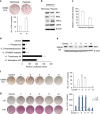
Hypoxia decreases Mfn1/2 expression, and chemicals that inhibit Mfn1 expression promote the acquisition and maintenance of stem cell fate. (a) MEFs were reprogrammed via the retroviral transduction of OSKM under normoxia (N) and hypoxia (H, 5% O2). Representative images of AP+ colonies (top). The total number of AP+ colonies was counted on D11 (bottom). (b) Western blot analysis of Mfn1/2, HIF1α, and LDHA on D7 of reprogramming in cultures under normoxia and hypoxia. (c) Mfn1 promoter activity was determined in Mfn1−/− MEFs stably expressing an Mfn1 promoter reporter construct after 48 h of normoxic or hypoxic culture. (d) Relative luciferase activity was determined in Mfn1−/− MEFs carrying the Mfn1 promoter reporter, at 48 h after treatment with 84 redox library compounds. The top three downregulated (−1, −2, and −3) and the top two upregulated (+1 and +2) hits controlling Mfn1 promoter activity are presented. (e) MEFs were reprogrammed via retroviral transduction of OSKM together with treatment with the selected top hits, and Mfn1 protein expression was determined on D9 of reprogramming by western blot analysis. (f) Representative images of AP+ colonies were obtained (left) and the total number of AP+ colonies was counted (right) on D11 of reprogramming under treatment with the selected top hits. (g) J1 mESCs were cultured with (+ self-renewing condition, bottom left) or without LIF (− non-self-renewing condition, top left). Representative images of AP+ colonies were obtained (left) and the total number of AP+ colonies was counted (right) on D5 under treatment with the selected top hits. β-actin was used as an internal control. The data are presented as the mean±S.E. (n=3). *P<0.05; **P<0.01 (Student's t-test)
Figure 8
Figure 8
Proposed model for the control of cellular stability and plasticity via the reciprocal interaction of the p53/p21 and Mfn1/2 pathways. Depletion of Mfn1/2 promoted somatic cell reprogramming (top), which increases plasticity, allowing reprogramming barriers, such as mitochondrial fusion, cell cycle arrest, and/or failure of metabolic reprogramming, to be overcome (bottom)

References

    1. Takahashi K, Yamanaka S. Induction of pluripotent stem cells from mouse embryonic and adult fibroblast cultures by defined factors. Cell 2006; 126: 663–676. - PubMed
    1. Theunissen TW, Jaenisch R. Molecular Control of Induced Pluripotency. Cell Stem Cell 2014; 14: 720–734. - PMC - PubMed
    1. Buganim Y, Faddah DA, Cheng AW, Itskovich E, Markoulaki S, Ganz K et al. Single-cell expression analyses during cellular reprogramming reveal an early stochastic and a late hierarchic phase. Cell 2012; 150: 1209–1222. - PMC - PubMed
    1. Kawamura T, Suzuki J, Wang YV, Menendez S, Morera LB, Raya A et al. Linking the p53 tumour suppressor pathway to somatic cell reprogramming. Nature 2009; 460: 1140–1144. - V体育平台登录 - PMC - PubMed
    1. Utikal J, Polo JM, Stadtfeld M, Maherali N, Kulalert W, Walsh RM et al. Immortalization eliminates a roadblock during cellular reprogramming into iPS cells. Nature 2009; 460: 1145–1148. - PMC - PubMed

"V体育2025版" Publication types

MeSH terms

V体育官网 - Substances