Skip to main page content
U.S. flag

An official website of the United States government

Dot gov

The . gov means it’s official. Federal government websites often end in . gov or . mil VSports app下载. Before sharing sensitive information, make sure you’re on a federal government site. .

Https

The site is secure. The https:// ensures that you are connecting to the official website and that any information you provide is encrypted and transmitted securely V体育官网. .

. 2015 Apr 28;112(17):5425-30.
doi: 10.1073/pnas.1501555112. Epub 2015 Apr 13.

VSports app下载 - Metabolic reprogramming in triple-negative breast cancer through Myc suppression of TXNIP

Affiliations

Metabolic reprogramming in triple-negative breast cancer through Myc suppression of TXNIP

Liangliang Shen et al. Proc Natl Acad Sci U S A. .

Abstract

Triple-negative breast cancers (TNBCs) are aggressive and lack targeted therapies. Understanding how nutrients are used in TNBCs may provide new targets for therapeutic intervention VSports手机版. We demonstrate that the transcription factor c-Myc drives glucose metabolism in TNBC cells but does so by a previously unappreciated mechanism that involves direct repression of thioredoxin-interacting protein (TXNIP). TXNIP is a potent negative regulator of glucose uptake, aerobic glycolysis, and glycolytic gene expression; thus its repression by c-Myc provides an alternate route to c-Myc-driven glucose metabolism. c-Myc reduces TXNIP gene expression by binding to an E-box-containing region in the TXNIP promoter, possibly competing with the related transcription factor MondoA. TXNIP suppression increases glucose uptake and drives a dependence on glycolysis. Ectopic TXNIP expression decreases glucose uptake, reduces cell proliferation, and increases apoptosis. Supporting the biological significance of the reciprocal relationship between c-Myc and TXNIP, a Mychigh/TXNIPlow gene signature correlates with decreased overall survival and decreased metastasis-free survival in breast cancer. The correlation between the Mychigh/TXNIPlow gene signature and poor clinical outcome is evident only in TNBC, not in other breast cancer subclasses. Mutation of TP53, which is a defining molecular feature of TNBC, enhances the correlation between the Mychigh/TXNIPlow gene signature and death from breast cancer. Because Myc drives nutrient utilization and TXNIP restricts glucose availability, we propose that the Mychigh/TXNIPlow gene signature coordinates nutrient utilization with nutrient availability. Further, our data suggest that loss of the p53 tumor suppressor cooperates with Mychigh/TXNIPlow-driven metabolic dysregulation to drive the aggressive clinical behavior of TNBC. .

Keywords: MondoA; Myc; glycolysis; thioredoxin-interacting protein; triple-negative breast cancer V体育安卓版. .


"V体育官网入口" Conflict of interest statement

The authors declare no conflict of interest.

Figures
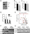
Fig. 1.
Fig. 1.
Myc regulates glycolysis and represses TXNIP in TNBC. (AD) c-Myc levels were reduced in MDA-MB-157 cells by siRNA transfection. siC, control siRNA transfection. (A) Levels of the indicated proteins were determined by Western blotting 72 h after transfection. (B and C) The rates of glucose (B) and glutamine (C) uptake were determined in control or c-Myc–knockdown cells. (D) Levels of the indicated mRNAs were determined from control or c-Myc–knockdown cells by qPCR. (E) Myc activity as determined by gene-pathway signature is plotted against TXNIP mRNA level in 56 breast cancer cell lines, and the Pearson’s correlation coefficient was determined. Basal breast cancers are shown as red circles.(F) MDA-MB-157 cells with or without Myc overexpression were treated with the indicated amount of JQ1 for 24 h, and levels of the indicated proteins were determined by Western blotting. (G) Breast tumor explant cells HCI-010, HCI-014, and HCI-007 were treated for 24 h with the indicated dose of JQ1, and the levels of the indicated proteins were determined by Western blotting. *P < 0.05 as measured using unpaired t test; n, number of independent biological replicates. Values are reported as mean ± SEM.
Fig. 2.
Fig. 2.
TXNIP up-regulation following Myc suppression requires MondoA. (AC) c-Myc levels were decreased by transfection of a c-Myc–specific siRNA pool. Representative experiments performed in triplicate are shown. (A and B) MDA-MB-157 (A) or MDA-MB-231 (B) cells were transfected with either a wild-type TXNIP luciferase reporter construct or a TXNIP promoter construct carrying mutations in the ChoRE MondoA:Mlx binding site. (C) Wild-type MEFs or MondoA-null (Mon−/−) MEFs were transfected with a wild-type TXNIP luciferase reporter construct. (D) Wild-type or MondoA-null MEFs were treated for 24 h with the indicated concentrations of JQ1, and the levels of the indicated proteins were determined by Western blotting. n, number of independent biological replicates. Values are reported as mean ± SEM.
Fig. 3.
Fig. 3.
Myc represses TXNIP expression directly. (A) c-Myc levels were decreased in MDA-MB-157 cells using a c-Myc–specific siRNA pool. The amount of MondoA bound to the TXNIP promoter was determined by ChIP 72 h after transfection. (B) MDA-MB-157 cells were serum starved for 72 h with Myc levels being reduced for 48 h with a c-Myc–specific siRNA pool before serum treatment. The different cell populations were then serum stimulated for the indicated number of hours. Levels of the indicated proteins were determined by Western blotting. (C and D) The amount of MondoA (C) or Myc (D) bound to the TXNIP promoter in quiescent (G0) or early G1 (4 h after serum release) in MDA-MB-157 cells was determined by ChIP. ***P < 0.001; ****P < 0.0001 as determined using paired t tests. n, number of independent biological replicates. Values are reported as mean ± SEM.
Fig. 4.
Fig. 4.
TXNIP is a suppressor of TNBC glucose metabolism. We reduced TXNIP levels in MDA-MB-157 cells using a lentivirus expressing a TXNIP-specific shRNA. We used transfection of a c-Myc–specific siRNA pool to reduce c-Myc levels in each cell population. (A) Levels of the indicated proteins were determined by Western blotting. (B) Rates of glucose uptake in the different cell populations were determined. (C) We determined the number of viable cells in control or TXNIP-knockdown cells following 3 d of growth in glucose-free medium that contained 20 mM 2-deoxyglucose. The number of viable cells is expressed relative to the number of cells seeded on day 1. (D) Control (Vec) or TXNIP-inducible MDA-MB-157 cells were treated with 5 ng/µL doxycycline for 48 h, and the levels of the indicated proteins were determined by Western blotting. (EG) We determined the rates of glucose uptake (E) and ECAR (F) and the relative number of viable cells or percentage apoptotic cells (G) in control (Vec) or TXNIP-induced cells. In G, the different cell populations were grown in the indicated amounts of serum for 3 d. *P < 0.05; **P < 0.01; ***P < 0.001; ****P < 0.0001. In B, statistical significance was determined using ordinary one-way ANOVA. In C, E, F, and G, statistical significance was determined using t tests. n, number of independent biological replicates. Values are reported as mean ± SEM in B and G and as mean ± SD in C, E, and F.
Fig. 5.
Fig. 5.
Low TXNIP expression and a Mychigh/TXNIPlow gene-expression signature correlate with poor patient outcome. Kaplan–Meier plots indicate the clinical outcomes for the gene-expression patterns given at the top of each panel. For TXNIP and Myc, high expression indicates expression above the mean, calculated across all samples. Conversely, low expression indicates expression below the mean. Mychigh/TXNIPlow indicates Myc expression above the mean in combination with TXNIP expression below the mean. n indicates the number of patient samples evaluated in each analysis. (AC) Data available from the Netherlands Cancer Institute were analyzed (24). (DF) Clinical outcomes for the Mychigh/TXNIPlow signature were evaluated in the METABRIC dataset (41). (D and E) The signature was evaluated in TNBCs (D) and non-TNBCs (E). (F) The Mychigh/TXNIPlow signature was correlated with clinical outcome based on p53 mutation status. In F, the 95% confidence interval of the hazard ratio is 1.91–5.70. P values were calculated using the Mantel–Cox log-rank test.

References

    1. Foulkes WD, Smith IE, Reis-Filho JS. Triple-negative breast cancer. N Engl J Med. 2010;363(20):1938–1948. - PubMed
    1. DeBerardinis RJ, Lum JJ, Hatzivassiliou G, Thompson CB. The biology of cancer: Metabolic reprogramming fuels cell growth and proliferation. Cell Metab. 2008;7(1):11–20. - "V体育官网" PubMed
    1. Palaskas N, et al. 18F-fluorodeoxy-glucose positron emission tomography marks MYC-overexpressing human basal-like breast cancers. Cancer Res. 2011;71(15):5164–5174. - "V体育官网入口" PMC - PubMed
    1. Timmerman LA, et al. Glutamine sensitivity analysis identifies the xCT antiporter as a common triple-negative breast tumor therapeutic target. Cancer Cell. 2013;24(4):450–465. - PMC - PubMed
    1. Alles MC, et al. Meta-analysis and gene set enrichment relative to er status reveal elevated activity of MYC and E2F in the “basal” breast cancer subgroup. PLoS ONE. 2009;4(3):e4710. - PMC (VSports) - PubMed

V体育官网 - Publication types

MeSH terms