Skip to main page content
U.S. flag

An official website of the United States government

Dot gov

The . gov means it’s official VSports app下载. Federal government websites often end in . gov or . mil. Before sharing sensitive information, make sure you’re on a federal government site. .

Https

The site is secure. The https:// ensures that you are connecting to the official website and that any information you provide is encrypted and transmitted securely. V体育官网.

. 2015 Aug;64(8):2847-58.
doi: 10.2337/db14-1916. Epub 2015 Apr 6.

Dietary Polyphenols Promote Growth of the Gut Bacterium Akkermansia muciniphila and Attenuate High-Fat Diet-Induced Metabolic Syndrome

Affiliations

Dietary Polyphenols Promote Growth of the Gut Bacterium Akkermansia muciniphila and Attenuate High-Fat Diet-Induced Metabolic Syndrome

Diana E Roopchand et al. Diabetes. 2015 Aug.

Abstract

Dietary polyphenols protect against metabolic syndrome, despite limited absorption and digestion, raising questions about their mechanism of action. We hypothesized that one mechanism may involve the gut microbiota. To test this hypothesis, C57BL/6J mice were fed a high-fat diet (HFD) containing 1% Concord grape polyphenols (GP). Relative to vehicle controls, GP attenuated several effects of HFD feeding, including weight gain, adiposity, serum inflammatory markers (tumor necrosis factor [TNF]α, interleukin [IL]-6, and lipopolysaccharide), and glucose intolerance. GP lowered intestinal expression of inflammatory markers (TNFα, IL-6, inducible nitric oxide synthase) and a gene for glucose absorption (Glut2) VSports手机版. GP increased intestinal expression of genes involved in barrier function (occludin) and limiting triglyceride storage (fasting-induced adipocyte factor). GP also increased intestinal gene expression of proglucagon, a precursor of proteins that promote insulin production and gut barrier integrity. 16S rRNA gene sequencing and quantitative PCR of cecal and fecal samples demonstrated that GP dramatically increased the growth of Akkermansia muciniphila and decreased the proportion of Firmicutes to Bacteroidetes, consistent with prior reports that similar changes in microbial community structure can protect from diet-induced obesity and metabolic disease. These data suggest that GP act in the intestine to modify gut microbial community structure, resulting in lower intestinal and systemic inflammation and improved metabolic outcomes. The gut microbiota may thus provide the missing link in the mechanism of action of poorly absorbed dietary polyphenols. .


Figures

Figure 1
Figure 1
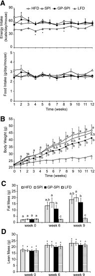
GP-SPI diet reduces weight gain and adiposity of mice but not food intake or lean mass. A: Calorie consumption (mean ± SD) of HFD-based groups was not significantly different during intervention period, but LFD group showed lower calorie intake that was significant at weeks indicated by the asterisks (one-way ANOVA followed by unequal honestly significantly different [HSD] post hoc test, P < 0.05) (top). Food intake (mean ± SD) of mice on indicated diets during the intervention period was not significantly different between groups (one-way ANOVA followed by unequal HSD post hoc test) (bottom). B: Body weights (g) of mice (mean ± SD) consuming the indicated diets for the 12-week intervention period. One-way ANOVA followed by Tukey HSD post hoc test was performed on data at each time point for HFD, SPI diet, and GP-SPI diet groups. LFD group is shown as reference. C: Echo MRI data showing percentage of whole-body fat mass (mean ± SD) for each group. D: Echo MRI data showing percentage of whole-body lean mass (mean ± SD) for each group. C and D: One-way ANOVA followed by unequal HSD post hoc test was performed on data from the four diet groups at each time point. Significant difference between groups for each week is signified by letter a, b, or c; different letters indicate significant difference (P < 0.05) between groups, while the same letter indicates no difference.
Figure 2
Figure 2
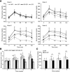
Mice fed GP-SPI diet show improved fasting glucose and oral glucose tolerance. A: Blood glucose concentrations (mg/dL) expressed as mean ± SD (n = 15 for HFD, SPI diet, and GP-SPI diet; n = 10 for LFD) were measured at the indicated time points (0–120 min) after administration of 2 g/kg glucose to mice after they had consumed HFD, SPI diet, GP-SPI diet, or LFD for 0, 3, 6, or 9 weeks. LFD and HFD groups are shown for reference, and main analyses were performed on SPI and GP-SPI groups to assess the effect of GP supplementation. At each time point, a two-tailed t test was performed to evaluate the significance of differences between SPI and GP-SPI groups: *P < 0.05; **P < 0.01; ***P < 0.001. B: AUC representation of HFD, SPI, GP-SPI, and LFD group data in A; each bar represents the mean ± SD (n = 15 for HFD, SPI, and GP-SPI; n = 10 for LFD) for each group at indicated weeks. Between-group analyses were performed with one-way ANOVA followed by unequal honestly significantly different post hoc test. Significant differences between diet groups at the indicated week are signified by letters, where different letters indicate difference (P < 0.05) between groups, while the same letter indicates no difference. C: Blood glucose measurements (mg/dL) taken after a 4-h fast at indicated weeks. Each bar represents mean ± SD (n = 15) of SPI (white bars) and GP-SPI (black bars) diet groups. Within-group analyses comparing data at 0, 3, 6, and 9 weeks were performed with one-way ANOVA followed by Tukey post hoc test. Significant differences within group are signified by letters, where different letters indicate difference (P < 0.05) within group, while the same letter indicates no difference. Between-group differences at each week were determined by t test (two tailed): *P < 0.05; ***P < 0.001.
Figure 3
Figure 3
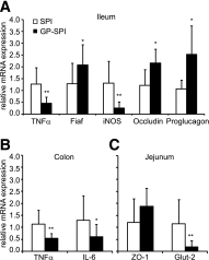
Intestinal tissues of mice fed GP-SPI diet show gene expression changes consistent with attenuated inflammation, increased gut barrier integrity, and improved metabolic function. A: Ileum tissues of mice fed GP-SPI diet have significantly decreased gene expression of TNFα and iNOS and increased Fiaf, occludin, and proglucagon (GCG) expression compared with control. B: Colon tissues of GP-SPI diet–fed mice have significantly higher expression of TNFα and IL-6. C: Jejunum tissues of GP-SPI diet–fed mice have significantly lower levels of Glut2. Data are means ± SD (n = 10 samples per group) for each RT-PCR experiment and were attained using the average of technical duplicates for each sample. Between-group differences were determined by t test (two tailed): *P < 0.05; **P < 0.01.
Figure 4
Figure 4
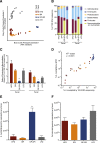
GP supplementation has a dramatic impact on the gut microbiota. A: Bray-Curtis principal coordinate analysis of the 16S rRNA gene in cecal samples and fecal samples collected from mice after 13 weeks of consuming HFD, SPI diet, GP-SPI diet, or LFD. Note the strong separation of GP-SPI samples along PC1, which explains 46% of variation in the sample pool. Diets with a high-fat base (HFD, SPI diet, GP-SPI diet) separate from LFD along PC2, which explains an additional 18% of variation. B: Relative abundance of bacterial phyla. In both cecal and fecal samples, the GP-SPI diet is associated with a significantly higher abundance of Verrucomicrobia, specifically, A. muciniphila (Kruskal-Wallis ANOVA with Dunn correction for multiple comparisons; all GP-SPI pairwise P < 0.05). C: Ratio of the percentage of 16S rRNA gene sequences assigned to Firmicutes versus Bacteroidetes. In both cecal and fecal samples, the GP-SPI diet is associated with a significantly lower ratio (mean ± SEM) (Kruskal-Wallis ANOVA with Dunn correction for multiple comparisons; all GP-SPI pairwise P < 0.05). D: Strong relationship between the relative abundance of A. muciniphila in fecal samples analyzed by 16S rRNA gene sequencing (x-axis) and by qPCR (y-axis), where qPCR relative abundance was quantified by amplifying fecal DNA with primers specific for A. muciniphila (AM1/AM2) and universal bacterial V4 primers (515F/806R). R2 and P value reflect linear regression on log10-transformed data. E: Absolute abundance of A. muciniphila per gram of feces was higher in the GP-SPI group, with significant differences observed between GP-SPI and all other diets (mean ± SEM) (Kruskal-Wallis ANOVA with Dunn correction for multiple comparisons; all GP-SPI pairwise P < 0.05). F: qPCR revealed a trend toward lower microbial DNA on HFD-based diets (HFD, SPI diet, and GP-SPI diet) versus LFD, but total microbial DNA per gram of feces did not differ significantly among groups (mean ± SEM) (Kruskal-Wallis ANOVA; P = 0.2634). *P < 0.05; **P < 0.01; ***P < 0.001.

Comment in

References

    1. Wilson PWF, D’Agostino RB, Parise H, Sullivan L, Meigs JB. Metabolic syndrome as a precursor of cardiovascular disease and type 2 diabetes mellitus. Circulation 2005;112:3066–3072 - PubMed
    1. Cani PD, Osto M, Geurts L, Everard A. Involvement of gut microbiota in the development of low-grade inflammation and type 2 diabetes associated with obesity. Gut Microbes 2012;3:279–288 - VSports在线直播 - PMC - PubMed
    1. Ding S, Lund PK. Role of intestinal inflammation as an early event in obesity and insulin resistance. Curr Opin Clin Nutr Metab Care 2011;14:328–333 - PMC - PubMed
    1. Lam YY, Ha CW, Campbell CR, et al. . Increased gut permeability and microbiota change associate with mesenteric fat inflammation and metabolic dysfunction in diet-induced obese mice. PLoS ONE 2012;7:e34233. - PMC - PubMed
    1. Ding S, Chi MM, Scull BP, et al. . High-fat diet: bacteria interactions promote intestinal inflammation which precedes and correlates with obesity and insulin resistance in mouse. PLoS ONE 2010;5:e12191. - PMC - PubMed

Publication types

MeSH terms