Skip to main page content
U.S. flag

An official website of the United States government

Dot gov

The . gov means it’s official. Federal government websites often end in . gov or . mil VSports app下载. Before sharing sensitive information, make sure you’re on a federal government site. .

Https

The site is secure. The https:// ensures that you are connecting to the official website and that any information you provide is encrypted and transmitted securely V体育官网. .

. 2015 Jun:421:89-95.
doi: 10.1016/j.jim.2015.03.014. Epub 2015 Apr 2.

A 3-D enteroid-based model to study T-cell and epithelial cell interaction

Affiliations

A 3-D enteroid-based model to study T-cell and epithelial cell interaction

Aneta Rogoz (VSports注册入口) et al. J Immunol Methods. 2015 Jun.

"V体育官网入口" Abstract

The constant interaction between intestinal epithelial cells (IECs) and intraepithelial lymphocytes (IELs) is thought to regulate mucosal barrier function and immune responses against invading pathogens. IELs represent a heterogeneous population of mostly activated and antigen-experienced T cells, but the biological function of IELs and their relationship with IECs is still poorly understood. Here, we describe a method to study T-cell-epithelial cell interactions using a recently established long-term intestinal "enteroid" culture system. This system allowed the study of peripheral T cell survival, proliferation, differentiation and behavior during long-term co-cultures with crypt-derived 3-D enteroids. Peripheral T cells activated in the presence of enteroids acquire several features of IELs, including morphology, membrane markers and movement in the epithelial layer. This co-culture system may facilitate the investigation of complex interactions between intestinal epithelial cells and immune cells, particularly allowing long term-cultures and studies targeting specific pathways in IEC or immune cell compartments VSports手机版. .

Keywords: 3-D cultures; Intestinal epithelial cells; Intraepithelial lymphocytes; T cells V体育安卓版. .


Figures (VSports注册入口)

Figure 1
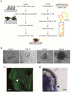
Figure 1. T cell-enteroid co-culture
(A) Schematic view of enteroid and T cell isolation for co-cultures (see Methods for detailed explanation).(B) 20× objective light microscopy image of enteroid cultures taken on days 0, 1, 3 and 6 (scale bar=50μm).(C) Confocal tri-dimensional reconstruction of a CD4+ T cell (in red, white arrow) inserted in an enteroid (in green) after 14 days of co-culture (two passages). Enteroids were derived from UBC-GFP mice and in vitro activated CD4+ T cells from DsRed mice (scale bar=25μm).(D) CellVoyager microscopy image of T cells (blue) and enteroid taken on day 14 of the co-culture. Enteroids were derived from WT mice and co-cultured with pre-activated (OVA peptide plus DCs) OTI CD8+ T cells. Black arrows indicate CellTrace-labeled OTI CD8+ T cells (scale bar=5μm). Data are representative of at least three experiments.
Figure 2
Figure 2. T cell proliferation and differentiation in enteroid co-culture
(A-D) CellTrace-V labeled naiïve and activated OTI CD8+ T cells were co-cultured with WT or iFABP-tOVA enteroids for 14 days in the presence of OTI specific peptide SIINFEKL, where indicated. (A) Frequency of proliferating cells among gated (CD45.1+CD8α+Vα2+) OTI cells based on CellTrace-V dilution. As control OTI cells were culture without enteroides. (B) Expression of CD103 and CellTrace-V by OTI CD8+ T cells co-cultured with WT or iFABP-tOVA enteroids. (C,D) Expression of CD103 (C) and CCR9 (D) by gated CellTrace-Vlow (CD45.1+CD8α+Vα2+) OTI cells. Data are representative of at least three experiments.
Figure 3
Figure 3. T cell movement in enteroids co-culture
OTI CD8+ T cells were pre- activated with OVA peptide (SIINFEKL) plus DCs in the presence or not of RA for 3 days and co-cultured with WT enteroids. T cell-enteroid co-cultures were recorded by CellVoyager microscopy every 5 minutes for 18 hours. Time-lapse images of co- cultures at 0, 6, 12 and 18 hours are depicted (scale bar=40μm). For movies refer to supplemental material.

References

    1. Cheroutre H, Lambolez F, Mucida D. The light and dark sides of intestinal intraepithelial lymphocytes. Nat Rev Immunol. 2011;11:445–56. - PMC - PubMed
    1. Dotan I, Allez M, Nakazawa A, Brimnes J, Schulder-Katz M, Mayer L. Intestinal epithelial cells from inflammatory bowel disease patients preferentially stimulate CD4+ T cells to proliferate and secrete interferon-gamma. American journal of physiology. Gastrointestinal and liver physiology. 2007;292:G1630–40. - PubMed
    1. Edelblum KL, Shen L, Weber CR, Marchiando AM, Clay BS, Wang Y, Prinz I, Malissen B, Sperling AI, Turner JR. Dynamic migration of gammadelta intraepithelial lymphocytes requires occludin. Proceedings of the National Academy of Sciences of the United States of America. 2012;109:7097–102. - PMC - PubMed
    1. Farache J, Koren I, Milo I, Gurevich I, Kim KW, Zigmond E, Furtado GC, Lira SA, Shakhar G. Luminal bacteria recruit CD103+ dendritic cells into the intestinal epithelium to sample bacterial antigens for presentation. Immunity. 2013;38:581–95. - PMC - PubMed
    1. Fuchs A, Vermi W, Lee JS, Lonardi S, Gilfillan S, Newberry RD, Cella M, Colonna M. Intraepithelial type 1 innate lymphoid cells are a unique subset of IL-12- and IL-15-responsive IFN-gamma-producing cells. Immunity. 2013;38:769–81. - PMC - PubMed

VSports在线直播 - Publication types

MeSH terms

VSports手机版 - LinkOut - more resources