<b draggable="iZBvYEbq"><bdo draggable="eUSpd"></bdo></b><area dropzone="ZVKjn"></area> Skip to main page content
U.S. flag

An official website of the United States government

Dot gov

The . gov means it’s official. Federal government websites often end in VSports app下载. gov or . mil. Before sharing sensitive information, make sure you’re on a federal government site. .

Https

The site is secure V体育官网. The https:// ensures that you are connecting to the official website and that any information you provide is encrypted and transmitted securely. .

. 2015 Mar 15;29(6):658-71.
doi: 10.1101/gad.256628.114.

Brg1 promotes both tumor-suppressive and oncogenic activities at distinct stages of pancreatic cancer formation

Affiliations

Brg1 promotes both tumor-suppressive and oncogenic activities at distinct stages of pancreatic cancer formation

Nilotpal Roy et al. Genes Dev. .

Abstract

Pancreatic ductal adenocarcinoma (PDA) develops predominantly through pancreatic intraepithelial neoplasia (PanIN) and intraductal papillary mucinous neoplasm (IPMN) precursor lesions VSports手机版. Pancreatic acinar cells are reprogrammed to a "ductal-like" state during PanIN-PDA formation. Here, we demonstrate a parallel mechanism operative in mature duct cells during which functional cells undergo "ductal retrogression" to form IPMN-PDA. We further identify critical antagonistic roles for Brahma-related gene 1 (Brg1), a catalytic subunit of the SWI/SNF complexes, during IPMN-PDA development. In mature duct cells, Brg1 inhibits the dedifferentiation that precedes neoplastic transformation, thus attenuating tumor initiation. In contrast, Brg1 promotes tumorigenesis in full-blown PDA by supporting a mesenchymal-like transcriptional landscape. We further show that JQ1, a drug that is currently being tested in clinical trials for hematological malignancies, impairs PDA tumorigenesis by both mimicking some and inhibiting other Brg1-mediated functions. In summary, our study demonstrates the context-dependent roles of Brg1 and points to potential therapeutic treatment options based on epigenetic regulation in PDA. .

Keywords: Brg1; EMT; IPMN; Kras; dedifferentiation; pancreatic cancer. V体育安卓版.


"V体育安卓版" Figures

Figure 1.
Figure 1.
PDCs expressing oncogenic Kras and loss of Brg1 undergo dedifferentiation. Quantitative PCR analysis of duct cell differentiation markers in PDCs isolated from LSL- KrasG12D mice (A), Brg1f/f mice (B), and LSL-KrasG12D; Brg1f/f mice (C) infected with adenovirus expressing either RFP or Cre recombinase. (D) FACS analysis of KrasG12D and KrasG12D; Brg1f/f PDCs using the Sca1 antibody. (E) Quantitative PCR analysis of duct cell differentiation markers in PDCs sorted from KrasG12D; Brg1f/f PDCs based on Sca1 expression. (F, left) Quantitative PCR analysis of duct cell differentiation markers in KrasG12D; Brg1f/f PDCs with ectopic Brg1 expression. (Right) FACS analysis of Sca1 in KrasG12D; Brg1f/f PDCs expressing empty vector or vector expressing wild-type Brg1.
Figure 2.
Figure 2.
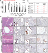
PDCs undergo dedifferentiation en route to IPMN formation. (A) Brg1, Krt19, Hnf4a, Pdx1, and Sox9 immunohistochemistry analysis of either control or 3-, 6-, and 9-wk-old PKB animals. Bars: Brg1, Hnf4a, Pdx1, and Sox9, 100 μm; Krt19, 250 μm. (B) Quantitative PCR analysis of duct cell differentiation markers in DBA lectin-sorted PDCs from either control or 3- and 6-wk-old PKB mice.
Figure 3.
Figure 3.
Ectopic expression of Sox9 in PDCs leads to changes in cellular identity and reduced IPMN occurrence. (A) Quantitative PCR analysis of duct cell differentiation markers in PDCs isolated from LSL-KrasG12D; Brg1f/f; Sox9OE mice infected with adenovirus expressing either RFP or Cre recombinase. (B) Analysis of IPMN or duct atypia occurrence for the indicated genotypes. (C) H&E, Brg1, Pdx1, and HA immunohistochemistry analysis of Hnf1bCreER; LSL- KrasG12D; Brg1f/+, Hnf1bCreER; LSL- KrasG12D; Brg1f/f, Hnf1bCreER; LSL- KrasG12D; Brg1f/+; Sox9OE, or Hnf1bCreER; LSL- KrasG12D; Brg1f/f; Sox9OE mice 4 wk after tamoxifen administration. Bars: H&E, 250 μm; HA, Brg1, and Pdx1, 50 μm.
Figure 4.
Figure 4.
KrasG12D; Brg1f/f PDCs have a distinct transcriptome signature. (A) Heat map showing differential expression signature between wild-type, KrasG12D, and KrasG12D; Brg1f/f PDCs. (B) Heat map showing expression of genes implicated in pancreas development between wild-type, KrasG12D, and KrasG12D; Brg1f/f PDCs. (C) Pathway enrichment analysis for differentially expressed genes between wild-type PDCs and KrasG12D or KrasG12D; Brg1f/f PDCs. Uniquely activated pathways are marked in red. Each colored bar represents the number of genes that belong to a particular pathway. (D) Heat map demonstrating expression of genes that belong to the indicated pathways between wild-type and KrasG12D; Brg1f/f PDCs.
Figure 5.
Figure 5.
Re-expression of Brg1 in KrasG12D; Brg1f/f PDCs and IPMN-derived PDA tumor cells enhances their tumorigenic properties. (A) Western blot showing ectopic expression of Brg1 in IPMN-derived PDA. (B) Brg1 put backs in KrasG12D; Brg1f/f PDCs and IPMN-derived PDA show increased anchorage-independent growth compared with Brg1-null cells. (C, left) Western blot depicting ectopic expression of Brg1 in the Brg1-null human PDA cell line Tu8988T. (Right) Soft agar colony formation assay showing that Brg1-null human PDA cell line Tu8988T forms more colonies on soft agar upon ectopic expression of Brg1. Brg1 put backs in IPMN-derived PDA lines (D) and KrasG12D; Brg1f/f PDCs (E) were subcutaneously injected into NOD scidγ (NSG) mice and monitored for tumor growth. Mice were sacrificed 4 wk after inoculation, and tumors were excised. Tumor mass and volume are significantly higher in Brg1 put backs. (F) Costaining of Ki67, smooth muscle actin (SMA), cleaved caspase 3, and DAPI of tumors obtained in D. Bars: H&E, 250 μm; Ki67, cleaved caspase 3, and SMA, 100 μm.
Figure 6.
Figure 6.
Re-expression of Brg1 induces a mesenchymal phenotype in mouse and human Brg1-deficient pancreatic cancer cell lines. (A) Bright-field images of IPMN-derived KrasG12D; Brg1f/f PDA cells expressing a control empty vector and Brg1 put back. (B) Brg1 put backs in KrasG12D; Brg1f/f in IPMN-derived PDA lines and Tu8988T were probed for epithelial marker E-cadherin, EMT regulator Hmga2, and mesenchymal marker vimentin. (C) Heat map showing an induction of a pronounced mesenchymal gene signature in Brg1-overexpressing IPMN-derived KrasG12D; Brg1f/f PDA cells. (D) Correlation between expression of Brg1 and Hmga2 in human pancreatic cancer cell lines. Plots show signal intensities as measured by Affymetrix chip. Spearman correlation coefficient is shown. (E) Quantitative PCR analysis of Hmga2, Cdh1 (E-cadherin), Vimentin, and Fn1 (fibronectin). (F,G) Bright-field images (F) and crystal violet staining (G) in scrambled siRNA or Hmga2 siRNA transfected IPMN-derived KrasG12D; Brg1f/f PDA cells expressing wild-type Brg1. Lower levels of crystal violet in Hmga2 siRNA transfected cells indicate reduced proliferative capacity.
Figure 7.
Figure 7.
JQ1 can be exploited for the treatment of duct-derived pancreatic cancer. (A) Chromatin immunoprecipitation (ChIP) analysis of H3K27ac enrichment on the indicated promoter regions. Values are expressed as fold enrichment over IgG control. (B) Quantitative PCR analysis of duct cell differentiation markers in KrasG12D; Brg1f/f PDCs treated with DMSO or JQ1. (C) Quantitative PCR analysis of duct cell differentiation markers in IPMN-derived PDA cell line treated with DMSO or JQ1. (D) Quantitative PCR analysis of Hmga2 expression in DMSO- or JQ1-treated KrasG12D or KrasG12D; Brg1f/f PDCs. (E) Quantitation of soft agar assay of KrasG12D; Brg1f/f PDCs or IPMN-derived PDA cell lines treated with DMSO or JQ1. (F) NSG mice were subcutaneously injected with an IPMN-PDA cell line. Once the tumor reached 100 mm3, mice were randomized and treated with JQ1 or vehicle. The graph shows the percent change in tumor volume at the study end point. (G) H&E stainings of vehicle- or JQ1-treated tumors. Arrows point to necrotic lesions. Bar, 250 μm. (H, top) Costaining of Ki67, cleaved caspase 3, and DAPI of tumors treated with vehicle or JQ1. Bar, 100 μm. (Bottom) Percentage of Ki67-positive or cleaved caspase 3-positive cells in vehicle- or JQ1-treated tumors. Ten random fields from three mice in each group were chosen for quantification.

Comment in

References

    1. Bardeesy N, Cheng KH, Berger JH, Chu GC, Pahler J, Olson P, Hezel AF, Horner J, Lauwers GY, Hanahan D, et al.2006. Smad4 is dispensable for normal pancreas development yet critical in progression and tumor biology of pancreas cancer. Genes Dev 20: 3130–3146. - PMC (V体育ios版) - PubMed
    1. Buscarlet M, Krasteva V, Ho L, Simon C, Hebert J, Wilhelm B, Crabtree GR, Sauvageau G, Thibault P, Lessard JA. 2014. Essential role of BRG, the ATPase subunit of BAF chromatin remodeling complexes, in leukemia maintenance. Blood 123: 1720–1728. - PMC - PubMed
    1. Collisson EA, Sadanandam A, Olson P, Gibb WJ, Truitt M, Gu S, Cooc J, Weinkle J, Kim GE, Jakkula L, et al.2011. Subtypes of pancreatic ductal adenocarcinoma and their differing responses to therapy. Nat Med 17: 500–503. - PMC - PubMed
    1. Conroy T, Desseigne F, Ychou M, Bouche O, Guimbaud R, Becouarn Y, Adenis A, Raoul JL, Gourgou-Bourgade S, de la Fouchardiere C, et al.2011. FOLFIRINOX versus gemcitabine for metastatic pancreatic cancer. N Engl J Med 364: 1817–1825. - PubMed
    1. Dal Molin M, Hong SM, Hebbar S, Sharma R, Scrimieri F, de Wilde RF, Mayo SC, Goggins M, Wolfgang CL, Schulick RD, et al.2012. Loss of expression of the SWI/SNF chromatin remodeling subunit BRG1/SMARCA4 is frequently observed in intraductal papillary mucinous neoplasms of the pancreas. Hum Pathol 43: 585–591. - PMC - PubMed

"V体育2025版" Publication types

"VSports" MeSH terms