Skip to main page content
U.S. flag

An official website of the United States government

Dot gov

The . gov means it’s official. Federal government websites often end in . gov or . mil. Before sharing sensitive information, make sure you’re on a federal government site. VSports app下载.

Https

The site is secure. The https:// ensures that you are connecting to the official website and that any information you provide is encrypted and transmitted securely. V体育官网.

. 2015 Mar 24;112(12):3734-9.
doi: 10.1073/pnas.1411713112. Epub 2015 Mar 9.

The nucleolar ubiquitin-specific protease USP36 deubiquitinates and stabilizes c-Myc

Affiliations

The nucleolar ubiquitin-specific protease USP36 deubiquitinates and stabilizes c-Myc

Xiao-Xin Sun et al. Proc Natl Acad Sci U S A. .

Abstract

c-Myc protein stability and activity are tightly regulated by the ubiquitin-proteasome system. Aberrant stabilization of c-Myc contributes to many human cancers. c-Myc is ubiquitinated by SCF(Fbw7) (a SKP1-cullin-1-F-box complex that contains the F-box and WD repeat domain-containing 7, Fbw7, as the F-box protein) and several other ubiquitin ligases, whereas it is deubiquitinated and stabilized by ubiquitin-specific protease (USP) 28. The bulk of c-Myc degradation appears to occur in the nucleolus. However, whether c-Myc is regulated by deubiquitination in the nucleolus is not known. Here, we report that the nucleolar deubiquitinating enzyme USP36 is a novel c-Myc deubiquitinase. USP36 interacts with and deubiquitinates c-Myc in cells and in vitro, leading to the stabilization of c-Myc. This USP36 regulation of c-Myc occurs in the nucleolus. Interestingly, USP36 interacts with the nucleolar Fbw7γ but not the nucleoplasmic Fbw7α. However, it abolished c-Myc degradation mediated both by Fbw7γ and by Fbw7α. Consistently, knockdown of USP36 reduces the levels of c-Myc and suppresses cell proliferation. We further show that USP36 itself is a c-Myc target gene, suggesting that USP36 and c-Myc form a positive feedback regulatory loop. High expression levels of USP36 are found in a subset of human breast and lung cancers. Altogether, these results identified USP36 as a crucial and bono fide deubiquitinating enzyme controlling c-Myc's nucleolar degradation pathway VSports手机版. .

Keywords: USP36; c-Myc; deubiquitination; nucleolus; ubiquitination. V体育安卓版.


Conflict of interest statement

The authors declare no conflict of interest.

Figures

Fig. 1.
Fig. 1.
USP36 deubiquitinates and stabilizes c-Myc. (A) Overexpression of WT USP36, but not the C131A mutant, induces c-Myc levels. HeLa cells transfected with control or Flag-USP36 (WT or the C131A mutant) were assayed by IB. (B) USP36 stabilizes endogenous c-Myc. HeLa cells transfected with the indicated plasmids for 48 h were treated with 50 μg/mL cycloheximide (CHX). The cells were harvested at different time points and assayed by IB. (C) USP36 deubiquitinates c-Myc in cells. HEK293 cells transfected with the indicated plasmids were treated with MG132 for 6 h before harvesting. The cells were subjected to pulldown (PD) using the Ni2+-NTA bead under denaturing conditions, followed by IB. Asterisks indicate nonspecific PD of native c-Myc. (D) Knockdown of USP36 increases c-Myc ubiquitination. HeLa cells were transfected with scrambled or USP36 siRNA together with or without His-Ub for 48 h and treated with MG132 for 6 h. The cells were subjected to PD using the Ni2+-NTA bead, followed by IB. (E) USP36 deubiquitinates c-Myc in vitro. Ubiquitinated c-Myc was purified from 293 cells transfected with His-Ub and Flag–c-Myc using anti-Flag affinity purification. The ubiquitinated c-Myc was incubated with recombinant WT His-USP361-800 or its C131A mutant protein, followed by IB with anti-Flag antibody.
Fig. 2.
Fig. 2.
USP36 interacts with c-Myc in cells and in vitro. (A and B) Co-IP of ectopic USP36 with ectopic c-Myc. H1299 cells transfected with V5–c-Myc and Flag-USP36 (A) or V5-USP36 and Flag–c-Myc (B) individually or together were assayed by co-IP. (C) Co-IP between endogenous USP36 and c-Myc. Lysates from 293 cells were assayed by co-IP with anti–c-Myc (Y69) or control IgG. (D and E) The N-terminal USP domain of USP36 is required for binding to c-Myc. HEK293 cells transfected with V5–c-Myc together with control or Flag-USP36 (WT or deletion mutants) were assayed by co-IP with anti-Flag antibody (D). The diagram of the Flag-USP36 and its deletion mutants are shown in E. (F) USP36 interacts with c-Myc in vitro. Purified GST or GST-USP36 immobilized on glutathione beads was incubated with purified His–c-Myc. Bound proteins were assayed by IB with anti-Myc (Top) and anti-GST (Bottom).
Fig. 3.
Fig. 3.
USP36 interacts with Fbw7γ, but not Fbw7α; inhibits c-Myc degradation mediated by either Fbw7γ or Fbw7α; and deubiquitinates c-Myc in the nucleolus. (A) Immunofluorescence (IF) staining. HeLa cells transfected with Flag-USP36, Flag-Fbw7γ, or Flag-Fbw7α were immunostained with anti-Flag (green) and anti-nucleostemin (NS) (red). (B) USP36 interacts with Fbw7γ, but not Fbw7α. HeLa cells transfected with the indicated plasmids were assayed by co-IP. (C) USP36 forms a complex with c-Myc and Fbw7γ. HeLa cells transfected with Flag-Fbw7γ, V5-USP36, and c-Myc were subjected to IP with anti-Flag, followed by elution with the Flag peptide. The elution was then subjected to co-IP with control IgG or anti-V5. (D and E) USP36 inhibits c-Myc degradation mediated by Fbw7γ or Fbw7α. HeLa cells transfected with the indicated plasmids were assayed by IB. (F) USP36 interacts with c-Myc in the nucleolus. The nucleolar fraction isolated from HeLa cells expressing Flag-USP36 as in Fig. S3E was assayed by co-IP. (G) USP36 deubiquitinates c-Myc in the nucleolus. The nucleoli isolated from HeLa cells transfected with the indicated plasmids were assayed by Ni2+-NTA PD. (H) Overexpression of USP36 increases the levels of c-Myc in both the nucleoplasm and the nucleolus. HeLa–TO (tetracycline operator)–Flag-USP36 cells were induced without or with doxycycline (dox) for 12 h and assayed by cell fractionation and IB.
Fig. 4.
Fig. 4.
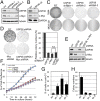
Knockdown of USP36 reduces c-Myc levels and suppresses cell proliferation. (A and B) Knockdown of USP36 reduces c-Myc levels. HeLa (A) and MCF10A (B) cells were infected with scrambled or USP36 shRNA-encoding lentiviruses. The cell lysates were assayed for the expression of c-Myc and USP36 by IB. (C) Knockdown of USP36 suppresses cell proliferation. HeLa or MDA-MB-231 cells were infected with control or USP36 shRNA-encoding lentiviruses. The cells were cultured for up to 3 wk. The colonies were visualized by staining with crystal violet. (D–F) Knockdown of c-Myc does not further increase the inhibitory effects of knockdown of USP36 on cell proliferation. HeLa cells infected with control, USP36 shRNA, or c-Myc shRNA-encoding lentiviruses alone or together were subjected to colony formation assay (D) or measurement of cell confluence over time using IncuCyte System (F). A representative IB detection of the expression of USP36 and c-Myc is shown in E. (G) Knockdown of USP36 induces apoptosis. HeLa cells infected with control or USP36 shRNA-encoding lentiviruses were stained with propidium iodide (PI) followed by flow cytometry analysis. The average percentages of cells in sub-G1 are shown. (H) Soft agar colony formation assays. MDA-MB-231 breast cancer cell lines were infected with control or USP36 shRNA-encoding lentiviruses, followed by colony formation assay in soft agar.
Fig. 5.
Fig. 5.
USP36 is a target gene of c-Myc. (A) c-Myc induces the expression of USP36. 293-TO-Myc cells were cultured in the presence of dox for different times as indicated, followed by IB. (B and C) Dynamic expression of USP36 and c-Myc in cells following serum stimulation. HeLa cells cultured in 0.2% FBS containing medium for 48 h were stimulated with 20% FBS and harvested at the indicated time points. The cells were assayed for USP36 expression by IB (B) or RT-qPCR assays (C). (D) Diagram of the USP36 gene exon–intron regions. Bars indicate the four regions amplified by ChIP–qPCR assays. Red boxes and asterisks indicate canonical and noncanonical E boxes, respectively. Filled boxes indicate exons. The numbers of exons are also indicated. Arrows indicate the transcription start. (E) c-Myc specifically binds to the E box-containing exon 1 and intron 1, but not intron 6, regions of the USP36 gene. HeLa cells were subjected to ChIP using control IgG or anti–c-Myc antibodies followed by qPCR amplification of the indicated genomic regions of the USP36 gene. (F) Serum stimulation increases the c-Myc binding to the USP36 gene. HeLa cells were serum starved for 48 h, followed by stimulation with 20% FBS for 2 h, as in B. The serum-starved and restimulated cells were subjected to ChIP–qPCR to detect the USP36 promoter DNA (exon 1). (G) Knockdown of endogenous c-Myc reduces USP36 levels. HeLa cells transfected with c-Myc or scrambled siRNA were assayed by IB.
Fig. 6.
Fig. 6.
USP36 is overexpressed in cancer cells. (A and B) USP36 is overexpressed in breast cancer cell lines. The expression of USP36 was examined in a total of 10 breast cancer cell lines by RT-qPCR (A) and IB (B) compared with the immortalized mammary epithelia MCF10A cells. (C and D) USP36 is overexpressed in a subset of primary breast cancers. Tissues from breast cancer patients as well as patient-matched normal adjacent tissues were assays for USP36 mRNA expression by RT-qPCR (C). The average 2.19-fold increase of USP36 expression is shown in D. (E) A schematic diagram illustrating the role of USP36 in regulating c-Myc in the nucleolus (see Discussion for details).

References

    1. Adhikary S, Eilers M. Transcriptional regulation and transformation by Myc proteins. Nat Rev Mol Cell Biol. 2005;6(8):635–645. - PubMed
    1. Meyer N, Penn LZ. Reflecting on 25 years with MYC. Nat Rev Cancer. 2008;8(12):976–990. - "VSports在线直播" PubMed
    1. van Riggelen J, Yetil A, Felsher DW. MYC as a regulator of ribosome biogenesis and protein synthesis. Nat Rev Cancer. 2010;10(4):301–309. - PubMed
    1. Nesbit CE, Tersak JM, Prochownik EV. MYC oncogenes and human neoplastic disease. Oncogene. 1999;18(19):3004–3016. - VSports - PubMed
    1. Farrell AS, Sears RC. MYC degradation. Cold Spring Harb Perspect Med. 2014;4(3):a014365. - PMC - PubMed

Publication types (VSports手机版)

MeSH terms