Skip to main page content
U.S. flag

An official website of the United States government

Dot gov

The . gov means it’s official. Federal government websites often end in . gov or . mil VSports app下载. Before sharing sensitive information, make sure you’re on a federal government site. .

Https

The site is secure. The https:// ensures that you are connecting to the official website and that any information you provide is encrypted and transmitted securely. V体育官网.

. 2015 Apr 15;194(8):3937-52.
doi: 10.4049/jimmunol.1402658. Epub 2015 Mar 11.

K+ efflux agonists induce NLRP3 inflammasome activation independently of Ca2+ signaling

Affiliations

K+ efflux agonists induce NLRP3 inflammasome activation independently of Ca2+ signaling

Michael A Katsnelson (VSports app下载) et al. J Immunol. .

Abstract

Perturbation of intracellular ion homeostasis is a major cellular stress signal for activation of NLRP3 inflammasome signaling that results in caspase-1-mediated production of IL-1β and pyroptosis. However, the relative contributions of decreased cytosolic K(+) concentration versus increased cytosolic Ca(2+) concentration ([Ca(2+)]) remain disputed and incompletely defined. We investigated roles for elevated cytosolic [Ca(2+)] in NLRP3 activation and downstream inflammasome signaling responses in primary murine dendritic cells and macrophages in response to two canonical NLRP3 agonists (ATP and nigericin) that facilitate primary K(+) efflux by mechanistically distinct pathways or the lysosome-destabilizing agonist Leu-Leu-O-methyl ester VSports手机版. The study provides three major findings relevant to this unresolved area of NLRP3 regulation. First, increased cytosolic [Ca(2+)] was neither a necessary nor sufficient signal for the NLRP3 inflammasome cascade during activation by endogenous ATP-gated P2X7 receptor channels, the exogenous bacterial ionophore nigericin, or the lysosomotropic agent Leu-Leu-O-methyl ester. Second, agonists for three Ca(2+)-mobilizing G protein-coupled receptors (formyl peptide receptor, P2Y2 purinergic receptor, and calcium-sensing receptor) expressed in murine dendritic cells were ineffective as activators of rapidly induced NLRP3 signaling when directly compared with the K(+) efflux agonists. Third, the intracellular Ca(2+) buffer, BAPTA, and the channel blocker, 2-aminoethoxydiphenyl borate, widely used reagents for disruption of Ca(2+)-dependent signaling pathways, strongly suppressed nigericin-induced NLRP3 inflammasome signaling via mechanisms dissociated from their canonical or expected effects on Ca(2+) homeostasis. The results indicate that the ability of K(+) efflux agonists to activate NLRP3 inflammasome signaling can be dissociated from changes in cytosolic [Ca(2+)] as a necessary or sufficient signal. .


Figures

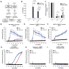
Figure 1
Figure 1. Nigericin-induced increases in cytosolic [Ca2+] in LPS-primed BMDC occur downstream of the NLRP3 inflammasome/ caspase-1/ pyroptotic signaling cascade
A, Diagram of changes in cation homeostasis occurring in myeloid cells in response to treatment with nigericin and ATP/P2X7. B, IL-1β release from LPS-primed WT, Nlrp3−/−, and Casp-1/11−/− BMDC in response to 10μM nigericin and 5mM ATP was measured by ELISA. Data represent mean of 2 independent experiments. C, LPS-primed WT, Nlrp3−/−, and Casp-1/11−/− BMDC were stimulated with 10μM nigericin or 5mM ATP for 30 min. BMDC were lysed with 10% nitric acid and lysates were analyzed by atomic absorption spectroscopy to measure cellular [K+]. Data represent mean of three independent experiments. D–F, WT, Nlrp3−/−, or Casp-1/11−/− BMDC were primed with LPS (1μg/ml) for 4h and loaded with 1μg/ml fluo-4 AM for 30 min. Baseline readings were taken for 5 min and 10μM nigericin (NG) or 5mM ATP were added at t=5 min. Cytosolic [Ca2+] was determined by measuring fluo-4 fluorescence. Data represent a mean of two independent experiments. G–I, LPS-primed WT, Nlrp3−/−, and Casp-1/11−/−BMDC were stimulated with 10μM nigericin or 5mM ATP for 30 min. Onset of pyroptosis was determined by measuring permeability of the cell membrane to propidium2+. Baseline readings were taken for 5 min and nigericin or ATP were added at t=5 min. Data represent a mean of two independent experiments.
Figure 2
Figure 2. NLRP3 inflammasome signaling responses to K+ efflux agonists are dissociated from influx of extracellular Ca2+
A, LPS-primed BMDC were treated with 10μM nigericin (NG) or 5mM ATP for 30 min in the presence/absence of 1.5mM extracellular [Ca2+]. Cytosolic [Ca2+] was determined as described in figure 1. Data represent a mean of three independent experiments. B–E, LPS-primed BMDC were treated for 30 min with 10μM nigericin or 5mM ATP in the presence/absence of 1.5mM extracellular [Ca2+]. B, IL-1β release was determined by ELISA. Data represent a mean of six independent experiments. C, Soluble lysate fraction (Lys) was probed for procaspase-1 and pro-IL-1β, insoluble lysate pellet was crosslinked with DSS and probed for oligomerized ASC, and extracellular medium fraction (ECM) was probed for mature caspase-1 and IL-1β. D, Onset of pyroptosis was determined by measuring permeability of the cell membrane to propidium2+. Baseline readings were taken for 5 min and nigericin or ATP were added at t=5 min. Data represent mean of four independent experiments. E, BMDC were lysed with 10% nitric acid and lysates were analyzed by atomic absorption spectroscopy to measure cellular [K+]. Data represent mean of three independent experiments.
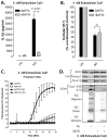
Figure 3
Figure 3. NLRP3 inflammasome signaling responses to lysosomal destabilization are dissociated from influx of extracellular Ca2+
A–C, LPS-primed BMDC were stimulated with 1mM LLME for 30 min. A, LPS-primed WT, Nlrp3−/−, and Casp1−/− BMDC were stimulated with LLME and IL-1β release was determined by ELISA. Data represent a mean of two independent experiments. B, LPS-primed WT, Nlrp3−/−, and Casp1−/− BMDC were stimulated with LLME, lysed with 10% nitric acid, and the lysates analyzed by atomic absorption spectroscopy to measure cellular [K+]. Data represent mean of three independent experiments C, LPS-primed WT BMDC were stimulated with LLME in the presence/absence of 1.5mM extracellular [Ca2+] and IL-1β release was determined by ELISA. Data represent a mean of two independent experiments. D, F, Cytosolic [Ca2+] was determined as described in figure 1 in WT (panels D, F) Nlrp3−/−, and Casp1−/− (Panel F) BMDC. Baseline readings were taken for 5 min and LLME was added at t=5 min. Data represent a mean of two independent experiments. E, LLME-induced propidium2+ influx was measured in LPS-primed BMDC in the presence/absence of 1.5mM extracellular [Ca2+]. Baseline readings were taken for 5 min and LLME was added at t=5 min. Data represent mean of two independent experiments..
Figure 4
Figure 4. NLRP3 inflammasome signaling responses to K+ efflux agonists are dissociated from release of thapsigargin-sensitive intracellular Ca2+ stores
A, LPS-primed BMDC were treated with 300nM thapsigargin (TG) at t=0 in the absence of extracellular Ca2+. BMDC were washed at t=30 min and 10 min of baseline readings were taken prior to addition of 10μM nigericin or 5mM ATP at t=40 min. Cytosolic [Ca2+] was determined as described in figure 1. Data represent a mean of three independent experiments. B and C, LPS-primed BMDC were treated with 300nM thapsigargin in the absence of extracellular Ca2+. BMDC were then stimulated with 10μM nigericin or 5mM ATP in the absence of extracellular Ca2+. B, Onset of pyroptosis was determined by measuring permeability of the cell membrane to propidium2+. Baseline readings were taken for 5 min and nigericin or ATP were added at t=5 min. Data represent a mean of three independent experiments. C, IL-1β release was determined by ELISA. Data represent a mean of six independent experiments.
Figure 5
Figure 5. NLRP3 inflammasome signaling responses to K+ efflux agonists are dissociated from changes in cytosolic [Ca2+] in BMDC primed with TLR2 or TNF receptor agonists
A and E, BMDC were primed with Pam3CSK4 (2μg/ml) or TNFα (100ng/ml) for 4h. Baseline fluorescence measurements were taken for 5 min prior to addition of 10μM nigericin or 5mM ATP at t=5 min. Cytosolic [Ca2+] was determined as described in figure 1. Data represent a mean of two independent experiments. B–C and F–G, Pam3CSK4-primed or TNFα-primed BMDC were treated with 10μM nigericin or 5mM ATP for 30 min in the presence or absence of 1.5mM extracellular [Ca2+]. B and F, Onset of pyroptosis was determined by measuring permeability of the cell membrane to propidium2+. Baseline readings were taken for 5 min and nigericin or ATP were added at t=5 min. Data represent mean of two independent experiments. C and G, IL-1β release from Pam3CSK4-primed BMDC or TNFα-primed BMDC was determined by ELISA. Data represent a mean of two independent experiments for each panel. D and H, Pam3CSK4-primed or TNFα-primed BMDC were pretreated with 300nM thapsigargin (TG) for 30 min in the absence of extracellular Ca2+. BMDC were washed and stimulated with 10μM nigericin or 5mM ATP for 30 min in the absence of extracellular Ca2+. IL-1β release from Pam3CSK4-primed BMDC or TNFα-primed BMDC was determined by ELISA. Data represent a mean of two independent experiments for each panel.
Figure 6
Figure 6. NLRP3 inflammasome signaling responses to K+ efflux are dissociated from changes in cytosolic [Ca2+] in murine macrophages
A, Murine BMDM were primed with LPS (1μg/ml) for 4h. Baseline readings were taken for 5 min and 10μM nigericin (NG) or 5mM ATP were added at t=5 min. Cytosolic [Ca2+] was determined as described in figure 1. Data represent a mean of two independent experiments. B and C, LPS-primed BMDM were treated for 30 min with 10μM nigericin or 5mM ATP in the presence/absence of 1.5mM extracellular [Ca2+]. B, Onset of pyroptosis was determined by measuring permeability of the cell membrane to propidium2+. Baseline readings were taken for 5 min and nigericin or ATP were added at t=5 min. Data represent mean of four independent experiments. C, IL-1β release was determined by ELISA. Data represent a mean of four independent experiments. D, LPS-primed BMDM were treated with 300nM thapsigargin for 30 min in the absence of extracellular Ca2+, and then stimulated with 10μM nigericin or 5mM ATP for 30 min in the absence of extracellular Ca2+. IL-1β release was determined by ELISA. Data represent a mean of four independent experiments.
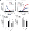
Figure 7
Figure 7. Increased cytosolic [Ca2+] induced by Ca2+ ionophore or Ca2+-mobilizing GPCRs is not a sufficient signal for NLRP3 inflammasome activation
A, Cytosolic [Ca2+] was determined as described in figure 1. Baseline readings were taken for 5 min and 5mM ATP or 3μM ionomycin were added at t=5 min. Data represent a mean of two independent experiments. B, LPS-primed BMDC were treated with 10μM NG, 5mM ATP or 3μM ionomycin for 30 min. IL-1β release was measured by ELISA. Data represent the mean of ten independent experiments. C, LPS-primed BMDC were treated with 30μM R568, 0.1mM ATP, 1μM N-formyl-Met-Leu-Phe (FMLP), or 0.1mM UTP for 30 min. Cytosolic calcium levels were determined as described in figure 1. Baseline readings were taken for 5 min and GPCR agonists were added at t=5 min. Data are representative of two independent experiments. D, LPS-primed BMDC were treated with calcium mobilizing agents for 30 min. IL-1β release was measured by ELISA. Data represent mean of five independent experiments. E, LPS-primed BMDC were treated with 10μM nigericin, 5mM ATP or 3μM ionomycin. Onset of pyroptosis was determined by measuring permeability of the cell membrane to propidium2+. Baseline readings were taken for 5 min and nigericin, ATP, or ionomycin were added at t=5 min. Data represent a mean of two independent experiments. F, LPS-primed BMDC were treated with 10μM nigericin, 5mM ATP or 3μM ionomycin. Cells were lysed with 10% nitric acid and lysates were analyzed by atomic absorption spectroscopy to measure cellular [K+]. Data represent mean of eight independent experiments. G, LPS-primed BMDC were stimulated with 10μM nigericin, 5mM ATP, 3μM ionomycin or 30μM R568 for 30 min. Soluble lysate fraction (Lys) was probed for procaspase-1 and pro-IL-1β, insoluble lysate pellet was crosslinked with DSS and probed for oligomerized ASC, and extracellular medium fraction (ECM) was probed for mature caspase-1 and IL-1β.
Figure 8
Figure 8. Suppression of nigericin-stimulated NLRP3 inflammasome signaling by BAPTA can be dissociated from perturbation of Ca2+ signaling
LPS-primed BMDC were loaded with 25μM BAPTA-AM for 45 min per manufacturer’s protocol. Cells were then treated with 10μM nigericin for 30 min in the absence of extracellular Ca2+. A, IL-1β release was measured by ELISA. Data represent a mean of three independent experiments. B, Efflux of cellular K+ was measured by atomic absorption spectroscopy. Data represent a mean of three independent experiments. C. Onset of pyroptosis was determined by measuring permeability of the cell membrane to propidium2+. Baseline readings were taken for 5 min and 10μM nigericin was added at t=5 min. Data represent a mean of four independent experiments. D, LPS-primed BMDC were stimulated with 10μM nigericin for 30 min in the presence/absence of BAPTA loading. Soluble lysate fraction (Lys) was probed for procaspase-1 and pro-IL-1β, insoluble lysate pellet was crosslinked with DSS and probed for oligomerized ASC, and extracellular medium fraction (ECM) was probed for mature caspase-1 and IL-1β.
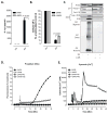
Figure 9
Figure 9. Suppression of nigericin-stimulated NLRP3 inflammasome signaling by 2-APB can be dissociated from perturbation of Ca2+ signaling
A–C, LPS-primed BMDC were incubated with 100μM 2-APB for 20 min prior to treatment with 10μM nigericin for 30 min in the presence of 1.5mM extracellular [Ca2+]. A, IL-1β release was measured by ELISA. Data represent a mean of three independent experiments. B, Efflux of cellular K+ was measured by atomic absorption spectroscopy. Data represent a mean of three independent experiments. C, Soluble lysate fraction (Lys) was probed for procaspase-1 and pro-IL-1β, insoluble lysate pellet was crosslinked with DSS and probed for oligomerized ASC, and extracellular medium fraction (ECM) was probed for mature caspase-1 and IL-1β. D, LPS-primed BMDC were preincubated with 100μM 2-APB for 20 min in the presence of 1.5mM extracellular [Ca2+] and onset of pyroptosis in response to nigericin was assayed by measuring permeability of cells to propidium2+. Baseline readings were taken for 5 min and 10μM nigericin was added at t=5 min. E, Cytosolic [Ca2+] in LPS-primed BMDC was measured using fluo-4 fluorescence in the presence of 1.5mM extracellular [Ca2+]. Baseline readings were taken for 5 min. Cells were treated with 100μM 2-APB at t=5 min (double arrow) and 10μM nigericin (single arrow) at t=15 min. Data are representative of two independent experiments.

References

    1. Warren J. Williams Hematology. 8. McGraw Hill Companies; 2010. The Inflammatory Response. Access Medicine. Web. 7 August 2014.
    1. Schroder K, Tschopp J. The Inflammasomes. Cell. 2010;140(6):821–832. - PubMed
    1. Gross O, Thomas CJ, Guarda G, Tschopp J. The inflammasome: an integrated view. Immunological Reviews. 2011;243(1):136–151. - VSports在线直播 - PubMed
    1. Lu A, Magupalli V, Ruan J, Yin Q, Atianand M, Vos M, Schröder G, Fitzgerald K, Wu H, Egelman E. Unified polymerization mechanism for the assembly of ASC-dependent inflammasomes. Cell. 2014;156(6):1193–1206. - PMC - PubMed
    1. Cai X, Chen J, Xu H, Liu S, Jiang Q, Halfmann R, Chen Z. Prion-like polymerization underlies signal transduction in antiviral immune defense and inflammasome activation. Cell. 2014;156(6):1207–1222. - VSports app下载 - PMC - PubMed

Publication types

MeSH terms

Substances