Skip to main page content
U.S. flag

An official website of the United States government

Dot gov

The . gov means it’s official. Federal government websites often end in . gov or . mil. Before sharing sensitive information, make sure you’re on a federal government site VSports app下载. .

Https

The site is secure V体育官网. The https:// ensures that you are connecting to the official website and that any information you provide is encrypted and transmitted securely. .

. 2015 Mar 11;11(3):e1004712.
doi: 10.1371/journal.ppat.1004712. eCollection 2015 Mar.

Dependence of intracellular and exosomal microRNAs on viral E6/E7 oncogene expression in HPV-positive tumor cells

Affiliations

"VSports在线直播" Dependence of intracellular and exosomal microRNAs on viral E6/E7 oncogene expression in HPV-positive tumor cells

Anja Honegger et al. PLoS Pathog. .

V体育安卓版 - Abstract

Specific types of human papillomaviruses (HPVs) cause cervical cancer. Cervical cancers exhibit aberrant cellular microRNA (miRNA) expression patterns. By genome-wide analyses, we investigate whether the intracellular and exosomal miRNA compositions of HPV-positive cancer cells are dependent on endogenous E6/E7 oncogene expression. Deep sequencing studies combined with qRT-PCR analyses show that E6/E7 silencing significantly affects ten of the 52 most abundant intracellular miRNAs in HPV18-positive HeLa cells, downregulating miR-17-5p, miR-186-5p, miR-378a-3p, miR-378f, miR-629-5p and miR-7-5p, and upregulating miR-143-3p, miR-23a-3p, miR-23b-3p and miR-27b-3p. The effects of E6/E7 silencing on miRNA levels are mainly not dependent on p53 and similarly observed in HPV16-positive SiHa cells. The E6/E7-regulated miRNAs are enriched for species involved in the control of cell proliferation, senescence and apoptosis, suggesting that they contribute to the growth of HPV-positive cancer cells. Consistently, we show that sustained E6/E7 expression is required to maintain the intracellular levels of members of the miR-17~92 cluster, which reduce expression of the anti-proliferative p21 gene in HPV-positive cancer cells. In exosomes secreted by HeLa cells, a distinct seven-miRNA-signature was identified among the most abundant miRNAs, with significant downregulation of let-7d-5p, miR-20a-5p, miR-378a-3p, miR-423-3p, miR-7-5p, miR-92a-3p and upregulation of miR-21-5p, upon E6/E7 silencing. Several of the E6/E7-dependent exosomal miRNAs have also been linked to the control of cell proliferation and apoptosis. This study represents the first global analysis of intracellular and exosomal miRNAs and shows that viral oncogene expression affects the abundance of multiple miRNAs likely contributing to the E6/E7-dependent growth of HPV-positive cancer cells. VSports手机版.


Conflict of interest statement

The authors have declared that no competing interests exist.

Figures

Fig 1
Fig 1. Silencing of HPV18 E6/E7 expression by RNA interference.
(A) qRT-PCR analysis of HPV18 E6/E7 (left panel) and p21 (right panel) mRNA expression, 72 h after transfection of HeLa cells with si18E6/E7, control siRNA siContr-1, or upon mock treatment. mRNA levels were normalized to ACTB and calculated relative to the mock control. Data represent mean ± SEM (n = 4). Asterisks above columns indicate statistically significant differences from siContr-1-treated cells (p ≤ 0.05 (*), p ≤ 0.001 (***)). (B) Immunoblot analysis of HPV18 E6, p53, and p21 protein levels, 72 h after transfection of HeLa cells with si18E6/E7 or siContr-1. α-Tubulin: loading control. (C) Immunoblot analysis of HPV18 E7, total pRb (pRb), phosphorylated pRb (pRb-P), and Cyclin A1 protein levels, 72 h after transfection of HeLa cells with si18E6/E7 or siContr-1. α-Tubulin: loading control.
Fig 2
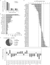
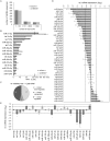
Fig 2. Inhibition of endogenous HPV18 E6/E7 expression: Effects on the intracellular miRNA composition of cervical cancer cells.
Small RNA deep sequencing (A—D) and qRT-PCR analyses (E) of cellular miRNAs, 72 h after transfection of HeLa cells with si18E6/E7 or control siRNA siContr-1. (A) Mean read count distribution of mature miRNA sequences in si18E6/E7- and siContr-1-transfected cells (n = 2). Only miRNAs with a mean read count > 1 were considered. (B) The 15 most frequently sequenced cellular miRNAs. Selection based on siContr-1 samples, respective values for the si18E6/E7-treatment are indicated. Data represent mean ± SEM (n = 2). Interrupted x-Axis. (C) Overview on differentially affected (> 1.5-fold) cellular miRNAs, determined by small RNA deep sequencing. RPM values of si18E6/E7-treated samples were calculated relative to the control treatment (siContr-1). Only miRNAs with > 1,000 RPM in each sample were considered (n = 2). (D) Relative quantification of miRNAs in si18E6/E7- versus siContr-1-treated cells as assessed by small RNA deep sequencing (log2 display). Dashed lines: 1.5-fold up- or downregulation (log2(1.5) = 0.585). Only miRNAs with > 1,000 RPM in each sample were considered. Data represent mean ± SEM (n = 2). (E) qRT-PCR analyses of E6/E7-dependent cellular miRNAs identified by small RNA deep sequencing. Cellular miRNA levels were normalized to snRNA RNU6–2 and calculated relative to siContr-1 (log2 display). Dashed lines: 1.5-fold up- or downregulation (log2(1.5) = 0.585). The column color shows regulation in the same (dark grey) or opposite (light grey) direction compared to the small RNA deep sequencing data of the individual miRNAs. Data represent mean ± SEM (n = 2 or 3). Asterisks indicate statistically significant differences (p ≤ 0.05 (*), p ≤ 0.01 (**) and p ≤ 0.001 (***)).
Fig 3
Fig 3. Inhibition of endogenous HPV16 E6/E7 expression: Effects on selected intracellular miRNAs.
(A) Immunoblot analysis of HPV16 E7, HPV16 E6, p53 and p21 protein levels, 72 h after transfection of SiHa cells with si16E6/E7 or control siRNA (siContr-1), or upon mock treatment. α-Tubulin: loading control. (B) qRT-PCR analyses of ten selected cellular miRNAs, 72 h after transfection of SiHa cells with si16E6/E7 or siContr-1. Cellular miRNA levels were normalized to the snRNA RNU6–2 and calculated relative to siContr-1 (log2 display). Dashed lines: 1.5-fold up- or downregulation (log2(1.5) = 0.585). Data represent mean ± SEM (n = 3). Asterisks indicate statistically significant differences (p ≤ 0.05 (*) and p ≤ 0.01 (**)).
Fig 4
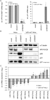
Fig 4. Effects of the p53 status on the E6/E7-dependent modulation of intracellular miRNAs.
(A) qRT-PCR analysis of HPV18 E6/E7 (left panel) and p21 (right panel) mRNA expression, 72 h after transfection of parental or “p53-null” HeLa cells with si18E6/E7, control siRNA (siContr-1), or upon mock treatment. mRNA levels were normalized to ACTB and calculated relative to the mock control (mock). Data represent mean ± SEM (n = 3). Asterisks above columns indicate statistically significant differences from siContr-1-treated cells (p ≤ 0.05 (*), p ≤ 0.001 (***)). (B) Immunoblot analysis of HPV18 E6, p53 and p21 protein levels, 72 h after transfection of parental or “p53-null” HeLa cells with si18E6/E7 or siContr-1, or upon mock treatment. α-Tubulin: loading control. (C) qRT-PCR analyses of selected cellular miRNAs, 72 h after transfection of parental or “p53-null” HeLa cells with si18E6/E7 or siContr-1. miR-34a-3p, positive control miRNA (p53-inducible). Cellular miRNA levels were normalized to snRNA RNU6–2 and calculated relative to siContr-1 (log2 display). Dashed lines: 1.5-fold up- or downregulation (log2(1.5) = 0.585). Data represent mean ± SEM (n = 3). Asterisks indicate statistically significant differences (p ≤ 0.05 (*), p ≤ 0.01 (**) and p ≤ 0.001 (***)).
Fig 5
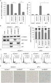
Fig 5. Influence of combined silencing of p21 and HPV18 E6/E7 expression on the senescent phenotype of HPV-positive cancer cells.
(A) qRT-PCR analysis of HPV18 E6/E7 (left panel) and p21 (right panel) mRNA expression, 72 h after transfection of HeLa cells with the indicated siRNAs or in mock-treated cells. mRNA levels were normalized to ACTB and calculated relative to the mock control. Data represent mean ± SEM (n = 2 or 3). Asterisks above columns indicate statistically significant differences between the indicated treatments (p ≤ 0.05 (*), p ≤ 0.01 (**)). (B) Immunoblot analysis of HPV18 E7, p53, and p21 protein levels, 72 h after transfection of HeLa cells with the indicated siRNAs or upon mock-treatment. α-Tubulin: loading control. (C + D) Cell cycle distribution analyzed by FACS, 72 h after transfection of HeLa cells with the indicated siRNAs or upon mock treatment. Percentage of cells in the G1, S and G2 cell cycle phases are indicated. Representative samples of one experiment are shown as well as a summary of multiple biological replicates. Data represent mean ± SEM (n = 3). (E) HeLa cells were stained for expression of the senescence marker SA-β-Gal, 168 h after transfection with the indicated siRNAs. Visualization by bright field microscopy.
Fig 6
Fig 6. Effects of miRNAs of the miR-17~92 cluster on p21 expression in HeLa cells.
(A) qRT-PCR analyses of cellular miRNA levels, 72 h after transfection of HeLa cells with the indicated vectors or upon mock treatment. miR-17~92: vector coding for the mir-17~92 cluster; “control”: repective empty expression vector. miRNA levels were normalized to snRNA RNU6–2 and calculated relative to the mock control. miR-17–5p, miR-20a-5p, miR-19b-3p, miR-92a-3p: encoded by the mir-17~92 expression vector; miR-34a-5p: negative control (not encoded by the vector). Data represent mean ± SEM (n = 3). Asterisks above columns indicate statistically significant differences from vector control-treated cells (p ≤ 0.05 (*)). (B) qRT-PCR analysis of p21 mRNA expression, 72 h after transfection of HeLa cells with the indicated vectors or upon mock treatment. mRNA levels were normalized to ACTB and calculated relative to the mock control. Data represent mean ± SEM (n = 4). Asterisks above columns indicate statistically significant differences from vector control-treated cells (p ≤ 0.05 (*)). (C) Immunoblot analysis of p53 and p21 protein levels, 72 h after transfection with the indicated vectors. α-Tubulin: loading control. A representative image is shown with corresponding densitometrically quantified band intensities of p21, normalized to α-Tubulin and calculated relative to mock. (D) miRNA inhibitors against miR-17–5p and miR-20a-5p increase the expression of p21 in HeLa cells. Left panel: Immunoblot analysis of p53 and p21 protein levels, 72 h after transfection of HeLa cells with the indicated miRNA inhibitors, an inhibitor control (‘Inhib. control’), or upon mock treatment. α-Tubulin: loading control. A representative image is shown. Numbers below individual lanes correspond to densitometrically quantified band intensities for p21, normalized to α-Tubulin and calculated relative to the Inhib. control. Right panel: Summary of densitometric quantification of p21 protein signal intensities. Data represent mean ± SEM (n = 3). Asterisks above columns indicate statistically significant differences from Inhib. control-treated cells (p ≤ 0.05 (*), p ≤ 0.01 (**)).
Fig 7
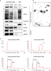
Fig 7. Characterization of exosomes secreted by HeLa cells used for small RNA deep sequencing.
(A) Immunoblot analysis of total cellular extract (30, 10 and 1 μg) from exosome-producing cells, and of 1 μg protein from exosome preparations. Hsc70, CD63, Annexin-1, CD9 and β-Actin: exosomal markers; EEA1: early endosome marker; GRP78: ER marker. (B) Visualization of exosomes by electron microscopy. Bar corresponds to 100 nm. (C) Characterization of cellular and exosomal RNA. Electropherograms of total RNA isolated from HeLa cells and from RNAse A-treated exosomes. Upper panel: total RNA contents; lower panel: small RNA contents. M = marker. Shown are representative images for siContr-1-treated samples.
Fig 8
Fig 8. Inhibition of endogenous HPV18 E6/E7 expression: Effects on the miRNA composition of exosomes secreted by cervical cancer cells.
Small RNA deep sequencing (A—D) and qRT-PCR analyses (E) of exosomal miRNAs, 72 h after transfection of HeLa cells with si18E6/E7 or control siRNA (siContr-1), and subsequent exosome purification from the cell culture supernatant. (A) Mean read count distribution of mature miRNA sequences in exosomes released from si18E6/E7- and siContr-1-treated HeLa cells (n = 3). Only miRNAs with a mean read count > 1 were considered. (B) The 15 most frequently sequenced exosomal miRNAs. Selection based on siContr-1 samples, respective values for si18E6/E7-treatment are indicated. Data represent mean ± SEM (n = 3). Interrupted x-Axis. (C) Overview on differentially deregulated (> 1.5-fold) exosomal miRNAs determined by small RNA deep sequencing. RPM values of si18E6/E7-treated samples were calculated relative to the control treatment (siContr-1). Only miRNAs with > 1,000 RPM in each sample were considered (n = 2). (D) Relative quantification of miRNAs in exosomes released from si18E6/E7- versus siContr-1-treated cells, as assessed by small RNA deep sequencing (log2 display). Dashed lines: 1.5-fold up- or downregulation (log2(1.5) = 0.585). Only miRNAs with > 1,000 RPM in each sample were considered. Data represent mean ± SEM (n = 3). (E) qRT-PCR analysis of E6/E7-dependent exosomal miRNAs identified by small RNA deep sequencing. Exosomal miRNA levels were normalized to miR-452–5p and miR-183–5p and calculated relative to siContr-1 (log2 display). Dashed lines: 1.5-fold up- or downregulation (log2(1.5) = 0.585). The column color shows regulation in the same (dark grey) or opposite (light grey) direction compared to the small RNA deep sequencing data of the individual miRNAs. Ct-values > 35 were considered as not detected (n.d.). Data represent mean ± SEM (n = 2 or 3). Asterisks indicate statistically significant differences (p ≤ 0.05 (*), p ≤ 0.01 (**)).
Fig 9
Fig 9. Inhibition of endogenous HPV16 E6/E7 expression: Effects on selected exosomal miRNAs.
qRT-PCR analysis of selected exosomal miRNAs, 72 h after transfection of SiHa cells with si16E6/E7 or control siRNA (siContr-1), and subsequent exosome purification from the cell culture supernatant. Exosomal miRNA levels were normalized to miR-452–5p and miR-183–5p and calculated relative to siContr-1 (log2 display). Dashed lines: 1.5-fold up- or downregulation (log2(1.5) = 0.585). Data represent mean ± SEM (n = 3). Asterisks indicate statistically significant differences (p ≤ 0.05 (*), p ≤ 0.01 (**) and p ≤ 0.001 (***)).
Fig 10
Fig 10. HPV oncogenes control p21 expression at multiple levels.
E6 can repress p21 transcription at the promoter level by inducing the degradation of the p21 transcriptional activator p53; sustained E6/E7 expression maintains the concentration of miR-17 family members in HPV-positive cancer cells which repress p21 expression by targeting the p21 mRNA; the E7 protein can directly bind to the p21 protein and inhibit its function.

References

    1. zur Hausen H (2002) Papillomaviruses and cancer: from basic studies to clinical application. Nat Rev Cancer 2: 342–350. - PubMed
    1. von Knebel Doeberitz M, Oltersdorf T, Schwarz E, Gissmann L (1988) Correlation of modified human papilloma virus early gene expression with altered growth properties in C4–1 cervical carcinoma cells. Cancer Res 48: 3780–3786. - "VSports在线直播" PubMed
    1. McLaughlin-Drubin ME, Munger K (2009) Oncogenic activities of human papillomaviruses. Virus Res 143: 195–208. 10.1016/j.virusres.2009.06.008 - "V体育ios版" DOI - PMC - PubMed
    1. Scheffner M, Werness BA, Huibregtse JM, Levine AJ, Howley PM (1990) The E6 oncoprotein encoded by human papillomavirus types 16 and 18 promotes the degradation of p53. Cell 63: 1129–1136. - PubMed
    1. Klingelhutz AJ, Foster SA, McDougall JK (1996) Telomerase activation by the E6 gene product of human papillomavirus type 16. Nature 380: 79–82. - PubMed

Publication types

"VSports在线直播" MeSH terms