Skip to main page content
U.S. flag

An official website of the United States government

Dot gov

The . gov means it’s official. Federal government websites often end in . gov or . mil. Before sharing sensitive information, make sure you’re on a federal government site. VSports app下载.

Https

The site is secure. The https:// ensures that you are connecting to the official website and that any information you provide is encrypted and transmitted securely. V体育官网.

. 2015 May;25(5):561-73.
doi: 10.1038/cr.2015.30. Epub 2015 Mar 10.

"V体育官网入口" ERK kinase phosphorylates and destabilizes the tumor suppressor FBW7 in pancreatic cancer

Affiliations

ERK kinase phosphorylates and destabilizes the tumor suppressor FBW7 in pancreatic cancer

Shunrong Ji et al. Cell Res. 2015 May.

Abstract

F-box and WD repeat domain-containing 7 (FBW7) is the substrate recognition component of the Skp1-Cul1-F-box (SCF) ubiquitin ligase complex and functions as a major tumor suppressor by targeting various oncoproteins for degradation VSports手机版. Genomic deletion or mutation of FBW7 has frequently been identified in many human cancers but not in pancreatic ductal adenocarcinoma. Thus it is important to know how the tumor suppressive function of FBW7 is impaired in pancreatic cancer. In this study, we first observed that low FBW7 expression correlated significantly with ERK activation in pancreatic cancer clinical samples, primarily due to KRAS mutations in pancreatic cancer. We further showed that ERK directly interacted with FBW7 and phosphorylated FBW7 at Thr205, which sequentially promoted FBW7 ubiquitination and proteasomal degradation. Furthermore, the phospho-deficient T205A FBW7 mutant is resistant to ERK activation and could significantly suppress pancreatic cancer cell proliferation and tumorigenesis. These results collectively demonstrate how the oncogenic KRAS mutation inhibits the tumor suppressor FBW7, thus revealing an important function of KRAS mutations in promoting pancreatic cancer progression. .


Figures

Figure 1
Figure 1
FBW7 is downregulated in pancreatic cancer. (A) Immunoblot analysis of the indicated human pancreatic cancer cell lines. The HPDE cell line was included as a positive control for the detection of endogenous FBW7 expression, and α-tubulin was used as a loading control. (B) IHC staining of human pancreatic adenocarcinoma tissue arrays using specific antibodies for FBW7. T, tumor; ANT, adjacent normal tissues; scale bar, 200 μm; magnification scale bar, 40 μm. (C) Kaplan-Meier analysis of overall survival rate of pancreatic cancer patients according to the expression of FBW7 (n = 86, P = 0.029, log-rank test).
Figure 2
Figure 2
Expression of FBW7 inversely correlates with ERK activation. (A) Representative IHC staining of FBW7 and p-ERK in pancreatic adenocarcinoma tissue arrays (scale bar, 200 μm; inset scale bar, 40 μm). (B) Statistical analysis of the correlation between the levels of FBW7 and p-ERK (P = 0.027; P value was obtained using a Pearson χ2 test). (C) Representative IHC staining of Fbw7 and p-Erk in KrasG12D transgenic mice. Control slides were obtained from the same mouse strain, which was kept in the same conditions and sacrificed at the same age as the transgenic mice (scale bar, 100 μm).
Figure 3
Figure 3
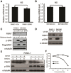
ERK negatively regulates FBW7 stability. (A, B) qPCR analysis of FBW7 expression in PANC-1 and SW1990 cells infected with empty vector or ERK1-expressing virus. GAPDH mRNA expression was used as an internal control (n = 3, independent t-test, P > 0.05). (C) Western blot analysis of FBW7 expression in PANC-1 and SW1990 cells infected with empty vector or ERK1-expressing virus. The band intensity was quantified with Image J software. (D) Western blot analysis of FBW7 expression in PANC-1 and SW1990 cells in the presence and absence of the MEK-1 inhibitor (UO126, 10 μM). DMSO was used as the control. (E) PANC-1 cells were treated with 20 μg/ml CHX in the presence or absence of 10 μM UO126, and whole-cell lysates (WCL) were collected at the indicated time points for immunoblot analysis. (F) Semi-quantification with α-tubulin as a loading control and relative FBW7 levels at time 0 were set as 100%.
Figure 4
Figure 4
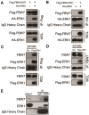
ERK interacts with FBW7. (A, B) Immunoblot (IB) analysis of whole-cell lysates (WCL) and immunoprecipitates (IP) from 293T cells transfected with Flag-WT-FBW7, or HA-ERK1 or together. Thirty hours post-transfection, cells were pretreated with 15 μM MG132 for 10 h before harvesting. (C, D) Immunoblot analysis of WCL and immunoprecipitates from the indicated SW1990 cells stably expressing the Flag-ERK1 construct. Cells were pretreated with 15 μM MG132 for 10 h before harvesting. (E) SW1990 cell extracts were immunoprecipitated with an antibody against FBW7 or control IgG and analyzed by immunoblot analysis.
Figure 5
Figure 5
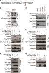
ERK phosphorylates FBW7 at T205. (A) Detection of in vivo FBW7 T205 phosphorylation by mass spectrometry analysis. (B) PANC-1 cells were pretreated with the proteasome inhibitor MG132 and trametinib, as indicated, overnight before harvest. Endogenous FBW7 phosphorylation status was examined by immunoblot analysis after immunoprecipitates (IP). (C) 293T cells were transfected with Flag-WT-FBW7. Thirty hours posttransfection, cells were pretreated with MG132 and various MEK/ERK inhibitors overnight before harvesting. FBW7 phosphorylation status was examined by immunoblot analysis after immunoprecipitation. (D-F) 293T cells were transfected with Flag-WT-FBW7 and Flag-T205A-FBW7, together with HA-MEK1 and HA-ERK1 constructs, respectively. Wild-type (WT) and the constitutively active (CA) form of MEK1 were used as indicated. Thirty hours post-transfection, cells were pretreated with MG132 overnight before harvesting. FBW7 phosphorylation status was examined by immunoblot analysis after immunoprecipitation. Immunoprecipitates were treated with CIP for 20 min at room temperature, and the reaction was stopped by the addition of SDS loading buffer (F). (G) Recombinant GST-WT-FBW7 and GST-T205A-FBW7 proteins were incubated with HA-ERK1 kinase in the presence of ATP and the kinase reaction buffer. Thirty minutes later, the reaction was stopped by the addition of the loading buffer. The kinase reaction products were resolved by SDS-PAGE, and FBW7 phosphorylation was detected by the specific p-FBW7 antibody.
Figure 6
Figure 6
ERK destabilizes FBW7 by promoting its ubiquitination. (A) Immunoblot (IB) analysis of whole-cell lysates (WCL) and anti-Flag immunoprecipitates of 293T cells transfected with HA-ERK1 together with Myc-Ub and various Flag-FBW7 constructs. Thirty hours posttransfection, cells were treated with MG132 overnight before harvest. (B) Immunoblot (IB) analysis of WCL and anti-Flag immunoprecipitates of 293T cells transfected with wild-type HA-ERK1 or kinase-dead (KD) mutant together with Myc-Ub and Flag-WT-FBW7 constructs. Thirty hours posttransfection, cells were treated with MG132 overnight before harvest. (C) Immunoblot analysis of WCL and anti-Flag immunoprecipitates of Hela cells transfected with Flag-WT-FBW7 together with Myc-Ub and siRNA against ERK1/2. Thirty hours posttransfection, cells were treated with MG132 overnight before harvest. (D) Immunoblot analysis of WCL and anti-Flag immunoprecipitates of 293T cells transfected with Flag-WT-FBW7 and Myc-Ub constructs. Thirty hours posttransfection, cells were treated with MG132 and trametinib (where indicated) overnight before harvest. (E) Immunoblot analysis of WCL and anti-Flag immunoprecipitates of 293T cells transfected with HA-KRASG12D together with Myc-Ub and Flag-WT-FBW7 constructs. Thirty hours posttransfection, cells were treated with MG132 overnight before harvest. (F) Immunoblot analysis of WCL and anti-Flag immunoprecipitates of 293T cells transfected with Flag-WT-FBW7 and HA-ERK1 together with Myc-Ub and siRNA oligos against Pin1. Thirty hours posttransfection, cells were treated with MG132 overnight before harvest.
Figure 7
Figure 7
Phosphorylation at the T205 site inhibits FBW7 tumor suppressor function in pancreatic cancer. (A, B) Representative images of clone formation on plastic plates with SW1990 cell lines expressing FBW7 (wild type and T205A mutant) or empty vector. Quantification of cell growth is shown in B (n = 3, independent t-test, **P < 0.01). (C) Cell growth curve assay with SW1990 cell lines expressing FBW7 (wild type and T205A mutant) or empty vector (n = 3, independent t-test, **P < 0.01). (D, E) SW1990 cells stably expressing WT-FBW7, T205A-FBW7, or empty vector were injected into nude mice. At the indicated times, tumors were measured with Vernier calipers (mean ± SEM; n = 5, independent t-test, *P < 0.05). (F) Western blot analysis for c-Myc in SW1990 cell lines stably expressing Flag-WT-FBW7 or Flag-T205A-FBW7 (with empty vector as a negative control). (G) Representative images of tumor tissue from staining with antibodies against FBW7 and c-Myc. Scale bar, 100 μm. (H) Proposed model of how the Ras/Raf/MEK/ERK pathway regulates FBW7 stability to influence its tumor suppressive functions in pancreatic cancer.

References

    1. Wolfgang CL, Herman JM, Laheru DA, et al. Recent progress in pancreatic cancer. CA Cancer J Clin. 2013;63:318–348. - PMC - PubMed
    1. Vincent A, Herman J, Schulick R, Hruban RH, Goggins M. Pancreatic cancer. Lancet. 2011;378:607–620. - PMC - PubMed
    1. Poruk KE, Firpo MA, Adler DG, Mulvihill SJ. Screening for pancreatic cancer: why, how, and who. Ann Surg. 2013;257:17–26. - PMC - PubMed
    1. Winter JM, Cameron JL, Campbell KA, et al. 1423 pancreaticoduodenectomies for pancreatic cancer: a single-institution experience. J Gastrointest Surg. 2006;10:1199–1210. - PubMed
    1. Iacobuzio-Donahue CA, Velculescu VE, Wolfgang CL, Hruban RH. Genetic basis of pancreas cancer development and progression: insights from whole-exome and whole-genome sequencing. Clin Cancer Res. 2012;18:4257–4265. - PMC - PubMed

Publication types

MeSH terms (V体育2025版)