Skip to main page content
U.S. flag

An official website of the United States government

Dot gov

The . gov means it’s official. Federal government websites often end in . gov or . mil. Before sharing sensitive information, make sure you’re on a federal government site VSports app下载. .

Https

The site is secure. The https:// ensures that you are connecting to the official website and that any information you provide is encrypted and transmitted securely. V体育官网.

. 2015 Mar;17(3):322-332.
doi: 10.1038/ncb3121.

FBXW7 modulates cellular stress response and metastatic potential through ​HSF1 post-translational modification

Affiliations

FBXW7 modulates cellular stress response and metastatic potential through ​HSF1 post-translational modification

Nikos Kourtis et al. Nat Cell Biol. 2015 Mar.

Abstract

​Heat-shock factor 1 (​HSF1) orchestrates the heat-shock response in eukaryotes. Although this pathway has evolved to help cells adapt in the presence of challenging conditions, it is co-opted in cancer to support malignancy. However, the mechanisms that regulate ​HSF1 and thus cellular stress response are poorly understood. Here we show that the ubiquitin ligase ​FBXW7α interacts with ​HSF1 through a conserved motif phosphorylated by ​GSK3β and ​ERK1 VSports手机版. ​FBXW7α ubiquitylates ​HSF1 and loss of ​FBXW7α results in impaired degradation of nuclear ​HSF1 and defective heat-shock response attenuation. ​FBXW7α is either mutated or transcriptionally downregulated in melanoma and ​HSF1 nuclear stabilization correlates with increased metastatic potential and disease progression. ​FBXW7α deficiency and subsequent ​HSF1 accumulation activates an invasion-supportive transcriptional program and enhances the metastatic potential of human melanoma cells. These findings identify a post-translational mechanism of regulation of the ​HSF1 transcriptional program both in the presence of exogenous stress and in cancer. .


Figures (VSports在线直播)

Figure 1
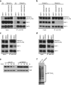
Figure 1. HSF1 is a substrate of the FBXW7α ubiquitin ligase
(a) Network of FBXW7α-interacting partners. Serial immunoprecipitation experiments from HEK293 cells coupled to mass-spectrometry based analysis revealed a large number of known substrates (NFKB2, MYC, MED13L, MED13), already characterized members of the Cullin 1 complex (SKP1, CUL1) and putative interactors (MED1, HSF1). The FBXW7 degrons on various substrates are indicated. (b) FBXW7α binds to HSF1 through specific residues in the WD40 domain. HEK293T cells were transfected with constructs encoding FLAG tagged HSF1, and FLAG-HA tagged empty vector (EV), or FLAG-HA tagged FBXW7α or FLAG-HA tagged FBXW7α (WD40), a substrate binding mutant, in which three residues within one of the seven WD40 repeats of FBXW7α have been mutated. HA-tagged FBXW7α was immunoprecipitated (IP) from cell extracts with anti-HA resin, followed by immunoblotting as indicated. The left panel shows inputs. (c) Alignment of the human HSF1 protein region containing the putative degron with HSF1 from various organisms. Conserved phospho-amino acids (amino acids 303 and 307 in human sequence) are highlighted. Uncropped blots are shown in Supplementary Fig. 8.
Figure 2
Figure 2. HSF1 interacts with FBXW7α through a conserved degron sequence phosphorylated by GSK3β and ERK1
(a) HSF1 binds FBXW7α through a conserved degron. HEK293T cells were transfected with FLAG-HA tagged FBXW7α and constructs encoding FLAG tagged HSF1 or HSF1(Ser303/307Ala) or HSF1(Ser363/367Ala). HA-tagged FBXW7α was immunoprecipitated (IP) from cell extracts with anti-HA resin, followed by immunoblotting as indicated. The left panel shows inputs. (b) Both Ser303 and Ser307 in HSF1 are required for the interaction with FBXW7α. HEK293T cells were transfected with FLAG-HA tagged FBXW7α and constructs encoding FLAG tagged HSF1 or HSF1(Ser303/307Ala) or HSF1(Ser303Ala) or HSF1(Ser307Ala). HA-tagged FBXW7α was immunoprecipitated (IP) from cell extracts with anti-HA resin, followed by immunoblotting as indicated. (c) Interaction between HSF1 and FBXW7α depends on GSK3β activity. HEK293T cells were transfected with constructs encoding FLAG tagged HSF1 and FLAG-HA tagged FBXW7α. Cells were treated with GSK3i IX (10 μM for 10 h) or DMSO. HA-tagged FBXW7α was immunoprecipitated (IP) from cell extracts with anti-HA resin, followed by immunoblotting as indicated. (d) Interaction between HSF1 and FBXW7α depends on ERK1 activity. HEK293T cells were transfected with constructs encoding FLAG tagged HSF1 and FLAG-HA tagged FBXW7α. Cells were treated with MEK1 inhibitor U0126 (10 μM for 2 h) or DMSO. HA-tagged FBXW7α was immunoprecipitated (IP) from cell extracts with anti-HA resin, followed by immunoblotting as indicated. (e) FBXW7α controls the half-life of nuclear HSF1. HEK293T cells were infected with the indicated shRNA-encoding lentiviruses. Cells were treated with 2 μg/ml cycloheximide for the indicated length of time. Nuclear fractions were analyzed by immunoblotting as indicated. TATA-binding protein (TBP) was used as loading control. (f) FBXW7α depletion abolishes HSF1 ubiquitylation in vivo. HEK293T cells were transfected with constructs encoding FLAG tagged HSF1 and Histidine-Myc tagged ubiquitin and infected with the indicated shRNA-encoding lentiviruses. Cells were heat shocked at 42°C for 1 h to induce ubiquitylation. Histidine tagged proteins were immunoprecipitated from whole cell extracts with nickel (Ni)-NTA beads, followed by immunoblotting for HSF1. Uncropped blots are shown in Supplementary Fig. 8.
Figure 3
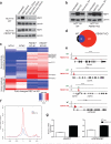
Figure 3. FBXW7 deficiency results in nuclear HSF1 accumulation and prolonged heat-shock response upon exposure to exogenous stress
(a) Loss of FBXW7 results in accumulation of nuclear HSF1 during recovery from heat shock. HCT116 WT and FBXW7 KO cells were heat shocked (42°C for 1 h) following recovery for the indicated time. Nuclear fractions were analyzed by immunoblotting as indicated. (b) Loss of FBXW7 results in accumulation of nuclear HSF1 during recovery from proteotoxic stress. HCT116 WT and FBXW7 KO cells were treated with MG132 (1 μM for 10 h) following recovery for 3 h. Nuclear fractions were analyzed by immunoblotting as indicated. (c) FBXW7 KO cells show defective attenuation of the heat-shock response pathway. Heat maps showing fold changes in expression of HSF1 targets comparing recovery (37°C for 2 h) to heat shock (42°C for 1 h) as determined by high-throughput RNA-Seq. The common targets of HSF1 in HCT116 WT and FBXW7 KO cells after heat shock, as revealed by ChIP-Seq analysis, are displayed on the heat map. Well-characterized genes, positively regulated by HSF1, are indicated. Each column represents a biological replicate. (d) Overlap of genes bound by HSF1 in HCT116 WT and FBXW7 KO cells, under basal conditions. (e) Representative ChIP-Seq tracks for common gene loci between WT and KO (HSPD1/E1, HSP90AB1) and unique for KO gene loci (EIF4A2, CCT6A). The scale corresponds to RPM. (f) Read density profile around TSS's of common HSF1 targets in WT and FBXW7 KO cells. (g) HSPD1 and HSP90AB1 mRNA expression in HCT116 WT and FBXW7 KO cells, under basal conditions (P<0.001 for WT versus KO; unpaired t-test). Error bars indicate mean ± SD, and n=3 independent experiments. Uncropped blots are shown in Supplementary Fig. 8.
Figure 4
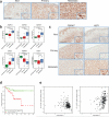
Figure 4. HSF1 protein levels and HSF1 targets expression are associated with metastasis and disease progression in melanoma
(a) IHC staining with anti-HSF1 antibody of the indicated tissue types (dysplastic nevi n=48, primary n=39 and metastatic n=45; P<0.001 for metastatic versus primary or nevi; unpaired t-test; scale bar, 200 μm). (b) Box plots showing expression of HSF1 targets (HSPD1, HSPE1, HSPH1, CKS2), FBXW7 and HSF1, derived from microarray analysis of normal skin (n=4), primary (n=42) and metastatic melanoma (n=40) samples. Whiskers represent the upper and the lower limits of the range. Boxes represent the first and third quartile, and the line represents the median (unpaired t-test). (c) IHC staining with anti-FBXW7 and anti-HSF1 antibodies of the indicated tissue types (dysplastic nevi n=59, primary n=53 and metastatic n=53; P<0.001 for nevi FBXW7 versus HSF1, P<0.01 for primary FBXW7 versus HSF1, P<0.05 for metastatic FBXW7 versus HSF1; unpaired t-test; scale bar, 200 μm). (d) Kaplan-Meier survival curves of patients with tumors expressing high (2D, n = 76) or low levels of nuclear HSF1 (including 0, 1F and 1D, n = 101; P= 0.01; Log rank test). Staining was scored according to the intensity (0-2) and distribution (Focal <50%, Diffuse ≥50%). (e) FBXW7 expression inversely correlates with HSF1 mRNA signature. FBXW7α (NM_033632) expression levels were measured by RNA-Seq in 325 Skin Cutaneous Melanoma (SKCM) samples from TCGA. HSF1 mRNA signature was defined as 1,864 mRNA expressions that were significantly correlated with HSF1 expression (NM_005526) with Spearman Correlation Coefficient > 0.3 and FDR < 5% in TCGA samples. Among them, 830 mRNAs with positive correlations were defined as the positive signature, while 1,034 mRNAs with negative correlations were defined as the negative signature. The first Principal Component (PC1) of the HSF1 positive signature was negatively correlated with FBXW7α expression (Spearman Correlation Coefficient = -0.4; P< 0.001); while the PC1 of the HSF1 negative signature was positively correlated with FBXW7α expression (Spearman Correlation Coefficient = 0.42; P< 0.001).
Figure 5
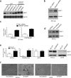
Figure 5. FBXW7 regulates nuclear HSF1 levels and invasion ability in human melanoma
(a) FBXW7 deficiency stabilizes nuclear HSF1 in melanoma. Nuclear fractions from wild type (COLO829, SKMEL28, SKMEL5, SKMEL24) and deficient for FBXW7 (WC00125, WM3862, WM39) melanoma cell lines were analyzed by immunoblotting as indicated. (b) FBXW7 knockdown results in nuclear HSF1 accumulation. 501mel cells were treated with non-coding (NC) siRNA or siRNA against FBXW7. Nuclear fractions were analyzed by immunoblotting as indicated. (c) FBXW7 knockdown results in increased HSF1 targets expression in melanoma. DNAJB1 and HSP90AB1 mRNA expression in 501mel cells treated with non-coding (NC) siRNA or siRNA against FBXW7 (P<0.001 for NC siRNA versus FBXW7 siRNA; unpaired t-test, and n=3 independent experiments) (d) 451Lu cells were treated with the BRAF inhibitor Vemurafenib (2 μM, 9 h) and MEK inhibitor Trametinib (50 nM, 9 h). Nuclear fractions were analyzed by immunoblotting as indicated. (e) FBXW7 depletion results in increased invasion in melanoma. 451Lu, SKMEL239 and A375 cells were infected with the indicated shRNA-encoding lentiviruses. One week after transduction, trans-well invasion assay was performed (P<0.05 for 451Lu shLUC versus shFBXW7, P<0.001 for SKMEL239 scrambled versus shFBXW7, P>0.05 for A375 shLUC versus shFBXW7; n= 10 fields per biological replicate; 4 biological replicates; unpaired t-test). Nuclear fractions were analyzed by immunoblotting as indicated. (f) Representative microphotographs of 451Lu and A375 cells after 8 days of infection with the indicated shRNA-encoding lentiviruses (scale bar, 200 μm). Error bars indicate mean ± SD.. Uncropped blots are shown in Supplementary Fig. 8.
Figure 6
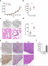
Figure 6. Nuclear HSF1 accumulation upon FBXW7 depletion results in increased metastasis in vivo
In vivo metastasis assay with 451Lu cells transduced with control (shLUC) or FBXW7 shRNA injected subcutaneously in NOG/SCID mice. (a) FBXW7 knockdown does not affect tumor growth or (b) tumor mass (shLUC: n=8; shFBXW7: n=7; n corresponds to number of mice per condition; ns: non significant; unpaired t-test) Error bars indicate mean ± SD. (c) Macroscopic pictures of mouse lungs and H&E-stained sections of lung metastases at termination of the experiment. Black circles mark metastatic foci (scale bar, 100 μm). (d) Whisker plots show the number of metastases per lung. Mean ± SEM is depicted shLUC: n=8; shFBXW7: n=7; n corresponds to number of mice per condition).(****P<0.0001; unpaired t-test). (e) HSF1, Ki67 and H&E-stained sections of subcutaneous tumors resected at termination of the experiment show that FBXW7 knockdown induces HSF1 nuclear accumulation but not proliferation (scale bar, 100 μm). (f) HSF1 is necessary for the increased metastatic potential of melanoma cells upon FBXW7 depletion. 451Lu cells were transduced with control (shLUC) or FBXW7 shRNA. Melanoma cells that acquired increased metastatic potential were subsequently transduced with the indicated shRNA lentiviruses. Transwell invasion assay was performed 3 days after transduction (P<0.001 for shScr versus shHSF1; n= 10 fields per biological replicate; 4 biological replicates; unpaired t-test). Error bars indicate mean ± SD.
Figure 7
Figure 7. HSF1 drives a metastatic-supportive transcriptional program that is affected by FBXW7 expression levels
(a) Genomic distribution of the regions of HSF1 occupancy (promoter, intragenic or intergenic). GSEA enrichment plot showing significant enrichment of the top 600 HSF1 targets for genes linked to metastasis in melanoma (Normalized Enrichment Score, NES=1.72; P=0.006). (b) Representative ChIP-Seq tracks of HSF1 metastasis-related targets. The scale corresponds to Reads Per Million (RPM). (c) mRNA expression analysis of HSF1 metastasis-related targets upon depletion of FBXW7 (P<0.01 for shLUC versus shFBXW7 for ADAM17 or ADAM22 or MTHFD2 and P<0.001 for shLUC versus shFBXW7 for HMGB1 or ITGB3BP; unpaired t-test). Error bars indicate mean ± SD, and n=3 independent experiments.

References

    1. Pelham HR. A regulatory upstream promoter element in the Drosophila hsp 70 heat-shock gene. Cell. 1982;30:517–528. - V体育官网 - PubMed
    1. Westerheide SD, Morimoto RI. Heat shock response modulators as therapeutic tools for diseases of protein conformation. The Journal of biological chemistry. 2005;280:33097–33100. - PubMed
    1. Whitesell L, Lindquist SL. HSP90 and the chaperoning of cancer. Nature reviews. Cancer. 2005;5:761–772. - PubMed
    1. Jolly C, Morimoto RI. Role of the heat shock response and molecular chaperones in oncogenesis and cell death. Journal of the National Cancer Institute. 2000;92:1564–1572. - PubMed
    1. Jin X, Moskophidis D, Mivechi NF. Heat shock transcription factor 1 is a key determinant of HCC development by regulating hepatic steatosis and metabolic syndrome. Cell metabolism. 2011;14:91–103. - V体育官网 - PMC - PubMed

Publication types

"VSports" MeSH terms

Substances