Skip to main page content
U.S. flag

An official website of the United States government

Dot gov

The . gov means it’s official. Federal government websites often end in . gov or . mil VSports app下载. Before sharing sensitive information, make sure you’re on a federal government site. .

Https

The site is secure. The https:// ensures that you are connecting to the official website and that any information you provide is encrypted and transmitted securely. V体育官网.

. 2015 Feb 1:13:49.
doi: 10.1186/s12967-015-0417-0.

"VSports手机版" Exosomes released from human induced pluripotent stem cells-derived MSCs facilitate cutaneous wound healing by promoting collagen synthesis and angiogenesis

Affiliations

"V体育安卓版" Exosomes released from human induced pluripotent stem cells-derived MSCs facilitate cutaneous wound healing by promoting collagen synthesis and angiogenesis

Jieyuan Zhang et al. J Transl Med. .

Abstract

Background: Human induced pluripotent stem cell-derived mesenchymal stem cells (hiPSC-MSCs) have emerged as a promising alternative for stem cell transplantation therapy. Exosomes derived from mesenchymal stem cells (MSC-Exos) can play important roles in repairing injured tissues. However, to date, no reports have demonstrated the use of hiPSC-MSC-Exos in cutaneous wound healing, and little is known regarding their underlying mechanisms in tissue repair VSports手机版. .

Methods: hiPSC-MSC-Exos were injected subcutaneously around wound sites in a rat model and the efficacy of hiPSC-MSC-Exos was assessed by measuring wound closure areas, by histological and immunofluorescence examinations V体育安卓版. We also evaluated the in vitro effects of hiPSC-MSC-Exos on both the proliferation and migration of human dermal fibroblasts and human umbilical vein endothelial cells (HUVECs) by cell-counting and scratch assays, respectively. The effects of exosomes on fibroblast collagen and elastin secretion were studied in enzyme-linked immunosorbent assays and quantitative reverse-transcriptase-polymerase chain reaction (qRT-PCR). In vitro capillary network formation was determined in tube-formation assays. .

Results: Transplanting hiPSC-MSC-Exos to wound sites resulted in accelerated re-epithelialization, reduced scar widths, and the promotion of collagen maturity V体育ios版. Moreover, hiPSC-MSC-Exos not only promoted the generation of newly formed vessels, but also accelerated their maturation in wound sites. We found that hiPSC-MSC-Exos stimulated the proliferation and migration of human dermal fibroblasts and HUVECs in a dose-dependent manner in vitro. Similarly, Type I, III collagen and elastin secretion and mRNA expression by fibroblasts and tube formation by HUVECs were also increased with increasing hiPSC-MSC-Exos concentrations. .

Conclusions: Our findings suggest that hiPSC-MSC-Exos can facilitate cutaneous wound healing by promoting collagen synthesis and angiogenesis. These data provide the first evidence for the potential of hiPSC-MSC-Exos in treating cutaneous wounds VSports最新版本. .


Figures
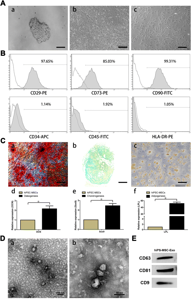
Figure 1
Figure 1
Characterization of human induced pluripotent stem cell-derived mesenchymal stem cells (hiPSC-MSCs) and hiPSC-MSC-derived exosomes (hiPSC-MSC-Exos). (A) Light microscopy images demonstrating morphological changes occurring during hiPSCs differentiation into fibroblast-like cells. (a) Representative cell morphology of hiPSCs before differentiation. (b) Intermediate phase of differentiating the hiPSCs into MSCs. (c) Typical fibroblast-like morphology of cells. (B) Flow cytometric analysis of the surface markers in hiPSC-MSCs. (C) Assessment of the tri-lineage differentiation capacity of iPSC-MSC-like cells. (a) Alizarin Red staining for osteocytes after 3 weeks in culture with osteogenic medium. (b) Alcian Blue staining for chondrocytes after 4 weeks in culture with chondrogenic medium. (c) Oil Red O staining for adipocytes after 2 weeks in culture with adipogenic medium. The qRT-PCR results for OCN (d), Sox9 (e), and LPL (f) after 7 days in culture with osteo-, chondro-, and adipogenic mediun. (D) Transmission electron microscope images of hiPSC-MSC-Exos morphology. Scale bars = 100 nm and 50 nm, respectively. (E) Detection of CD9, CD63, and CD81 incorporation into hiPSC-MSC-Exos by western blotting.
Figure 2
Figure 2
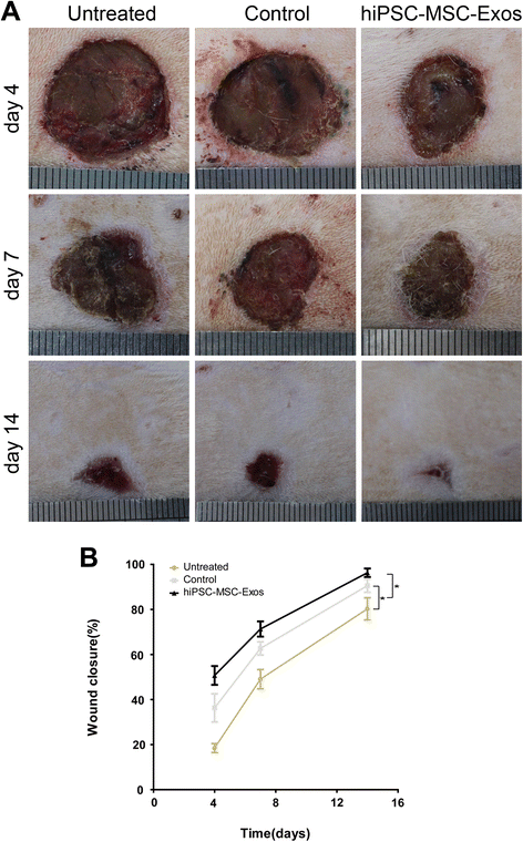
Rats macroscopic appearances of cutaneous wounds treated with PBS, MesenGro hMSC medium, or hiPSC-MSC-Exos. (A) Gross view of wounds treated with PBS, MesenGro hMSC medium, or hiPSC-MSC-Exos at 4, 7, and 14 days. (B) The effects of treatment with PBS, MesenGro hMSC medium, or hiPSC-MSC-Exos on wound closure at 4, 7, and 14 days. *P < 0.05.
Figure 3
Figure 3
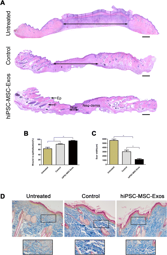
Rats histological analyses of cutaneous wounds treated with PBS, MesenGro hMSC medium, or hiPSC-MSC-Exos. (A) H&E staining of wound sections following treatment with PBS, MesenGro hMSC medium, or hiPSC-MSC-Exos at 14 days post-wounding. The double-headed arrows indicate the edges of the scar. The effects of PBS, MesenGro hMSC medium, or hiPSC-MSC-Exos on wound re-epithelialization (B) and scar widths (C) at 14 days post-wounding. (D) Evaluation of collagen maturity by Masson’s trichrome staining of wounds following treatment with PBS, MesenGro hMSC medium, or hiPSC-MSC-Exos at 14 days post-wounding. Scale bar = 500 μm. *P < 0.05; Ep, Epithelium; F, Follicle.
Figure 4
Figure 4
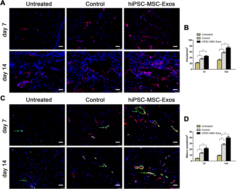
Immunofluorescence analyses of newly formed vessels and mature vessels. (A) CD31 (red arrows) Immunofluorescence staining of wound sections treated with PBS, MesenGro hMSC medium, or hiPSC-MSC-Exos at 7 and 14 days post-wounding. Scale bar = 50 μm. (B) Enumeration of newly formed vessels in wounds after treatment with PBS, MesenGro hMSC medium, or hiPSC-MSC-Exos at 7 and 14 days post-wounding. (C) Immunofluorescent triple staining of wound sections treated with PBS, MesenGro hMSC medium or hiPSC-MSC-Exos at 7 and 14 days post-wounding. Endothelial cells (CD31), smooth muscle cells (α-SMA), and cell nuclei (DAPI) fluoresced with red, green, and blue colours, respectively. Mature vessels (green arrows) were dually positive for CD31 and α-SMA. Scale bar = 50 μm. (D) Enumeration of mature vessels in wounds after treatment with PBS, MesenGro hMSC medium, or hiPSC-MSC-Exos at 7 and 14 days post-wounding. *P < 0.05.
Figure 5
Figure 5
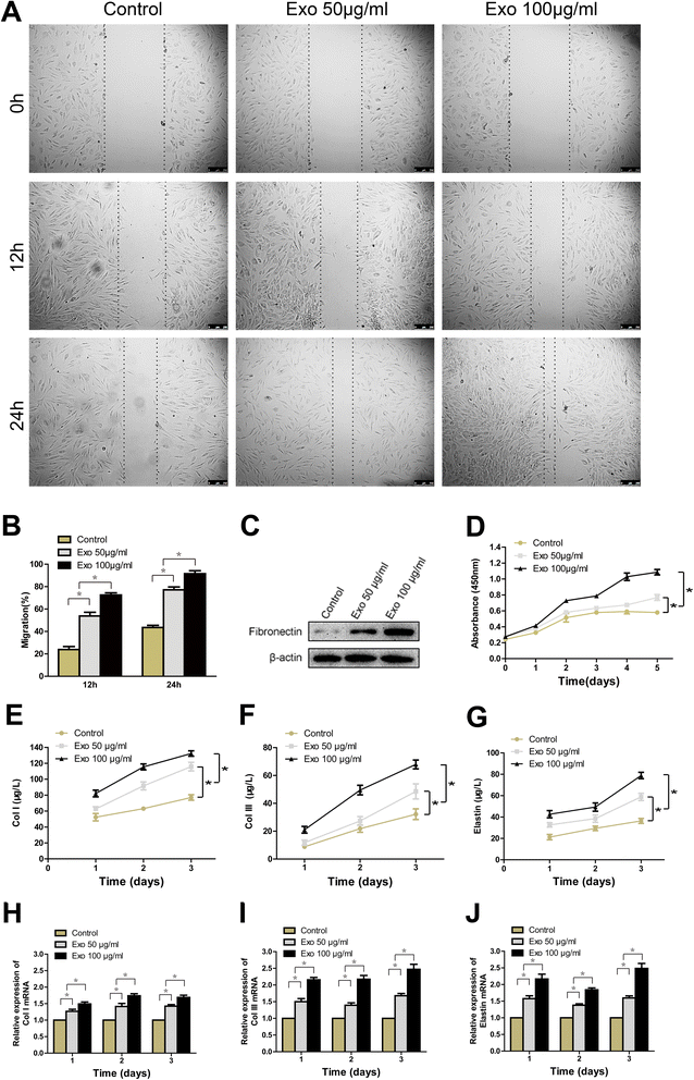
The effects of exosomes on the proliferation, migration, and collagen, elastin secretion of human fibroblasts. The light microscopy images (A) and migration rates (B) of human fibroblasts into scratch sites following growth in MesenGro hMSC medium containing 0, 50, or 100 μg/mL hiPSC-MSC-Exos for 12 or 24 h. Scale bar = 250 μm. (C) Fibronectin protein expression of human fibroblasts treated with MesenGro hMSC medium containing 0, 50, or 100 μg/mL hiPSC-MSC-Exos for 24 h. (D) Human fibroblasts proliferation after growth in MesenGro hMSC medium containing 0, 50, or 100 μg/mL hiPSC-MSC-Exos was detected with a CCK-8 kit over 5 days. Secretion of Col I (E), III (F) and elastin (G) by human fibroblasts after growth in MesenGro hMSC medium containing 0, 50, or 100 μg/mL hiPSC-MSC-Exos over 3 days. (G) The Col I (H), III (I) and elastin (J) mRNA expression of human fibroblasts treated with MesenGro hMSC medium containing 0, 50, or 100 μg/mL hiPSC-MSC-Exos over 3 days. *P < 0.05.
Figure 6
Figure 6
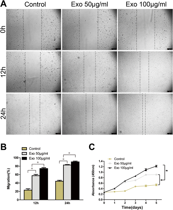
The effects of exosomes on HUVECs proliferation and migration. The light microscopy images (A) and migration rates (B) of HUVECs into the scratched area of monolayers following growth in M200 medium containing 0, 50, or 100 μg/mL hiPSC-MSC-Exos for 12 or 24 h. Scale bar = 250 μm. (C) The proliferation of HUVECs grown in M200 containing 0, 50, or 100 μg/mL hiPSC-MSC-Exos was detected over 5 days, using a cell-counting kit.*P < 0.05.
Figure 7
Figure 7
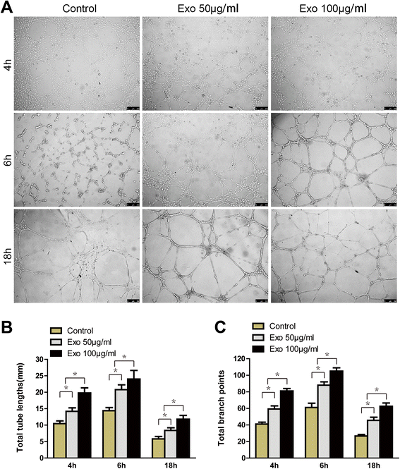
The effects of exosomes on tube formation by HUVECs. (A) HUVECs tube formation was studied by growing cells in Matrigel in M200 medium containing 0, 50, or 100 μg/mL hiPSC-MSC-Exos. Scale bar = 250 μm. Total tube lengths (B) and branch points (C) of HUVECs following growth in M200 medium containing 0, 50, or, 100 μg/mL hiPSC-MSC-Exos for 4, 6, or 18 h. * P < 0.05.

References

    1. Nakamura Y, Ishikawa H, Kawai K, Tabata Y, Suzuki S. Enhanced wound healing by topical administration of mesenchymal stem cells transfected with stromal cell-derived factor-1. Biomaterials. 2013;34:9393–400. doi: 10.1016/j.biomaterials.2013.08.053. - DOI - PubMed
    1. Lee KB, Choi J, Cho SB, Chung JY, Moon ES, Kim NS, et al. Topical embryonic stem cells enhance wound healing in diabetic rats. J Orthop Res. 2011;29:1554–62. doi: 10.1002/jor.21385. - DOI - PubMed
    1. Biazar E, Keshel SH. The healing effect of stem cells loaded in nanofibrous scaffolds on full thickness skin defects. J Biomed Nanotechnol. 2013;9:1471–82. doi: 10.1166/jbn.2013.1639. - DOI - PubMed
    1. Xin Y, Wang YM, Zhang H, Li J, Wang W, Wei YJ, et al. Aging adversely impacts biological properties of human bone marrow-derived mesenchymal stem cells: implications for tissue engineering heart valve construction. Artif Organs. 2010;34:215–22. doi: 10.1111/j.1525-1594.2009.00824.x. - DOI - PubMed
    1. Zaim M, Karaman S, Cetin G, Isik S. Donor age and long-term culture affect differentiation and proliferation of human bone marrow mesenchymal stem cells. Ann Hematol. 2012;91:1175–86. doi: 10.1007/s00277-012-1438-x. - DOI - PubMed

Publication types

MeSH terms

"VSports手机版" LinkOut - more resources