"VSports app下载" Skip to main page content
U.S. flag

An official website of the United States government

Dot gov

The . gov means it’s official. Federal government websites often end in . gov or . mil. Before sharing sensitive information, make sure you’re on a federal government site VSports app下载. .

Https

The site is secure. The https:// ensures that you are connecting to the official website and that any information you provide is encrypted and transmitted securely V体育官网. .

. 2015;13(1):64-79.
doi: 10.2174/1570162x13666150121111548.

Didehydro-cortistatin A inhibits HIV-1 Tat mediated neuroinflammation and prevents potentiation of cocaine reward in Tat transgenic mice

Affiliations

VSports最新版本 - Didehydro-cortistatin A inhibits HIV-1 Tat mediated neuroinflammation and prevents potentiation of cocaine reward in Tat transgenic mice

Sonia Mediouni (V体育安卓版) et al. Curr HIV Res. 2015.

Abstract

HIV-1 Tat protein has been shown to have a crucial role in HIV-1-associated neurocognitive disorders (HAND), which includes a group of syndromes ranging from undetectable neurocognitive impairment to dementia. The abuse of psychostimulants, such as cocaine, by HIV infected individuals, may accelerate and intensify neurological damage. On the other hand, exposure to Tat potentiates cocaine-mediated reward mechanisms, which further promotes HAND. Here, we show that didehydro-Cortistatin A (dCA), an analog of a natural steroidal alkaloid, crosses the blood-brain barrier, cross-neutralizes Tat activity from several HIV-1 clades and decreases Tat uptake by glial cell lines. In addition, dCA potently inhibits Tat mediated dysregulation of IL-1β, TNF-α and MCP-1, key neuroinflammatory signaling proteins. Importantly, using a mouse model where doxycycline induces Tat expression, we demonstrate that dCA reverses the potentiation of cocaine-mediated reward VSports手机版. Our results suggest that adding a Tat inhibitor, such as dCA, to current antiretroviral therapy may reduce HIV-1-related neuropathogenesis. .


Conflict of interest statement

Conflict of interest

The authors declare no conflict of interest.

Figures

Figure 1
Figure 1
Schematic representation of Tat protein’s domains. NLS: nuclear localization signal; RGD: Arg-gly-Asp.
Figure 2
Figure 2
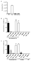
dCA inhibits Tat induced HIV-1 LTR activation. (A) Tat activates the HIV-LTR. HeLa-CD4-LTR-Luc cells were treated with recombinant Tat protein and luciferase activity was measured 24 h later. Results are represented as relative light units (RLU, luciferase activity per protein concentration). DN: denatured; DN B: denatured buffer. (B, C) dCA inhibits Tat transactivation of the HIV-1 LTR. dCA prevents transactivation of the HIV-1 promoter from HeLa-CD4-LTR-Luc cells by recombinant Tat (B) or Transfected Tat (C). Raltegravir (Ralt.) was used as negative control. Tat mutated in the basic domain (Tat Mut), does not transactivate the promoter, and is used as negative control. Results obtained with Tat in the presence of DMSO were set as 100 %. EV: Empty vector control. Results are representative of three independent experiments.
Figure 3
Figure 3
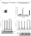
dCA interacts with the basic domain of Tat. (A) Interaction of Tat with Bio-dCA by Dot blot. Tat or BSA were spotted onto a nitrocellulose membrane, incubated with Bio-dCA and the interaction revealed using an anti-biotin antibody. (B) Schematic depiction of the sequences encompassed by Tat peptides. (C) Bio-dCA recognizes peptides of the basic domain of Tat and the full-length Tat in ELISA. Tat or Tat peptides were coated onto 96-well plates, and incubated with Bio-dCA, the interaction was revealed using anti-biotin antibody. (D, E) Inhibition of Tat from different HIV subtypes by dCA. (D) Upper: HeLa cells stably expressing firefly luciferase driven by the HIV-1 LTR from subtype B were transfected with Flag-Tat from subtypes A through E or GFP as a control or (E) HeLa cells were transfected with firefly luciferase reporters driven by the HIV-1 LTR from subtypes A through E with or without Flag-Tat from matching subtypes. At 24 h post transfection, cells were treated with DMSO or 100 nM dCA and 48 h later, firefly luciferase activity and protein concentration were determined. Data are plotted as relative luciferase units (RLU) as percent of DMSO set to 100 %. Lower: Cell lysates from above were subjected to western blot detection of Flag-Tat in the presence or absence of 100 nM dCA. Actin serves as a loading control. Results are representative of three independent experiments.
Figure 4
Figure 4
dCA reduces the extracellular uptake of Tat. Recombinant Tat was added to the PMA differentiated monocyte cell line U937, used as microglia-like cells, and to the astrocyte cell line U87MG, in the presence of DMSO, dCA or Raltegravir (Ralt.). After 4 h incubation, cells were thoroughly washed and intracellular Tat detected by Western blot, with actin serving as loading control. Results are represented as the ratio of cellular up-taken Tat per total Tat input. Shown is the mean ± SD between two independent experiments.
Figure 5
Figure 5
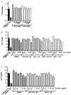
Effect of dCA and Tat on the cellular viability of the astrocytic cell line U87MG. A) MTT assay on U87MG cells incubated with increasing concentrations of dCA, Raltegravir (Ralt.) and DMSO for 48 h. B, C) MTT assay on U87MG cells incubated with dCA and Tat. Cells were incubated with Tat (B) or Tat mut (C) in presence of dCA or Ralt. at 250 nM or 1 μM for 24 h. Raltegravir and DMSO (0.1%) were used as controls. DMSO 10% was used as positive control. Results are representative of three independent experiments.
Figure 6
Figure 6
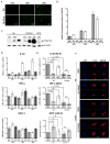
dCA reverses Tat-induced cytokines and chemokine dysregulation and alters Tat’s nucleolar localization. (A) Flag-Tat or Flag-Tat Mut DNA were co-transfected with eGFP DNA into U87MG cells. Transfection efficiency was assessed by measuring GFP fluorescence at 24 h, 36 h and 48 h post-transfection. (B) Analysis of mRNA Tat and Tat Mut expression after transfection at different time points. Total RNA was extracted at each time point and converted to cDNA. Quantification was performed by RT-qPCR using primers recognizing Tat and Tat Mut. Results were normalized as copies of Tat per copy of GAPDH mRNAs. The arbitrary value of 1 was assigned to the amount of Tat Mut generated after 24 h. RNA samples not reverse transcribed were used as negative control. (C) Analysis of Tat and Tat Mut protein expression at different time points. Lysates from cells were subjected to western blot analysis. Anti-Flag antibody was used to reveal Tat and Tat Mut. GFP transfected in astrocyte cells and expressed for 48 h was used as negative control. Actin was used as loading control. (D) Analysis of IL1-β, TNF-α, and MCP-1 mRNA expression induced by Tat and Tat Mut transfection at different time points. Results were normalized to copies of GAPDH mRNA, with a value of 1 assigned to the Tat Mut 24 h. (E) Analysis of IL-1β, TNF-α and MCP-1 mRNA induced by Tat and Tat Mut at 48 h post transfection upon dCA treatment. Results were normalized to copies of GAPDH and Tat (or Tat Mut) mRNA, with value of 1 assigned to the Tat alone. Raltegravir (Ralt.) was used as negative control. A two-tailed paired t-test was used for statistical comparisons of the different conditions. NS: not significant. Shown is the mean ± SD between three independent experiments. (F) dCA excludes Tat from the nucleolus U87MG cells. Confocal microscopy analysis of the sub-cellular localization of transfected Flag-Tat or Flag-Tat Mut after 24 h in presence of DMSO or dCA in astrocyte cells. Flag-Tat was recognized with anti-flag and Alexa Fluor 568 anti-IgG. Magnification 60X.
Figure 7
Figure 7
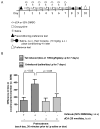
Tat potentiation of cocaine-conditioned place preference is reverted by dCA. (A) Experimental design for cocaine-conditioned place preference testing. GT-tg bigenic mice were treated daily for 7 days with the vehicle (50 % DMSO, i.c.v.) or dCA (20 nmol, i.c.v.) for 30 min before i.p injecting saline or Dox (100 mg/kg/d) to induce Tat expression. This was followed by 2 days of cocaine place-conditioning before the final conditioning place preference test. (B) dCA reverses the cocaine-conditioned place preference induced by Tat. All mice treated with dCA demonstrated cocaine CPP responses that did not significantly differ from vehicle-treated, uninduced mice (p>0.05, Tukey’s post hoc test). However, dCA-pretreatment (n=10) prevented Tat-induced potentiation of cocaine-CPP (p<0.05; Tukey’s post hoc test). Bars = 16–20 mice

References

    1. Kaul M. HIV-1 associated dementia: update on pathological mechanisms and therapeutic approaches. Curr Opin Neurol. 2009;22(3):315–20. - PMC - PubMed
    1. Kaul M, Zheng J, Okamoto S, et al. HIV-1 infection and AIDS: consequences for the central nervous system. Cell Death Differ. 2005;12(Suppl 1):878–92. - PubMed
    1. Bagashev A, Sawaya BE. Roles and functions of HIV-1 Tat protein in the CNS: an overview. Virol J. 2013;10:358. - PMC (V体育安卓版) - PubMed
    1. Gonzalez-Scarano F, Martin-Garcia J. The neuropathogenesis of AIDS. Nat Rev Immunol. 2005;5(1):69–81. - PubMed
    1. Gorry PR, Howard JL, Churchill MJ, et al. Diminished production of human immunodeficiency virus type 1 in astrocytes results from inefficient translation of gag, env, and nef mRNAs despite efficient expression of Tat and Rev. J Virol. 1999;73(1):352–61. - "VSports手机版" PMC - PubMed

Publication types

MeSH terms

Substances