Skip to main page content
U.S. flag

An official website of the United States government

Dot gov

The VSports app下载. gov means it’s official. Federal government websites often end in . gov or . mil. Before sharing sensitive information, make sure you’re on a federal government site. .

Https

The site is secure. The https:// ensures that you are connecting to the official website and that any information you provide is encrypted and transmitted securely V体育官网. .

. 2015 Jan 15;11(1):e1004593.
doi: 10.1371/journal.ppat.1004593. eCollection 2015 Jan.

DNA is an antimicrobial component of neutrophil extracellular traps

Affiliations

DNA is an antimicrobial component of neutrophil extracellular traps

Tyler W R Halverson et al. PLoS Pathog. .

"VSports" Abstract

Neutrophil extracellular traps (NETs) comprise an ejected lattice of chromatin enmeshed with granular and nuclear proteins that are capable of capturing and killing microbial invaders. Although widely employed to combat infection, the antimicrobial mechanism of NETs remains enigmatic VSports手机版. Efforts to elucidate the bactericidal component of NETs have focused on the role of NET-bound proteins including histones, calprotectin and cathepsin G protease; however, exogenous and microbial derived deoxyribonuclease (DNase) remains the most potent inhibitor of NET function. DNA possesses a rapid bactericidal activity due to its ability to sequester surface bound cations, disrupt membrane integrity and lyse bacterial cells. Here we demonstrate that direct contact and the phosphodiester backbone are required for the cation chelating, antimicrobial property of DNA. By treating NETs with excess cations or phosphatase enzyme, the antimicrobial activity of NETs is neutralized, but NET structure, including the localization and function of NET-bound proteins, is maintained. Using intravital microscopy, we visualized NET-like structures in the skin of a mouse during infection with Pseudomonas aeruginosa. Relative to other bacteria, P. aeruginosa is a weak inducer of NETosis and is more resistant to NETs. During NET exposure, we demonstrate that P. aeruginosa responds by inducing the expression of surface modifications to defend against DNA-induced membrane destabilization and NET-mediated killing. Further, we show induction of this bacterial response to NETs is largely due to the bacterial detection of DNA. Therefore, we conclude that the DNA backbone contributes both to the antibacterial nature of NETs and as a signal perceived by microbes to elicit host-resistance strategies. .


Conflict of interest statement

The authors have declared that no competing interests exist.

V体育官网入口 - Figures
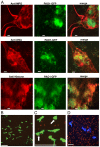
Figure 1
Figure 1. P. aeruginosa PAO1 is trapped by human and mouse neutrophil extracellular traps.
(A) PMA-induced NETs trapped P. aeruginosa Tn7::gfp and contained myeloperoxidase (MPO), DNA and histones when visualized by immunofluorescence with antibodies from autoimmune patient sera (see Methods). Representative images of the NET components (left), Gfp-tagged PAO1 (middle), and the merged (right) are presented. (B) NETosis in the skin of mice infected with ChFP-labeled P. aeruginosa as visualized by Sytox green stained extracellular DNA structures. Scale bar: 40 μm. (C) Arrows indicate ChFP-labeled P. aeruginosa trapped by Sytox green-stained NETs in vivo. (D) ChFP-labeled P. aeruginosa is phagocytosed by neutrophils during a skin infection model. Neutrophils (blue) are visualized with anti-mouse GR-1 antibody. Scale bar: 25 μm.
Figure 2
Figure 2. Quantification of NETosis in human neutrophils ex vivo and mouse neutrophils during an infection.
(A) Human neutrophils were stimulated with PMA, P. aeruginosa, E. coli and S. aureus and Sytox green fluorescence was measured as an indicator of DNA release by NETosis after 1 hr stimulation. Exogenous DNase was added as a control to confirm extracellular DNA presence in NETs, indicated by a plus sign (+). Asterisks denote a significant difference in extracellular DNA release between stimulated and unstimulated neutrophils (white bar) (**P<0.01, ***P<0.001). Each value shown is an average from 6 replicates with error bars representing the standard error. (B) The total number of NETs in uninfected mice, or infected mice with P. aeruginosa PAO1 or S. aureus and (C) the total NET area quantified. # denotes a significant difference in NET area and number in P. aeruginosa PAO1 infected mice compared to the uninfected control (#P<0.05). Asterisks denote a significant difference in NET area and number in mice infected with P. aeruginosa compared to S. aureus infected mice (***P<0.001).
Figure 3
Figure 3. P. aeruginosa, E. coli and S. aureus differ in their ability to tolerate the bactericidal effects of NETs.
(A) Survival analysis of 1 × 10⁷ CFU P. aeruginosa, S. aureus and E. coli upon exposure to NETs (MOI 10:1). Bacterial viability was determined by direct plate counts (CFU/ml) before and after 4 hour incubation with PMA-activated neutrophils and was normalized to bacterial counts in the absence of neutrophils. DNase I was added exogenously 0.5 hour prior to the end of the experiment to degrade NETs and ensure accurate counts of recoverable colonies. Results are representative of three independent replicates. ***P<0.001 versus P. aeruginosa PAO1. °°°P<0.001 versus non-DNase condition by one-way ANOVA with Bonferroni post tests. (B) Bacterial viability was determined by measuring luminescence from 1 × 10⁷ CFU lux-tagged PAO1::p16Slux or E. coli DH5α/pσ70-lux in the absence or presence of PMA-induced NETs (MOI 10:1). Errors bars represent SEM from six replicates. All experiments were performed at least three times.
Figure 4
Figure 4. Extracellular DNA exerts bactericidal activity through cation chelation-mediated disruption of the bacterial outer membrane.
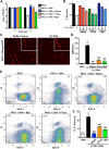
(A) Survival analysis of 1 × 10⁷ CFU P. aeruginosa PAO1 coincubated with 0.125% (w/v) DNA or DNA pretreated with DNase I, PTase or 5 mM Mg2+. Bacterial counts were performed before (0) and after two hours treatment with DNA (2). Results are representative of three independent experiments. Error bars represent the standard deviation (SD) from eight replicates. (B) High, medium and low concentrations of DNase (430kU, 43kU, 4.3kU), PTase (50 U, 10 U, 1 U) or excess Mg2+ (5 mM, 500 μM, 5 μM) leads to increased levels of protection from killing with 0.15% DNA (w/v). (C) Visualization of the outer membrane integrity of P. aeruginosa PAO1::OM-lipoChFP expressing an outer membrane-localized mCherry fluorescent lipoprotein [38] immediately after 2% (w/v) DNA-exposure. Insets represent increased magnification of presented micrographs. Scale bar: 10 μM. (D) Quantification of ChFP-rich OMV generation in the field of view from 6 representative images generated from bacteria-DNA coincubation as described in (C) or DNA pretreated with DNase, PTase and Mg2+. Error bars represent SD from 6 fields of view. (E) Flow cytometry of DNA-exposed P. aeruginosa PAO1 using SYTO9-PI dual staining as a measure of membrane-compromised bacteria [28, 32]. 2.5 × 10⁷ CFU P. aeruginosa PAO1 were exposed to 0.0125% DNA alone or pretreated as in (A) then immediately analyzed by the collection of positive events (N = 50 000) by BD LSRII. Numbers in corners represent the % of 50 000 events that fall into each quadrant gate. (F) Quantification of membrane-compromised, PI-stained P. aeruginosa PAO1 as measured by flow cytometry. Mean percent PI stained was derived from the average of three replicates (each with N = 50 000 for each plot) in each exposure condition as in (E). *** denotes a significant difference between the control and 0.125% DNA sample. ### and # denote a statistically significant difference, P<0.01 and P<0.05, respectively, between DNA alone sample and pretreated samples. Two-tailed student t-tests were performed to test for significant differences.
Figure 5
Figure 5. Neutralizing the cation chelating activity of the DNA backbone of NETs protects bacteria.
(A) Percent survival of P. aeruginosa PAO1 and E. coli DH5α as determined by direct plate counts (CFU/ml) before and after 4 hour incubation with PMA-activated neutrophils or combined treatment of NETS with DNase, PTase or Mg2+. Error bars are SEM from 6 replicates. ** or *** denotes a statistically significant difference (P<0.05 or P<0.01, respectively) between NET-alone versus NET and enzymatic or excess cation treatments, as determined by one-way ANOVA with Bonferroni post tests. (B) Luminescence-based viability as a real-time measure of P. aeruginosa PAO1::p16Slux survival in the presence of NETs alone, or combined treatment of NETS with DNase I, PTase or Mg2+. ### denotes a statistically significant difference of P<0.001 between NET-challenged PAO1 versus PAO1 alone (white). ***P<0.001 versus NET killed samples (black). (C) Flow cytometry of P. aeruginosa PAO1 (2 × 10⁷ CFU) coincubated for four hours with PMA-stimulated neutrophils alone (1 × 10⁶; MOI: 10) or with the addition of DNase I, PTase and 5 mM Mg2+. N = 50 000 for each plot. Numbers in each corner represent the % of 50 000 events that fall into each quadrant gate.
Figure 6
Figure 6. Structurally intact NETs still possess histones and MPO after treatment with excess Mg2+ and PTase.
Neutrophil extracellular traps were visualized with (A) Sytox Green staining of DNA and anti-histone primary antibodies. (B) Neutrophil extracellular traps were visualized with DAPI staining of DNA and anti-MPO primary antibodies. NETs were not observed in unactivated neutrophils, but were present in PMA-induced NETs that were treated with either excess 5 mM Mg2+ or exogenous PTase. Alexa Fluor 647-conjugated secondary antibodies were used to visualize histone H1 and MPO. Representative immunofluorescence images were merged to show overlap of histone H1 and MPO with structurally intact NETs. Scale bars, 10 µm.
Figure 7
Figure 7. Pseudomonas aeruginosa responds to DNA in neutrophil extracellular traps and induces protective bacterial surface modifications.
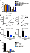
(A) Survival analysis of 1 × 10⁷ CFU wild-type P. aeruginosa PAO1 or mutants with defects in the aminoarabinose-LPS modification (PA3553::lux) or in spermidine synthesis (PA4774::lux) after coincubation with 0.125% DNA. *** denotes a statistically significant difference between time 0 and 2 hours post coincubation with 0.125% DNA. ### denotes a statistically significant difference of P<0.001 between wild-type and mutant P. aeruginosa when exposed to 0.125% DNA. Two-tailed student t-tests were performed to test for significant differences. Error bars represent SD from eight replicates. (B) 2 × 10⁷ CFU P. aeruginosa PAO1 spermidine synthesis (PA4774::lux) and aminoarabinose modification (PA3553::lux) transcriptional reporter strains were incubated with PMA-activated neutrophils (MOI 10:1) and gene expression (luminescence as quantified by CPS) was measured every 20 minutes in the absence (empty squares) and presence (solid circles) of NETs. Error bars represent SEM from six replicates. (C) The effect of DNase or 2 mM Mg2+ treatment on NET-mediated gene induction of 2 × 10⁷ CFU PA4774::lux or PA3553::lux after four hours of coincubation (MOI 10:1). *P<0.05, ***P<0.001 versus bacteria alone (white bar), #P<0.05, ###P<0.001 versus NET exposure (black bar) as determined by one-way ANOVA with Bonferroni post tests. (D) Bacterial survival analysis of 2 × 10⁷ CFU NET-exposed P. aeruginosa PAO1 wild-type, aminoarabinose modification mutant PA3553::lux, or the spermidine synthesis mutants PA47743/4::lux, PA4774::lux (MOI 10:1) Error bars represent the SEM from 6 replicates. *** denotes a statistically significant difference of P<0.001 versus wild-type survival as determined by one-way ANOVA with Bonferroni post tests. All assays were conducted at least three times and representative data is presented.
Figure 8
Figure 8. Bacterial species differ in their susceptibility to killing by DNA and histones.
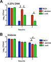
(A) P. aeruginosa PAO1, S. aureus and E. coli killing assay in the presence of 0.25% DNA (w/v). PAO1 was significantly more sensitive at both eDNA concentrations relative to the highly tolerant S. aureus. (B) P. aeruginosa PAO1, S. aureus and E. coli killing assay in presence of 1.25 µg/mL histones. Bacterial survival was quantified after 1 and 2 hours by colony count, and statistical significance assessed by 2-tailed student t-tests. * denotes statistical differences (P<0.05) between the indicated time point and the initial bacterial count, while # indicates significant differences (P<0.05) between bacterial species. The values shown are the mean plus standard deviation from 8 samples. Experiments were repeated three times and the data shown is from one representative experiment.

"VSports app下载" Comment in

V体育平台登录 - References

    1. Brinkmann V, Reichard U, Goosmann C, Fauler B, Uhlemann Y, et al. (2004) Neutrophil extracellular traps kill bacteria. Science 303(5663): 1532–1535. 10.1126/science.1092385 - "VSports app下载" DOI - PubMed
    1. Brinkmann V, Zychlinsky A. (2012) Neutrophil extracellular traps: Is immunity the second function of chromatin? J Cell Biol 198(5): 773–783. 10.1083/jcb.201203170 - DOI - PMC - PubMed
    1. Urban CF, Reichard U, Brinkmann V, Zychlinsky A. (2006) Neutrophil extracellular traps capture and kill Candida albicans yeast and hyphal forms. Cell Microbiol 8(4): 668–676. 10.1111/j.1462-5822.2005.00659.x - DOI - PubMed
    1. Clark SR, Ma AC, Tavener SA, McDonald B, Goodarzi Z, et al. (2007) Platelet TLR4 activates neutrophil extracellular traps to ensnare bacteria in septic blood. Nat Med 13(4): 463–469. 10.1038/nm1565 - DOI - PubMed
    1. Lu T, Kobayashi SD, Quinn MT, Deleo FR. (2012) A NET outcome. Front Immunol 3: 365 10.3389/fimmu.2012.00365 - DOI - PMC - PubMed

V体育2025版 - Publication types