V体育平台登录 - Skip to main page content
U.S. flag

An official website of the United States government

Dot gov

The . gov means it’s official. Federal government websites often end in . gov or . mil VSports app下载. Before sharing sensitive information, make sure you’re on a federal government site. .

Https

The site is secure. The https:// ensures that you are connecting to the official website and that any information you provide is encrypted and transmitted securely V体育官网. .

. 2015 Jun 2;112(22):6820-7.
doi: 10.1073/pnas.1411258112. Epub 2014 Dec 23.

ATRX tolerates activity-dependent histone H3 methyl/phos switching to maintain repetitive element silencing in neurons

Affiliations

ATRX tolerates activity-dependent histone H3 methyl/phos switching to maintain repetitive element silencing in neurons

Kyung-Min Noh et al. Proc Natl Acad Sci U S A. .

Erratum in

Abstract

ATRX (the alpha thalassemia/mental retardation syndrome X-linked protein) is a member of the switch2/sucrose nonfermentable2 (SWI2/SNF2) family of chromatin-remodeling proteins and primarily functions at heterochromatic loci via its recognition of "repressive" histone modifications [e. g. , histone H3 lysine 9 tri-methylation (H3K9me3)]. Despite significant roles for ATRX during normal neural development, as well as its relationship to human disease, ATRX function in the central nervous system is not well understood VSports手机版. Here, we describe ATRX's ability to recognize an activity-dependent combinatorial histone modification, histone H3 lysine 9 tri-methylation/serine 10 phosphorylation (H3K9me3S10ph), in postmitotic neurons. In neurons, this "methyl/phos" switch occurs exclusively after periods of stimulation and is highly enriched at heterochromatic repeats associated with centromeres. Using a multifaceted approach, we reveal that H3K9me3S10ph-bound Atrx represses noncoding transcription of centromeric minor satellite sequences during instances of heightened activity. Our results indicate an essential interaction between ATRX and a previously uncharacterized histone modification in the central nervous system and suggest a potential role for abnormal repetitive element transcription in pathological states manifested by ATRX dysfunction. .

Keywords: ATRX; H3K9me3S10ph; crystal structure; heterochromatin; neuron V体育安卓版. .


Conflict of interest statement

The authors declare no conflict of interest.

VSports app下载 - Figures
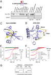
Fig. 1.
Fig. 1.
ADDATRX binding to the H3 N terminus is maintained in the presence or absence of H3S10ph. (A) Primary amino acid sequence of the histone H3 N-terminal tail (1–14). The H3K9methyl-S10phos switch is shown above the sequence. (B) IP assays examining binding interactions between the ADDATRX domain and modified histone H3 peptides, as indicated. High salt: IP assays were performed in buffer containing 500 mM NaCl. (C and D) Cocrystal structures of domain–peptide complexes for the CD of HP1ɑ (C, prepared from PDB ID code 1KNE) and the ADDATRX domain (D) bound to H3K9me3 (1–15) and H3K9me3S10ph (1–15) peptides, respectively. In both panels, H3 peptides are indicated in yellow, and amino acids of the polar residues comprising the S10ph recognition state are displayed in pink. (E and F) ITC curves for the HP1ɑ CD (E) and ADDATRX domain (F) with H3 (1–15) peptides containing various modifications. (G) Peptide IPs examining binding between the HP1ɑ CD/ADDATRX domain and modified histone H3 peptides in the presence or absence of phosphatase treatment.
Fig. 2.
Fig. 2.
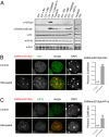
Activity-dependent H3K9me3S10ph colocalizes with Atrx, but not HP1ɑ, in primary neurons. (A) Western blot analysis of acid-extracted histones from primary cortical neurons in the presence or absence of PKA activation (Forskolin) or depolarization (KCl) in combination with numerous pharmacological inhibitors (Bis, Bisindolylmaleimide I, a PKC inhibitor; H89, H-89 dihydrochloride, a PKA inhibitor; PD98059, a MEK kinase inhibitor; Calyculin A, a potent PP1 and PP2A inhibitor; and EGTA, Ethylene glycol tetraacetic acid, a chelator for calcium ions). HP1α and Atrx protein expression were similarly examined. (B and C) Double immunofluorescence of neuronal nuclei ± stimulation (Forskolin) using antibodies reactive against H3K9me3S10ph (red) and Atrx (B) or HP1ɑ (C) (green). (Scale bar: 5 μm.) Neuronal nuclei were identified with DAPI. Percentages of colocalization between either Atrx or HP1α and H3K9me3S10ph are included to the right (n = 8 cells per group). Data are presented as mean ± SEM. N.D., not detectable.
Fig. 3.
Fig. 3.
Heterochromatic patterns of Atrx enrichment are maintained in the presence of H3K9me3S10ph after neuronal stimulation. (A) Genomic distribution of Atrx peaks in nonstimulated primary neurons. (B) Odds ratio analysis of correlations between Atrx peak enrichment and gene expression (binned as high, medium, and low) in nonstimulated neurons. P values were determined using the Fisher’s exact test and are labeled in red. (C) Genomic distribution of Atrx differential sites after neuronal stimulation (KCl depolarization). (D) Relative enrichment of Atrx and H3K9me3S10ph at different classes of repetitive sequences in response to neuronal stimulation (control enrichment values are normalized “1” represented by a dashed line). To do so, ChIP reads were directly aligned to a library of mouse consensus repetitive sequences (i.e., the repeatome). Data are represented as stimulation-induced fold enrichments over input in comparison with control neurons. (E) ChIP-qPCR validation (biological triplicates) of H3K9me3S10ph enrichment in response to neuronal stimulation at specific repetitive sequences, as well as at a highly active gene (Rps19), which served as a negative control. N.D., not detectable. Where appropriate, data are represented as mean ± SEM.
Fig. 4.
Fig. 4.
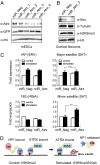
Atrx depletion increases minor satellite transcription in stimulated neurons. (A) Western blot analysis of GFP+ FACS-sorted mouse ESCs transfected with a negative control (miR_Neg) or Atrx-specific miR constructs coexpressing with GFP (sets 1–4 from Invitrogen). (B) Western blot analysis of primary cortical neurons transduced with Lenti-GFP-miR_Atrx_3 vs. miR_Neg. (C) RT-qPCR analysis of repeat element transcription in the presence or absence of KCl stimulation after infection with Lenti-GFP-miR_Neg vs. miR_Atrx_3. n = 3 biological replicates per group. Ct values were normalized to Gapdh. **P < 0.01; n.s., nonsignificant. (D) Model of H3K9me3S10ph associations with Atrx, but not HP1ɑ, at minor satellite sequences after periods of heightened neuronal activity. HP1ɑ is comprised of an N-terminal chromodomain (CD) and a C-terminal chromoshadow domain (CSD), separated by a hinge region. ATRX contains a conserved N-terminal ATRX-DNMT3-DNMT3L (ADD) domain and C-terminal collinear domains with ATPase activity found in the switch2/sucrose nonfermentable2 (SWI2/SNF2) family of proteins (SNF2h). Where appropriate, data are represented as mean ± SEM.

References

    1. Gibbons RJ, Picketts DJ, Villard L, Higgs DR. Mutations in a putative global transcriptional regulator cause X-linked mental retardation with α-thalassemia (ATR-X syndrome) Cell. 1995;80(6):837–845. - PubMed
    1. Argentaro A, et al. Structural consequences of disease-causing mutations in the ATRX-DNMT3-DNMT3L (ADD) domain of the chromatin-associated protein ATRX. Proc Natl Acad Sci USA. 2007;104(29):11939–11944. - PMC - PubMed
    1. Xie S, et al. Cloning, expression and chromosome locations of the human DNMT3 gene family. Gene. 1999;236(1):87–95. - PubMed
    1. Aapola U, et al. Isolation and initial characterization of a novel zinc finger gene, DNMT3L, on 21q22.3, related to the cytosine-5-methyltransferase 3 gene family. Genomics. 2000;65(3):293–298. - V体育安卓版 - PubMed
    1. Bérubé NG. ATRX in chromatin assembly and genome architecture during development and disease. Biochem Cell Biol. 2011;89(5):435–444. - "VSports在线直播" PubMed

Publication types

MeSH terms

Associated data