Skip to main page content
U.S. flag

An official website of the United States government

Dot gov

The . gov means it’s official VSports app下载. Federal government websites often end in . gov or . mil. Before sharing sensitive information, make sure you’re on a federal government site. .

Https

The site is secure. The https:// ensures that you are connecting to the official website and that any information you provide is encrypted and transmitted securely. V体育官网.

. 2014 Dec 18;159(7):1549-62.
doi: 10.1016/j.cell.2014.11.036.

Apoptotic caspases suppress mtDNA-induced STING-mediated type I IFN production

Affiliations

"VSports注册入口" Apoptotic caspases suppress mtDNA-induced STING-mediated type I IFN production

Michael J White et al. Cell. .

Abstract

Activated caspases are a hallmark of apoptosis induced by the intrinsic pathway, but they are dispensable for cell death and the apoptotic clearance of cells in vivo. This has led to the suggestion that caspases are activated not just to kill but to prevent dying cells from triggering a host immune response VSports手机版. Here, we show that the caspase cascade suppresses type I interferon (IFN) production by cells undergoing Bak/Bax-mediated apoptosis. Bak and Bax trigger the release of mitochondrial DNA. This is recognized by the cGAS/STING-dependent DNA sensing pathway, which initiates IFN production. Activated caspases attenuate this response. Pharmacological caspase inhibition or genetic deletion of caspase-9, Apaf-1, or caspase-3/7 causes dying cells to secrete IFN-β. In vivo, this precipitates an elevation in IFN-β levels and consequent hematopoietic stem cell dysfunction, which is corrected by loss of Bak and Bax. Thus, the apoptotic caspase cascade functions to render mitochondrial apoptosis immunologically silent. .


Figures

Figure 1
Figure 1. Deficiency of Caspase-9, but Not Bak and Bax, Impairs Hematopoietic Stem Cell function
(A) The intrinsic apoptosis pathway. (B) Representative plots of LSK cell frequency in E13.5 fetal livers (gates display percentage of Lin). (C) FACS analysis of LSKs in WT (n = 9), Bak−/− Bax+/+ (n = 5), Bak−/− Bax−/− (n = 7), and Casp9−/− (n = 10) E13.5 fetal livers. (D) Bone marrow cellularity in recipients of WT (n = 15), Bak−/− Bax−/−(n = 8), and Casp9−/− (n = 9) FLCs 12–16 weeks posttransplant. (E) Donor-derived LSK cells in WT (n = 15), Bak−/− Bax−/− (n = 8), and Casp9−/− (n = 9) bone marrow chimeras 12–16 weeks posttransplant. (F) Fetal liver competitive transplantation assay. (G) Proportion of CD45.2+ peripheral blood B lymphocytes, T lymphocytes, myeloid cells, and bone marrow LSK cells 16 weeks after competitive transplantation (n = 3–4 E13.5 test-CD45.2+ fetal livers per genotype and three recipients per donor mix). See also Figure S1. (H) Secondary transplantation assay. (I) Donor-CD45.2+ contribution to peripheral blood B lymphocyte, T lymphocyte and myeloid cells, and bone marrow LSK cells of 1° and 2° recipients 16 weeks posttransplant (n = 3–5 donor fetal livers per genotype and 2–3 recipients per donor bone marrow) See also Figure S1. Means were compared to WT using a one-way ANOVA with Bonferroni correction. Data represent the mean ± SEM. *p ≤ 0.05, **p ≤ 0.01, and ***p ≤ 0.005.
Figure 2
Figure 2. Loss of Caspase-9 Results in Elevated Type I Interferon In Vivo
(A) FACS analysis of mixed bone marrow chimeras 12–16 weeks posttransplant of WT (CD45.2+) or Casp9−/− (CD45.2+) with WT “bystander” (CD45.1 +CD45.2+) E13.5 FLCs into lethally irradiated CD45.1+ recipients. Number of donor-derived bone marrow LSK cells from both fractions is displayed (n = 8 mixed bone marrow chimeras per genotype from 3–4 fetal livers per genotype). Means of WT (CD45.1 +CD45.2+) “bystander” cells were compared using a two-tailed t test. (B) Sca1 expression on LinKit+ bone marrow cells from mixed bone marrow chimeras. (C) Scatterplot of differentially expressed probes in microarray analysis of WT, Casp9−/−, and Bak−/− Bax−/− LinKit+CD45.2+ bone marrow cells. See also Table S1. (D) Top ten gene sets (ranked by p value) from Gene Set Enrichment Analysis (GSEA) of Casp9−/− in (C) (gray indicates type I IFN signatures). (E) Top ten gene sets (ranked by p value) from GSEA of Bak−/− Bax−/− in (C). (F and G) Real-time qPCR analysis of type I ISGs in fetal liver (F) and bone marrow cells (G) (n = 3–4 E13.5 fetal livers and 3–4 bone marrow chimeras per genotype). (H) IFN-β protein in serum of WT (n = 6), Casp9−/− (n = 6), and Bak−/− Bax−/− (n = 8) bone marrow chimeras. Unless indicated, means were compared to WT using a one-way ANOVA with Bonferroni correction. Data represent the mean ± SEM. *p ≤ 0.05, **p ≤ 0.01, and ***p ≤ 0.005.
Figure 3
Figure 3. Type I Interferon Mediates the Hematopoietic Stem Cell Dysfunction Associated with Caspase-9 Loss
(A) Representative plots of LSK cell frequency in E13.5 fetal livers (gates display percentage of Lin). (B) FACS analysis of LSKs in Ifnar1−/− Casp9+/+ (n = 6), Ifnar1+/+Casp9−/− (n = 4), and Ifnar1−/− Casp9−/− (n = 6) E13.5 fetal liver. (C) Bone marrow cellularity from Ifnar1−/− Casp9+/+ (n = 8), Ifnar1+/+Casp9−/− (n = 4), and Ifnar1−/− Casp9−/− (n = 5) bone marrow chimeras, 16 weeks posttransplant. (D) Number of donor-derived LSK cells from Ifnar1−/− Casp9+/+ (n = 7), Ifnar1+/+Casp9−/− (n = 4), and Ifnar1−/− Casp9−/− (n = 5) bone marrow chimeras, 16 weeks posttransplant. (E) Donor-CD45.2+ contribution to the peripheral blood B lymphocyte, T lymphocyte, myeloid cells, and bone marrow LSK cells of 1° and 2° recipients at 16 weeks posttransplant. Ifnar1−/− Casp9+/+ (n = 4), Ifnar1+/+Casp9−/− (n = 3), and Ifnar1−/− Casp9−/− (n = 4) donor fetal livers per genotype and three recipients per donor bone marrow. (F) Plots of representative analysis of donor contribution to 2° recipient lymphoid lineages in (E). Means were compared to WT using a one-way ANOVA with Bonferroni correction. Data represent the mean ± SEM. *p ≤ 0.05, **p ≤ 0.01, and ***p ≤ 0.005.
Figure 4
Figure 4. Apoptotic Hematopoietic Cells Produce Type I Interferon When Caspases Are Inhibited
(A) Viability of murine splenocytes treated with ABT-737 ± 20–30 µM Q-VD-OPh (QVD) for 24 hr. (B and C) (B) Caspase activity and (C) IFN-β in culture supernatant after 24 hr of treatment (n = 4 mice). Means were compared using a two-tailed t test. Data represent the mean ± SEM. *p ≤ 0.05, **p ≤ 0.01, and ***p ≤ 0.005. (D) Percentage of viable human PBMCs as quantitated by ATP levels (Cell Titer Glo) after 24 hr of treatment with ABT-737 and coincubation with 20–30 µM Q-VD-OPh (QVD). (E) Bar graphs of caspase activity after 6 hr and (F) IFN-β in culture supernatant after 24 hr(n=5 healthy donor blood samples). Representative of two independent experiments.
Figure 5
Figure 5. Disabling Caspases Downstream of Bak and Bax Triggers Type IIFN Production
(A) Schematic diagram of the manipulation of intrinsic apoptosis in MEFs. (B–D) (B) Viability of MEFs treated with ABT-737 ± 20–30 µM of Q-VD-Oph (QVD) or z-VAD.fmk (zVAD) for 24 hr, (C) bar graphs of caspase activity after 6 hr, and IFN-β in supernatant after 24 hr (n = 9 independent MEF lines). Means were compared using a two-tailed t test. See also Figure S2. (E) Real-time qPCR analysis of Ifnb1 induction in Mcl1−/− MEFs (n = 3 independent MEF lines). Means were compared using a two-tailed t test. (F and G) (F) Viability of MEFs expressing Bims2A or Bims4E and treated with ABT-737 for 20–24 hr and (G) caspase activity after 6 hr. (H and I) (H) Immunoblot of lysates of MEFs treated with ABT-737 (1 µM) for 4 hr and (I) bar graph of IFN-β in supernatant after 20–24 hr (n = 3 independent MEF lines per genotype). See also Figures S2 and S3. (J) Bone marrow cellularity from WT (n = 22), Bak−/− Bax−/− (n = 8), Apaf1−/− (n = 5), Casp9−/− (n = 11), Casp3−/− (n = 13), Casp3−/− Casp7−/− (n = 7), and Bak−/− Bax−/− Casp9−/− (n = 9) bone marrow chimeras. (K) Number of donor-derived LSK cells from WT (n = 22),Bak−/− Bax−/−(n = 8), Apaf1−/− (n = 5), Casp9−/−(n = 11), Casp3−/−(n = 13), Casp3−/− Casp7−/− (n = 7), and Bak−/− Bax−/− Casp9−/− (n = 9) bone marrow chimeras. (L) IFN-β in serum of WT (n = 10), Casp9−/− (n = 5), Casp3−/− (n = 5), Casp3−/− Casp7−/− (n = 4), and Bak−/− Bax−/− Casp9−/− (n = 7) bone marrow chimeras. Not done, N.D. (M) Real-time qPCR analysis of type I ISGs in bone marrow cells (n = 3–4 bone marrow chimeras per genotype). Means were compared using a two-tailed t test. (N) Donor-CD45.2+ contribution to the peripheral blood B lymphocyte, T lymphocyte, myeloid cells, and bone marrow LSK cells of 1° and 2° recipients 16 weeks posttransplant (n = 3 donor fetal livers per genotype and 3 recipients per donor bone marrow). Unless otherwise indicated, means were compared to WT using a one-way ANOVA with Bonferroni correction. Data represent the mean ± SEM. *p ≤ 0.05, **p ≤ 0.01, and ***p ≤ 0.005.
Figure 6
Figure 6. mtDNA Triggers Type IIFN Production during Caspase-Inhibited Apoptosis
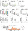
(A) Representative plots of infection efficiency of the MEFs used in (B–D). (B and C) Real-time qPCR analysis of nonapoptotic (expressing Bims4E, B) and apoptotic (expressing Bims2A, C) MEFs and their respective Ifnar1−/− bystander from cocultures after treatment with ABT-737 (500 nM) for 18–20 hr (data combined from three experiments, with two independent MEF lines per genotype). mRNA expression is shown relative to two independent housekeeping genes, tbp and gapdh. (D) Real-time qPCR analysis of WT bystanders from cocultures after treatment with ABT-737 for 18–20 hr (data combined from two experiments, with two independent MEF lines/genotype). (E) Real-time qPCR analysis of mtDNA content from Mcl1−/− MEFs cultured in ethidium bromide to generate mtDNA-depleted (ρ0) MEFs (used in F-J). Representative image of MEFs stained with PicoGreen nucleic acid stain. Arrows indicate mtDNA. Scale bars, 10 um. See also Figure S4. (F) Scatterplot of differentially expressed probes from microarray analysis of Mcl1−/−ρ0 MEFs compared to their respective parental Mcl1−/− MEF. (n = 3 independent MEF lines). Chromosome M, ChrM. See also Table S2. (G) Table of a selected set of type I IFN response genes from analysis in (F). (H) Bar graph of IFN-β in the supernatant of ρ0 and parental MEFs transfected with Poly(I:C)(HMW) (n = 3 independent MEF lines). (I–K) (I) Bar graphs of the viability of ρ0 and parental MEFs treated with ABT-737 ± 20–30 µM of Q-VD-Oph (QVD) for 24 hr, (J) caspase activity after 6 hr, and (K) IFN-β in supernatant after 24 hr (n = 4 independent MEF lines). Means were compared using a two-tailed t test. Data represent the mean ± SEM. *p ≤ 0.05, **p ≤ 0.01, and ***p ≤ 0.005.
Figure 7
Figure 7. mtDNA Release into the Cytosol Triggers cGAS-STING-Tbk1-Irf3-Mediated Type I Interferon Production
(A–C) (A) Viability of murine splenocytes treated with ABT-737 ± 20–30 µM Q-VD-OPh (QVD) for 24 hr, (B) caspase activity after 6 hr, and (C) IFN-β in culture supernatant after 24 hr (n = 4 mice per genotype). Means were compared to WT using a one-way ANOVA with Bonferroni correction. (D–F) (D) Viability of MEFs treated with ABT-737 ± 20–30 µM Q-VD-OPh (QVD) for 24 hr, (E) caspase activity after 6 hr, and (F) IFN-β in culture supernatant after 24 hr (n = 3 independent MEF lines per genotype, or 3 independent CRISPR/Cas9-targeted MEF clones). Means were compared to WT using a one-way ANOVA with Bonferroni correction. (G–I) (G) Bar graphs of the viability of Mcl1−/− MEFs pretreated for 1 hr with MRT-67307 (µM) followed by ABT-737 (500 nM) ± 20–30 µM Q-VD-OPh (QVD) for 24 hr, (H) caspase activity after 6 hr, and (I) IFN-β in culture supernatant after 24 hr (n = 3 independent MEF lines). (J) Immunoprecipitation (IP) followed by PCR. Left, immunoblot of lysates taken from Mcl1−/− MEFs transduced with an expression plasmid encoding FLAG- cGAS. Right, data represent the fold change (FC) in enrichment of DNA fragments using anti-FLAG or IgG (negative control) to coprecipitate DNA in untreated MEFs or MEFs treated with ABT-737 (1 µM) and Q-VD-OPh (QVD) (30 µM) for the indicated time. DNA fragments were amplified by real-time qPCR using eight primer pairs for mtDNA and two primer pairs for gDNA. The relative locations of the mtDNA amplicons are shown (data are combined from two, with three replicates). Means were compared between treated and untreated samples. See also Figures S5, S6, and S7. Unless otherwise stated, means were compared using a two-tailed t test. Data represent the mean ± SEM. *p ≤ 0.05, **p ≤ 0.01, and ***p ≤ 0.005.

V体育官网入口 - Comment in

References

    1. Adachi O, Kawai T, Takeda K, Matsumoto M, Tsutsui H, Sakagami M, Nakanishi K, Akira S. Targeted disruption of the MyD88 gene results in loss of IL-1- and IL-18-mediated function. Immunity. 1998;9:143–150. - PubMed
    1. Barber GN. STING-dependent cytosolic DNA sensing pathways. Trends Immunol. 2014;35:88–93. - PubMed
    1. Burguillos MA, Deierborg T, Kavanagh E, Persson A, Hajji N, Garcia-Quintanilla A, Cano J, Brundin P, Englund E, Venero JL, Joseph B. Caspase signalling controls microglia activation and neurotoxicity. Nature. 2011;472:319–324. - PubMed
    1. Cayrol C, Girard J-P. The IL-1-like cytokine IL-33 is inactivated after maturation by caspase-1. Proc. Natl. Acad. Sci. USA. 2009;106:9021–9026. - PMC - PubMed
    1. Chauvier D, Ankri S, Charriaut-Marlangue C, Casimir R, Jacotot E. Broad-spectrum caspase inhibitors: from myth to reality? Cell Death Differ. 2007;14:387–391. - PubMed

Publication types

V体育平台登录 - MeSH terms

Associated data