<u dropzone="XjGyig"></u> Skip to main page content
U.S. flag

An official website of the United States government

Dot gov

The . gov means it’s official. Federal government websites often end in VSports app下载. gov or . mil. Before sharing sensitive information, make sure you’re on a federal government site. .

Https

The site is secure. The https:// ensures that you are connecting to the official website and that any information you provide is encrypted and transmitted securely. V体育官网.

. 2014 Dec 30;111(52):18745-50.
doi: 10.1073/pnas.1404346111. Epub 2014 Dec 15.

Large-scale topology and the default mode network in the mouse connectome

Affiliations

"V体育2025版" Large-scale topology and the default mode network in the mouse connectome

James M Stafford et al. Proc Natl Acad Sci U S A. .

Abstract

Noninvasive functional imaging holds great promise for serving as a translational bridge between human and animal models of various neurological and psychiatric disorders. However, despite a depth of knowledge of the cellular and molecular underpinnings of atypical processes in mouse models, little is known about the large-scale functional architecture measured by functional brain imaging, limiting translation to human conditions. Here, we provide a robust processing pipeline to generate high-resolution, whole-brain resting-state functional connectivity MRI (rs-fcMRI) images in the mouse VSports手机版. Using a mesoscale structural connectome (i. e. , an anterograde tracer mapping of axonal projections across the mouse CNS), we show that rs-fcMRI in the mouse has strong structural underpinnings, validating our procedures. We next directly show that large-scale network properties previously identified in primates are present in rodents, although they differ in several ways. Last, we examine the existence of the so-called default mode network (DMN)--a distributed functional brain system identified in primates as being highly important for social cognition and overall brain function and atypically functionally connected across a multitude of disorders. We show the presence of a potential DMN in the mouse brain both structurally and functionally. Together, these studies confirm the presence of basic network properties and functional networks of high translational importance in structural and functional systems in the mouse brain. This work clears the way for an important bridge measurement between human and rodent models, enabling us to make stronger conclusions about how regionally specific cellular and molecular manipulations in mice relate back to humans. .

Keywords: connectivity; default mode network; mouse; resting-state functional MRI; structural connectivity. V体育安卓版.


Conflict of interest statement

The authors declare no conflict of interest.

Figures

Fig. 1.
Fig. 1.
Qualitative comparison of whole-brain mouse functional and structural data. Comparison of right primary motor cortex (MOp) and dorsal anterior cingulate (ACAd) seed regions. Note their strong similarities. The r label refers to correlation coefficient; s refers to the relative strength of the structural connections (30).
Fig. 2.
Fig. 2.
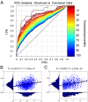
Quantitative comparison of mouse functional and structural data. (A) Full matrix comparison of the structural data with the functional data using receiver operator characteristic (ROC) analysis (details in the text and SI Methods). (B) Structural weights (i.e., nonzero connections) from the structural matrix (x axis) were compared with the corresponding functional weights of the functional connectivity data (y axis). Structural connection weights were log-transformed. A modest Pearson’s r value was observed with high significance (P < 0.00001). (C) The r value improved when the top 40% of the structural connections (i.e., top 10% of the total matrix) in B were considered (again with high significance; P < 0.00001). The structural data are presented on a lognormal scale because of the large range of connection strengths in the axonal structural data. TPR, true positive rate; FPR, false positive rate.
Fig. 3.
Fig. 3.
Rank–rank distribution and Rich Club topology of the mouse’s functional connectome. Upper Left shows the rank–rank distribution of the functional connections of the mouse connectome. Lower Left shows its fit to a bivariate distribution (details in the text and SI Methods). (Center) To determine if this topology is unique relative to what would be expected by chance, we generated a test distribution using the average of 10,000 random copies of the mouse connectome where the degree distribution was preserved. (Right) The functional and test distributions were compared by simple subtraction as shown.
Fig. 4.
Fig. 4.
Comparison between the mouse and macaque connectomes. Rank–rank distributions were compared between the mouse and macaque functional data using the same methodology as in Fig. 3.
Fig. 5.
Fig. 5.
Comparison between the mouse and human connectomes. Comparison of rank–rank distributions of the mouse and human functional connectomes using the same methodology as Fig. 3.
Fig. 6.
Fig. 6.
The DMN in humans and macaques. Using area 30 (RSP) as the seed region (red) reveals the presence of both the inferior (DMN) subsystem and superior DMN, similar to that first described by Andrews-Hanna et al. (40, 47). The critical components of this system are the parietal cortex (pink arrow), the orbitofrontal cortex (orange arrow), and the anterior cingulate cortex (yellow arrow). Note that the superior subsystem is present in both (A) humans and (B) macaques and includes area 23 (posterior cingulate cortex; black arrow). The r label refers to correlation coefficient.
Fig. 7.
Fig. 7.
Presence of the inferior DMN core in mouse functional and structural connectivity. The mouse seed region (red) used was the RSPagl, because it shows similar cytoarchitecture and location to that of area 30 in primates (45, 46). Both the (A) functional and (B) structural data in the mouse show hallmarks of the inferior DMN subsystem in primates, including the parietal cortex (pink arrow), the lateral/medial orbital cortex (orange arrow), and the cingulate area (yellow arrow). The r label refers to correlation coefficient; s refers to the relative strength of the structural connections (30).

"V体育平台登录" References

    1. Logothetis NK. Intracortical recordings and fMRI: An attempt to study operational modules and networks simultaneously. Neuroimage. 2012;62(2):962–969. - PubMed
    1. Fair DA, et al. The maturing architecture of the brain’s default network. Proc Natl Acad Sci USA. 2008;105(10):4028–4032. - "V体育平台登录" PMC - PubMed
    1. Hagmann P, et al. White matter maturation reshapes structural connectivity in the late developing human brain. Proc Natl Acad Sci USA. 2010;107(44):19067–19072. - "V体育安卓版" PMC - PubMed
    1. Grayson DS, et al. Structural and functional rich club organization of the brain in children and adults. PLoS ONE. 2014;9(2):e88297. - VSports注册入口 - PMC - PubMed
    1. Di Martino A, et al. Detection of functional connectivity in the resting mouse brain. Neuroimage. 2014;86:417–424. - "VSports手机版" PubMed

V体育官网 - Publication types