<big dropzone="yFx0ZrGt"><noframes id="tkQlJ7fW"> Skip to main page content
U.S. flag

An official website of the United States government

Dot gov

The . gov means it’s official. Federal government websites often end in . gov or . mil. Before sharing sensitive information, make sure you’re on a federal government site. VSports app下载.

Https

The site is secure. The https:// ensures that you are connecting to the official website and that any information you provide is encrypted and transmitted securely. V体育官网.

. 2014 Dec;24(12):1403-19.
doi: 10.1038/cr.2014.151. Epub 2014 Nov 21.

FTO-dependent demethylation of N6-methyladenosine regulates mRNA splicing and is required for adipogenesis

Affiliations

FTO-dependent demethylation of N6-methyladenosine regulates mRNA splicing and is required for adipogenesis

Xu Zhao et al. Cell Res. 2014 Dec.

Abstract

The role of Fat Mass and Obesity-associated protein (FTO) and its substrate N6-methyladenosine (m6A) in mRNA processing and adipogenesis remains largely unknown VSports手机版. We show that FTO expression and m6A levels are inversely correlated during adipogenesis. FTO depletion blocks differentiation and only catalytically active FTO restores adipogenesis. Transcriptome analyses in combination with m6A-seq revealed that gene expression and mRNA splicing of grouped genes are regulated by FTO. M6A is enriched in exonic regions flanking 5'- and 3'-splice sites, spatially overlapping with mRNA splicing regulatory serine/arginine-rich (SR) protein exonic splicing enhancer binding regions. Enhanced levels of m6A in response to FTO depletion promotes the RNA binding ability of SRSF2 protein, leading to increased inclusion of target exons. FTO controls exonic splicing of adipogenic regulatory factor RUNX1T1 by regulating m6A levels around splice sites and thereby modulates differentiation. These findings provide compelling evidence that FTO-dependent m6A demethylation functions as a novel regulatory mechanism of RNA processing and plays a critical role in the regulation of adipogenesis. .


Figures

Figure 1
Figure 1
FTO depletion interferes with adipogenesis. (A-C) 3T3-L1 pre-adipocytes (Day −2) were treated with FTO, ALKBH5, METTL3 or control siRNA. Forty-eight hours later, cells were lysed and subjected to immunoblotting with the indicated antibodies (A). Forty-eight hours following siRNA treatment differentiation was induced by incubation with the differentiation cocktail (IBMX/DEX/Insulin) on Day 0. Differentiation status was determined by Oil Red O staining (B) and triglyceride assay (C) on Day 0 and 10. (C) Triglyceride content was quantified and normalized to protein content. *P < 0.05 is considered significant. Results are shown as mean ± SD. (D) 3T3-L1 cells collected at different time points (D0/2/5/10) during adipogenesis were lysed and subjected to immunoblotting with the indicated antibodies. β-tublin was used as loading control. (E) RT-PCR detected the expression levels of FTO, METTL3, as well as adipogenic markers, including ADIPSIN and PREF-1, during adipocyte differentiation. β-Actin was used as loading control. (F) mRNA was isolated from multiple stages (D0/5/10) of adipogenesis and used in dot blot analyses with m6A antibody. mRNA was loaded by two-fold serial dilution. The m6A contents are shown in the upper panel. Equal loading of mRNA was verified by methylene blue staining (lower panel). See also Supplementary information, Figure S1.
Figure 2
Figure 2
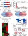
FTO affects RNA splicing via modulating m6A. (A) The percentage of m6A-mRNAs in control (6 011 multi-isoform/2 699 single-isoform, P = 2.26e-5, Fisher test) and FTO-knockdown cells (6 916 multi-isoform/3 221 single-isoform, P = 8.46e-11, Fisher test) derived from single-isoform or multi-isoform genes compared to the distribution predicted by the Ensembl-annotation reference (background). (B) The distribution of m6A-containing exons across different categories of splicing events in both control (orange) and FTO-depleted samples (red) compared to that Ensembl-annotation reference (Blue). P value for each category was calculated between siCTRL and siFTO samples. **P < 0.01 (Student's t-test) is considered significant (n = 2). Results are shown as mean ± SD. CNE, constitutive exon; CE, cassette exon; A5SS, alternative 5′ splice site; ALE, alternative last exon; II, intron isoform; A3SS, alternative 3′ splice site; IR, intron retention; MXE, mutually exclusive exons; AFE, alternative first exon; EI, exon isoform. (C) 1 491 isoforms (1 335 genes) are co-regulated by FTO and METTL3. Arrows pointing up/down indicates up/down-regulation. (D) FTO depletion resulted in increased m6A levels in 522 isoforms (452 genes) of the 1 491 isoforms (1 335 genes) co-regulated by FTO and METTL3 (shown in C). (E) The heatmap shows the expression levels of 522 reverse-regulated isoforms by FTO and METTL3, as well as the m6A modification in FTO-depleted cells. (F) The heatmap shows m6A peaks in 5′-UTR, CDS, and 3′-UTR (522 isoforms co-regulated by FTO and METTL3) in control and FTO-deficient cells. Blue lines represent m6A peaks. Each horizontal line represents one gene. The number of new m6A peaks upon FTO depletion and new m6A peaks within a RRACH motif are shown below the heatmap. (G) Function enrichment analysis of 522 isoforms (452 genes) based on the DAVID GO analysis result. The cutoff parameters for enrichment analysis with Cytoscape software are: P < 0.005, FDR q < 0.1, overlap cutoff > 0.5. See also Supplementary information, Figures S2-S4.
Figure 3
Figure 3
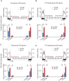
m6A is enriched at exonic splice sites. (A-D) Schematic analysis of the distribution of m6A peaks (upper panel in each figure; 100 nt upstream and 300 nt downstream from 5′SS; 300 nt upstream and 100 nt downstream from 3′SS) and m6A enrichment (lower panel in each figure; 100 nt up/downstream of the 5′ SS or 3′ SS) along 5′ exon-intron and 3′ intron-exon boundaries in control (blue) and FTO-deficient (red) cells. (A, C) m6A density in the vicinity of constitutive SS (A) or alternative SS (C) in the 5 326 genes showing changes in isoform expression upon FTO depletion. (B, D) Similar to A and C but here the m6A density was determined in the 452 FTO target genes showing increased m6A levels after FTO knockdown. The m6A levels in each region between control and FTO depletion are shown as mean ± SD and statistical analysis on their difference were performed by Student's t-test. **P < 0.01. See also Supplementary information, Figures S4 and S5.
Figure 4
Figure 4
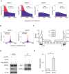
m6A modification influences RNA binding ability of SRSF2. (A) Density plots showing the relative distance (X-axis), calculated by BEDTools' closestBed, between m6A sites and SRSF1-4-binding sites. Randomly selected sites (blue color) within the same co-regulated genes were used as control. (B) The distance between the top-ranking SRSF1 and 2 ESEs predicted by ESEfinder and the nearest RRACH in identified m6A peak (red color) were calculated by BEDTools' closestBed (X axis). The distances of m6A-modified RRACH sites to the ESEs predicted by ESEfinder were calculated and their relative density distributions were shown as density plot. Randomly selected sites (blue color) within the same co-regulated genes were used as control. (C) Analysis of the distribution of SRSF1-4-binding clusters along 5′ exon-intron and 3′ intron-exon boundaries (100 nt upstream and 300 nt downstream of 5′SS; 300 nt upstream and 100 nt downstream of 3′SS). (D) PAR-CLIP of SRSF2 in control and FTO-depleted 3T3-L1 cells transfected with the pCS2-Flag-SRSF2 plasmid. SRSF2 protein-RNA complex was pulled down with Flag M2 Affinity Gel (mouse), and then RNA was labeled and detected following instruction of biotin labeling kit. The expression levels of SRSF2 protein in whole cell lysate (WCE) was detected by western blotting with Flag antibody (rabbit). Knockdown efficiency of FTO was detected with its specific antibody. β-tubulin was used as loading control. (E) RNA binding ability of SRSF2 was calculated by normalizing binding RNAs to the corresponding pull-downed proteins. Statistical analysis shows the relative binding ability of SRSF2 in FTO-depleted cells (P < 0.0001) to that in control cells (siCTRL). See also Supplementary information, Figures S6-S7.
Figure 5
Figure 5
FTO regulates splicing in a subset of SRSF2 target genes. (A) Splicing analysis of SRSF2 target genes by RT-PCR in FTO-depleted cells. Red boxes indicate the spliced internal exons. % exc (exclusion) indicates the exclusion level of the internal exons. The expression of Actin was detected to confirm the equal loading of PCR products. (B) Statistical analysis of the relative exclusion level of spliced exons in FTO-depleted 3T3-L1 cells to that in control cells. ***P < 0.0001, *P < 0.05. Results are shown as mean ± SD. (C) Detection of m6A levels around splicing sites of FTO target genes by m6A-IP and RT-qPCR. ***P < 0.0001. Results are shown as mean ± SD. (D) Effects of FTO depletion on SRSF2 binding ability to its target genes validated by PAR-CLIP and RT-qPCR. ***P < 0.0001. Results are shown as mean ± SD. See also Supplementary information, Figure S8.
Figure 6
Figure 6
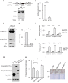
FTO regulates alternative splicing of adipogenensis-related factor RUNX1T1. (A) Alternative splicing of RUNX1T1 was identified by RT-PCR using the indicated PCR primer set (arrows). Depending on isoform, bands of either 496 bp or 245 bp can be detected. (B) Statistical analysis of the relative exclusion level of RUNX1T1 in FTO-depleted 3T3-L1 cells to that in control cells. **P < 0.01. Results are shown as mean ± SD. (C) RUNX1T1 detection in 3T3-L1 pre-adipocyte lysates shows the endogenous protein products of the two RUNX1T1 isoforms in control and FTO-deficient 3T3-L1 pre-adipocytes. An unspecific band was labeled by *. (D) Statistical analysis of RUNX1T1-S protein expression levels in FTO-depleted 3T3-L1 cells to that in control cells. **P < 0.01. Results are shown as mean ± SD. (E) Detection of m6A levels around splicing sites of RUNX1T1 gene by m6A-IP and RT-qPCR. ***P < 0.0001. Results are shown as mean ± SD. (F) Effect of FTO depletion on SRSF2 binding ability to exons around splicing sites validated by PAR-CLIP and RT-qPCR. ***P < 0.0001. Results are shown as mean ± SD. (G-I) 3T3-L1 pre-adipocytes were transfected with pEGFP-C1b, pEGFP-C1b-Runx1T1-L or pEGFP-C1b-Runx1T1-S. Fory-eight hours later, transfection cells were lysed and immunoblotted with the indicated antibodies (G). An unspecific band or degradation band was labeled by *. Transfected cells were induced to differentiation and subjected to triglyceride analysis (H) and Oil Red O staining (I) on D10. Triglyceride content was quantified and normalized to protein content. *P < 0.05 is considered significant. **P < 0.01. Results are shown as mean ± SD. See also Supplementary information, Figure S9.
Figure 7
Figure 7
Cooperative role of m6A in regulating SRSF2 function at splice sites. The splicing factor SRSF2 can recognize exon splicing enhancer (ESE), inducing exon inclusion. METTL3-mediated m6A modification enhances the recruitment of SRSF2 to its target ESE, promoting exon inclusion. On the other hand demethylation of m6A-RRACHs near ESEs by FTO prevents recognition of the ESEs by SRSF2, thereby inhibiting inclusion and instead promoting exon skipping of the specific exon. M6A co-regulated by methyltransferases and demthylases serves as a new RNA splicing exonic cis-regulatory element that in combination with ESEs regulates SRSF2 protein recruitment.

Comment in

References

    1. Gerken T, Girard CA, Tung YC, et al. The obesity-associated FTO gene encodes a 2-oxoglutarate-dependent nucleic acid demethylase. Science. 2007;318:1469–1472. - PMC - PubMed
    1. Loos RJ, Bouchard C. FTO: the first gene contributing to common forms of human obesity. Obes Rev. 2008;9:246–250. - "VSports app下载" PubMed
    1. Zabena C, Gonzalez-Sanchez JL, Martinez-Larrad MT, et al. The FTO obesity gene. Genotyping and gene expression analysis in morbidly obese patients. Obes Surg. 2009;19:87–95. - PubMed
    1. Loos RJ, Yeo GS. The bigger picture of FTO—the first GWAS-identified obesity gene. Nat Rev Endocrinol. 2014;10:51–61. - PMC - PubMed
    1. Fischer J, Koch L, Emmerling C, et al. Inactivation of the Fto gene protects from obesity. Nature. 2009;458:894–898. - V体育ios版 - PubMed

Publication types

MeSH terms

"VSports最新版本" Associated data