"V体育官网" Skip to main page content
U.S. flag

An official website of the United States government

Dot gov

The VSports app下载. gov means it’s official. Federal government websites often end in . gov or . mil. Before sharing sensitive information, make sure you’re on a federal government site. .

Https

The site is secure V体育官网. The https:// ensures that you are connecting to the official website and that any information you provide is encrypted and transmitted securely. .

. 2014 Nov 15;193(10):4962-70.
doi: 10.4049/jimmunol.1401613. Epub 2014 Oct 10.

"V体育官网入口" Identification of factor H-like protein 1 as the predominant complement regulator in Bruch's membrane: implications for age-related macular degeneration

Affiliations

"VSports" Identification of factor H-like protein 1 as the predominant complement regulator in Bruch's membrane: implications for age-related macular degeneration

Simon J Clark et al. J Immunol. .

Abstract

The tight regulation of innate immunity on extracellular matrix (ECM) is a vital part of immune homeostasis throughout the human body, and disruption to this regulation in the eye is thought to contribute directly to the progression of age-related macular degeneration (AMD) VSports手机版. The plasma complement regulator factor H (FH) is thought to be the main regulator that protects ECM against damaging complement activation. However, in the present study we demonstrate that a truncated form of FH, called FH-like protein 1 (FHL-1), is the main regulatory protein in the layer of ECM under human retina, called Bruch's membrane. Bruch's membrane is a major site of AMD disease pathogenesis and where drusen, the hallmark lesions of AMD, form. We show that FHL-1 can passively diffuse through Bruch's membrane, whereas the full sized, glycosylated, FH cannot. FHL-1 is largely bound to Bruch's membrane through interactions with heparan sulfate, and we show that the common Y402H polymorphism in the CFH gene, associated with an increased risk of AMD, reduces the binding of FHL-1 to this heparan sulfate. We also show that FHL-1 is retained in drusen whereas FH coats the periphery of the lesions, perhaps inhibiting their clearance. Our results identify a novel mechanism of complement regulation in the human eye, which highlights potential new avenues for therapeutic strategies. .


Figures

Figure 1
Figure 1. FHL-1 rather than FH is the predominant complement regulator in human Bruch’s membrane
(A) Schematic indicating the CCP regions of FH and FHL-1 recognised by the OX23, anti-FHL-1 and L20/3 antibodies. The AMD-associated Y402H polymorphism is located in CCP7 of both FH and FHL-1. (B) Grey-scale fluorescent staining of a human macula with anti-FHL-1. (C) Grey-scale staining of the same donor as shown in (B) but with the antibody L20/3. (D) Fluorescent staining of human macula with an equal mix of both anti-FHL-1 (green) and L20/3 (red). (E) Labeling of a druse with FHL-1 (green) and FH (red) antibodies. (F) Western blot of solubilised Bruch’s membrane from 4 donors stained with anti-FHL-1 (green) and OX23 (red): yellow staining is indicative of co-localisation of both antibodies. Blue color represents DAPI staining of cell nuclei. Images in (B)-(D) are representative of 6 individual donors: scale bar = 10 μm. Image (E) is representative of four donors: scale bar = 5 μm.
Figure 2
Figure 2. Detection of complement gene transcription by human RPE cells
Pooled RNA from five donors’ RPE cells was used to detect complement gene transcription. (A) RNA for a number of genes central to the alternative pathway of complement were detected in both liver and RPE cells, including FHL-1, FH, C3, FB and FI. (B) Two RPE-specific genes, Best-1 and RPE65, were used as positive controls for RPE cell RNA and β-actin and TATA binding protein (TBP) were selected as housekeeping gene controls.
Figure 3
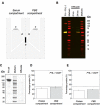
Figure 3. FHL-1 is able to diffuse through Bruch’s membrane from human serum
An Ussing chamber was used to compare the diffusion of FH and FHL-1 from human serum across enriched Bruch’s membrane from human donor eyes. (A) Schematic of the Ussing chamber layout where (a) the Bruch’s membrane, (b) sampling access points and (c) magnetic stirrer bars are shown. (B) Fluorescent western blot of one representative experiment from a total of three showing 0 and 24 hour samples from the PBS compartment: left hand lane shows a positive control sample containing 1μg each of FH and FHL-1. Red bands are recognised by OX23 alone, green by anti-FHL-1 alone and yellow by both antibodies. The Odyssey protein markers used here are visualized in the red channel. The Western shown here is representative of three separate experiments. (C) Purified FH was placed in one compartment and after 24 hours the entire protein content of the other ‘PBS’ compartment was concentrated, subjected to SDS-PAGE and the resultant gel stained with Coomassie Blue. (D)-(E) Diffusion experiments with purified recombinant FHL-1 proteins examined potential differences in the ability of the 402H and 402Y variants to cross Bruch’s membrane. Both 402H and 402Y forms were tested separately using three donor Bruch’s membranes and data are shown as percentage protein detected in each chamber after 24 hours at room temperature: the 50% mark is shown as a dashed line. The Donor tissues used in (C)-(E) are listed in Table 1.
Figure 4
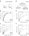
Figure 4. The 402H form of FHL-1 shows greater dependency on GAG sulfation for binding
Heparin is a highly sulfated model of heparan sulphate (HS). (A) Schematic showing the basic IdoUA – GlcN backbone disaccharide of heparin where all four possible sulfation positions are listed as: R1, 6-O sulfation; R2, N sulfation; R3, 2-O sulfation; and R4, 3-O sulfation. (B)-(C) Plate assays demonstrating the binding activities of FHL-1 402Y and 402H forms for selectively desulfated heparin. (D) Schematic diagram demonstrating the different disaccharide regions of an HS chain where: GlcNS = N-sulfated glucosamine; GlcNAc = N-acetylated glucosamine; GlcUA = glucuronic acid; IdoUA = iduronic acid; 6S = sulfation in the 6-O position; and 2S = sulfation in the 2-O position. (E)-(F) Plates assays demonstrating the AMD-associated 402H form of FHL-1 binding relatively poorly to two forms of HS compared to the 402Y form. Data in (B)-(C) and (E)-(F) are n=6, averaged from two independent experiments ± s.e.m.
Figure 5
Figure 5. Heparinase pre-treatment alters the pattern of FHL-1 and FH staining in the macula
Both FHL-1 and FH localisation were visualized in eye tissue before and after enzymatic pre-treatment with a heparinase I/II/III mix using the six normal donor eyes, and four drusen containing, AMD, eyes as listed in Table 1. In each case green staining represents FHL-1 and red staining FH. (A) Distribution of FHL-1 and FH before removal of HS, and (B) after enzymatic treatment in macular tissue without AMD pathology. FHL-1 and FH labelling of a druse without (C) and with heparinase treatment (D). Blue staining represents DAPI staining of cell nuclei. Scale bars represent 10 μm.

References

    1. Friedman DS, O’Colmain BJ, Muñoz B, Tomany SC, McCarty C, de Jong PTVM, Nemesure B, Mitchell P, Kempen J, E. D. P. R. Group Prevalence of age-related macular degeneration in the United States. Arch. Ophthalmol. 2004;122:564–572. - PubMed
    1. Coleman HR, Chan C-C, Ferris FL, III, Chew EY. Age-related macular degeneration. Lancet. 2008;372:1835–1845. - PMC - PubMed
    1. Anderson DH, Radeke MJ, Gallo NB, Chapin EA, Johnson PT, Curletti CR, Hancox LS, Hu J, Ebright JN, Malek G, Hauser MA, Rickman CB, Bok D, Hageman GS, Johnson LV. The pivotal role of the complement system in aging and age-related macular degeneration: Hypothesis re-visited. Prog. Retin. Eye Res. 2009;29:95–112. - PMC - PubMed
    1. Clark SJ, Bishop PN, Day AJ. Complement factor H and age-related macular degeneration: the role of glycosaminoglycan recognition in disease pathology. Biochem. Soc. Trans. 2010;38:1342–1348. - PubMed
    1. Whitcup SM, Sodhi A, Atkinson JP, Holers VM, Sinha D, Rohrer B, Dick AD. The Role of the Immune Response in Age-Related Macular Degeneration. Int. J. Inflam. 20132013:1–10. - "V体育平台登录" PMC - PubMed

Publication types

"V体育安卓版" MeSH terms

"V体育安卓版" Substances