Skip to main page content
U.S. flag

An official website of the United States government

Dot gov

The . gov means it’s official. Federal government websites often end in . gov or VSports app下载. mil. Before sharing sensitive information, make sure you’re on a federal government site. .

Https

The site is secure. The https:// ensures that you are connecting to the official website and that any information you provide is encrypted and transmitted securely V体育官网. .

. 2014 Dec 1;74(23):7037-47.
doi: 10.1158/0008-5472.CAN-14-1392. Epub 2014 Oct 8.

Inhibition of mTORC1/2 overcomes resistance to MAPK pathway inhibitors mediated by PGC1α and oxidative phosphorylation in melanoma

Affiliations

Inhibition of mTORC1/2 overcomes resistance to MAPK pathway inhibitors mediated by PGC1α and oxidative phosphorylation in melanoma (VSports app下载)

Y N Vashisht Gopal et al. Cancer Res. .

VSports在线直播 - Abstract

Metabolic heterogeneity is a key factor in cancer pathogenesis. We found that a subset of BRAF- and NRAS-mutant human melanomas resistant to the MEK inhibitor selumetinib displayed increased oxidative phosphorylation (OxPhos) mediated by the transcriptional coactivator PGC1α. Notably, all selumetinib-resistant cells with elevated OxPhos could be resensitized by cotreatment with the mTORC1/2 inhibitor AZD8055, whereas this combination was ineffective in resistant cell lines with low OxPhos. In both BRAF- and NRAS-mutant melanoma cells, MEK inhibition increased MITF expression, which in turn elevated levels of PGC1α. In contrast, mTORC1/2 inhibition triggered cytoplasmic localization of MITF, decreasing PGC1α expression and inhibiting OxPhos. Analysis of tumor biopsies from patients with BRAF-mutant melanoma progressing on BRAF inhibitor ± MEK inhibitor revealed that PGC1α levels were elevated in approximately half of the resistant tumors. Overall, our findings highlight the significance of OxPhos in melanoma and suggest that combined targeting of the MAPK and mTORC pathways may offer an effective therapeutic strategy to treat melanomas with this metabolic phenotype. VSports手机版.


Figures

Figure 1
Figure 1
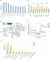
Cellular metabolism genes confer resistance to MEK inhibition by selumetinib. (A) Ingenuity Pathway Analysis (IPA) of cellular functions associated with the 164 genes that showed synthetic lethality (FDR corrected p<0.05) with selumetinib in a genome-wide siRNA screen in the MEL624 cell line. The bar graph shows the ten most significantly enriched cellular functions on the x-axis; y-axis, significance by the Fisher's exact test (p<0.05). (B) Netwalker analysis of the 164 selumetinib-synthetic lethal genes. Genes associated with mitochondrial activity are labeled with a red asterisk. Inset box shows the line colors of known gene interactions. (C) IPA analysis of upregulated KEGG canonical pathways by Fishers exact test (p< 0.05) in the genome-wide expression microarray data of selumetinib-sensitive (A375, WM35) and -resistant BRAF-mutant melanoma cell lines (MEL624, SKMEL5). (D) Synthetic lethal genes that were upregulated in the selumetinib-resistant lines following selumetinib treatment. Y-axis, change in mRNA expression from pre- to post-24 h treatment with 0.25μM selumetinib. (E) Seahorse extracellular flux analysis showing the basal, oligomycin-inhibited (“O”) and FCCP-activated (“F”) OCR in the sensitive and resistant cell lines. Data is average of quadruplicates.
Figure 2
Figure 2
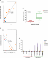
mTOR1/2 inhibition is synergistic in melanoma cell lines with de novo resistance to selumetinib and elevated OxPhos. (A) Scatter plot of basal OxPhos (OCR) and PGC1α transcript levels in a panel of 14 selumetinib-resistant melanoma cell lines that are BRAF-mutant (orange), NRAS-mutant (red), or BRAF/NRAS wild-type (blue). (B) Scatter plot showing correlation of the combination index (CI) of selumetinib and AZD8055 with basal OCR in the cell lines. CI < 1.0 indicates synergistic inhibition of cell proliferation by the combination. (C) Box plot showing of PGC1α expression in cell lines with CI>1.0 (Red) and CI<1.0 (Green) for selumetinib+AZD8055. (D) Sub-G1 cell populations detected by FACS following 72 h of the indicated treatments. The BRAF (“*”) mutant and NRAS (“**”) mutant cells were treated with 0.25μM of the inhibitors (alone and in combination). Data is average of 3 replicates; error bars, standard deviation.
Figure 3
Figure 3
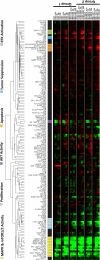
RPPA analysis of the effects of selumetinib + AZD8055 treatment on protein signaling networks. Supervised hierarchical clustering heatmap shows time-course analysis of three low OxPhos (Group 1) and three high OxPhos (Group 2) BRAF-mutant human melanoma cell lines treated with 0.25μM each of selumetinib+AZD8055 for 0, 3, 12, and 24 hrs. Data indicates fold changes in the inhibitor treated samples versus DMSO-treated controls in triplicates. Red, increased levels; green, decreased levels of proteins.
Figure 4
Figure 4
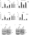
AZD8055 decreases PGC1α and OxPhos. qRT-PCR analysis of the fold changes in PGC1α and MITF transcripts (normalized by GAPDH) in MEL624 (A) and WM3854 (B) cells after 24 h treatment with DMSO, 0.25μM of selumetinib or AZD8055, or their combination. Data is average of triplicates. Asterisks indicate significant increases of MITF levels (p<0.05) in the AZD8055 and combination treatments compared to mock, as determined by t-tests. Western blot panels at the right show levels of the indicated proteins for the same treatments. (C) Relative luciferase units (RLU) of MEL624 (black bars) and WM3854 (gray bars) transfected with MITF promoter reporter following the indicated treatments for 24 h in triplicates. Asterisk indicates significant difference (p<0.05) of AZD8055 treatment compared to mock in both cell lines. (D) RLU in MEL624 cells transfected with a PGC1α (black bars) or TRPM1 (gray bars) reporter plasmid followed by the indicated treatments for 24 h in triplicate. Western blotting of cytoplasmic and nuclear extracts from MEL624 (E) and WM3854 (F) cells following treatment with the indicated inhibitors for 24h. Lamin A/C and Caveolin1 served as controls.
Figure 5
Figure 5
Comparative effects of inhibition of PI3K pathway components and in vivo efficacy of selumetinib+AZD8055. (A) Basal OCR levels in MEL624 (black bars) and WM3854 (white bars) cells after 24 h treatment with 0.25 μM selumetinib, 0.25 μM AZD8055, 0.1 μM Rapamycin, 1 μM of GDC0941, 1 μM BKM120 or 5 μM MK2206. Data is average of quadruplicates. (B) Western blotting following indicated treatments for 24 h. (C) Basal OCR in the MEL624 (black bars) and WM3854 (white bars) after siRNA-mediated knockdown of the indicated genes. OCR was determined 72 h after transfection with 20nM of siRNAs. Bars represent average of quadruplicates. (D) Cell viability in the MEL624 (gray bars) and WM3854 (black bars) following knockdown of PGC1α by siRNA with or without 0.25 μM selumetinib treatment. Selumetinib was added 24 h after siRNA transfection, cell viability was measured after 72 h with CTB. Data is average of triplicates. Asterisk indicates significant difference from siRisc+SEL by t-test (p<0.05). (E) The MEL624 and WM3854 cells were treated as in D, and the sub-G1 dead cell population was determined by FACS analysis (triplicates). (F) In vivo growth of MEL624 subcutaneous tumors treated with indicated inhibitors. Colored asterisks indicate significant difference (p<0.05) of a treatment from a different treatment represented by the respective line color. Western blot panel shows the levels of indicated proteins in tumor lysates on day 15.
Figure 6
Figure 6
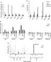
OxPhos and PGC1α in acquired resistance to MAPK pathway inhibitors. (A) Basal OCR and ECAR levels in the parental A375, WM35 cell lines and their selumetinib-resistant clones (“-R1”, “-R2”) determined by Seahorse flux analysis. Gray bars, OCR; Black bars, ECAR. Data is average of quadruplicates. (B) PGC1α and MITF mRNA levels in the A375 and WM35 cells and their resistant clones at 24 h following treatment with DMSO (mock), 0.25μM selumetinib, 0.25μM AZD8055, or selumetinib+AZD8055. qPCR was performed on triplicate samples, and GAPDH-normalized changes in mRNA levels in inhibitor treatments versus mocks were determined. (C) Sub-G1 dead cell populations of A375 and WM35 parental cells and their resistant clones following treatment with the indicated inhibitors for 72h. Data is average of triplicates. (D) Ratios of PGC1α and MITF gene expression at the time of disease progression versus pre-treatment in the MIA/WH patient cohort. Patients were treated with vemurafenib (*), dabrafenib (**), or dabrafenib+ trametinib (***). White bars, PGC1α; Black bars, MITF.

References (V体育2025版)

    1. Hocker T, Tsao H. Ultraviolet radiation and melanoma: a systematic review and analysis of reported sequence variants. Hum Mutat. 2007;28:578–88. - V体育平台登录 - PubMed
    1. Chapman PB, Hauschild A, Robert C, Haanen JB, Ascierto P, Larkin J, et al. Improved survival with vemurafenib in melanoma with BRAF V600E mutation. N Engl J Med. 2011;364:2507–16. - PMC - PubMed
    1. Hauschild A, Grob JJ, Demidov LV, Jouary T, Gutzmer R, Millward M, et al. Dabrafenib in BRAF-mutated metastatic melanoma: a multicentre, open-label, phase 3 randomised controlled trial. Lancet. 2012;380:358–65. - PubMed
    1. Flaherty KT, Robert C, Hersey P, Nathan P, Garbe C, Milhem M, et al. Improved survival with MEK inhibition in BRAF-mutated melanoma. N Engl J Med. 2012;367:107–14. - PubMed
    1. Sosman JA, Kim KB, Schuchter L, Gonzalez R, Pavlick AC, Weber JS, et al. Survival in BRAF V600-mutant advanced melanoma treated with vemurafenib. N Engl J Med. 2012;366:707–14. - PMC (V体育平台登录) - PubMed

Publication types

MeSH terms

Substances