Skip to main page content
U.S. flag

An official website of the United States government

Dot gov

The . gov means it’s official. Federal government websites often end in . gov or . mil VSports app下载. Before sharing sensitive information, make sure you’re on a federal government site. .

Https

The site is secure. The https:// ensures that you are connecting to the official website and that any information you provide is encrypted and transmitted securely. V体育官网.

. 2014 Oct 29;42(19):12070-81.
doi: 10.1093/nar/gku922. Epub 2014 Oct 7.

Regulation of BCL-X splicing reveals a role for the polypyrimidine tract binding protein (PTBP1/hnRNP I) in alternative 5' splice site selection

Affiliations

Regulation of BCL-X splicing reveals a role for the polypyrimidine tract binding protein (PTBP1/hnRNP I) in alternative 5' splice site selection

Pamela Bielli (V体育2025版) et al. Nucleic Acids Res. .

Abstract

Alternative splicing (AS) modulates many physiological and pathological processes. For instance, AS of the BCL-X gene balances cell survival and apoptosis in development and cancer. Herein, we identified the polypyrimidine tract binding protein (PTBP1) as a direct regulator of BCL-X AS. Overexpression of PTBP1 promotes selection of the distal 5' splice site in BCL-X exon 2, generating the pro-apoptotic BCL-Xs splice variant. Conversely, depletion of PTBP1 enhanced splicing of the anti-apoptotic BCL-XL variant VSports手机版. In vivo cross-linking experiments and site-directed mutagenesis restricted the PTBP1 binding site to a polypyrimidine tract located between the two alternative 5' splice sites. Binding of PTBP1 to this site was required for its effect on splicing. Notably, a similar function of PTBP1 in the selection of alternative 5' splice sites was confirmed using the USP5 gene as additional model. Mechanistically, PTBP1 displaces SRSF1 binding from the proximal 5' splice site, thus repressing its selection. Our study provides a novel mechanism of alternative 5' splice site selection by PTBP1 and indicates that the presence of a PTBP1 binding site between two alternative 5' splice sites promotes selection of the distal one, while repressing the proximal site by competing for binding of a positive regulator. .


Figures

Figure 1.
Figure 1.
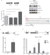
Identification of RNA-binding proteins involved in selection of distal 5′ splice site in BCL-X exon 2. (A) Schematic representation of BCL-X alternative splicing. Exons (boxes), introns (lines), and distal (S) and proximal (L) 5′ splice sites in exon 2 are indicated. (B) Scheme of BCL-X gene and mutant minigenes (upper panel). RT-PCR of in vivo splicing assay performed in HEK293T cells in the presence of indicated BCL-X minigenes (bottom panel). The bar graph shows the percentage of BCL-XL (mean ± SD, n = 3). (C) In the upper panel, sequence of BCL-X transcribed RNA using indicated primers (arrows) is shown. The sequences before the distal (S) 5′ splice sites (gray box) and deleted in ΔB2 minigene (underlined) are indicated. Western blot analysis of RNA-pulldown assay performed using biotin-labeled BCL-X B2 RNA or streptavidin (−), as negative control, and in the presence of commercial nuclear extracts from HeLa cells (bottom panel). (D) RT-PCR of in vivo splicing assay performed in HEK293T cells transfected with BCL-X minigene and the indicated splicing factors. The bar graph shows the percentage of BCL-XL (mean ± SD, n = 3). The P values of Student's t-test are reported. *P < 0.05; **, P < 0.01; ***, P < 0.001; n.s., not significant.
Figure 2.
Figure 2.
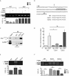
BCL-X is an RNA target of PTBP1 in live cells. (A) qPCR analysis of BCL-X isoforms in HEK293T cells transfected with scramble (CTRL), PTBP1 and PTBP2 (PTBP1/2), hnRNP F and SRSF1 siRNAs. The bar graph of fold variation of each sample was calculated by delta–delta Ct method as described in the Materials and Methods section (mean ± SD, n = 3). Scheme of exon junction primers used for qPCR (arrows) is also shown. Down-regulation of protein level was assessed by western blot analysis (right panel). (B) UV-crosslink and RNA immunoprecipitation (CLIP) of PTBP1 performed in HEK293T. Associated BCL-X RNA was quantified by qPCR using primers indicated in the upper BCL-X scheme and represented as fold enrichment relative to the IgG sample (mean ± SD, n = 3). (C) Schematic representation of distal (E2d; red arrows) and proximal (E2p; light blue arrows) exon 2 BCL-X primers used in (D). (D) CLIP of PTBP1 performed as in (B) in the presence of RNaseI (1:1000). (A, B and D) The P values of Student's t-test are reported. *P < 0.05, **, P < 0.01; ***, P < 0.001; n.s., not significant.
Figure 3.
Figure 3.
The B2 element is required for PTBP1-dependent BCL-Xs splicing. (A) RT-PCR analysis of the in vivo splicing assays performed in HEK293T cells transfected with WT or mutated (ΔB2) BCL-X minigenes and the indicated splicing factors. The bar graph of the percentage of BCL-XL is also shown (mean ± SD, n = 3). (B) Scheme of BCL-X exon 2 sequence. The putative PTBP1 binding site is highlighted (gray box). The mutated bases in E2m1 and E2m2 mutants are underlined. (C) Western blot analysis of RNA-pulldown assay using biotin-labeled BCL-X WT or mutant (E2m1 and E2m2) RNA. Streptavidin beads have been used as control (−). (D) CLIP experiments of PTBP1 performed in HEK293T transfected with the indicated minigenes. Associated BCL-X RNA was quantified by qPCR (primers used are indicated in Supplementary Table S1). Data are represented as fold enrichment relative to the IgG sample (mean ± SD, n = 3). (E and F) RT-PCR analysis of in vivo splicing assays performed in HEK293T cells transfected with wt and mutated (E2m1 and E2m2) minigenes in the presence (F) or absence (E) of PTBP1. The bar graph of the percentage of BCL-XL is shown (mean ± SD, n = 3). (A, D, E and F) The P values of Student's t-test are reported. *P < 0.05, **, P < 0.01; ***, P < 0.001; n.s., not significant.
Figure 4.
Figure 4.
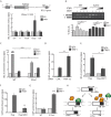
Binding of PTBP1 between two competing 5′ splice sites favors the selection of the distal one. (A) Schematic representation of USP5 alternative splicing. Exons (boxes), introns (lines), and distal (2) and proximal (1) 5′ splice sites in exon 15 are indicated. (B) RT-PCR of in vivo splicing assay performed in HEK293T cells transfected with control (CTRL) or PTBP1/2 siRNAs in the presence (right panel) or absence (left panel) of USP5 minigene. The bar graph shows the percentage of endogenous (left panel) or minigene-derived (right panel) USP52 variant (mean ± SD, n = 3). (C) Sequence of USP5 RNA transcribed using indicated primers (arrows). The sequence before the distal (2) 5′ splice site (gray box) and the putative PTBP1 binding site (bold) is indicated. The mutated bases in E15m1 mutant are underlined. Western blot analysis (bottom panel) of RNA-pulldown assay performed using biotin-labeled wt (WT) and mutated (E15m1) USP5 RNA, and streptavidin (−) as negative control. (D) CLIP experiments of PTBP1 performed in HEK293T transfected with wt (WT) or mutated (E15m1) minigenes. Associated USP5 RNA was quantified by qPCR (primers used are indicated in Supplementary Table S1). Data are represented as fold enrichment relative to the IgG sample (mean ± SD, n = 3). (E) RT-PCR of in vivo splicing assay performed in HEK293T cells transfected with WT or E15m1 mutant minigenes. The bar graph shows the percentage of USP52 isoform (mean ± SD, n = 3). (B, D and E) The P values of Student's t-test are reported. **, P < 0.01; ***, P < 0.001.
Figure 5.
Figure 5.
PTBP1 competes with SRSF1 for BCL-X and USP5 RNA binding. (A) CLIP experiment of SRSF1 performed in HEK293T in the presence of RNaseI (1:1000). Associated BCL-X RNA was quantified by qPCR using primers indicated in the upper BCL-X scheme (see also Supplementary Table S1). Data are represented as fold enrichment relative to the IgG sample (mean ± SD, n = 3). (B) RT-PCR of in vivo splicing assay performed in HEK293T transfected with the indicated minigenes, Flag-SRSF1 and increasing amounts of GFP-PTBP1. Bar graph (bottom panel) shows the percentage of BCL-XL (mean ± SD, n = 3). (C)–(G) CLIP experiment of SRSF1 (C, D and G) and PTBP1 (C, E and F) performed in HEK293T transfected with WT and E2m1 BCL-X minigenes (C), with WT (F and G) and E15m1 (G) USP5 minigenes, with the indicated siRNAs (D) or with Flag-SRSF1 (E and F). Associated BCL-X or USP5 RNAs were quantified by qPCR and represented as fold enrichment relative to the IgG sample (mean ± SD, n = 3). (A–G) The P values of Student's t-test are reported: *P < 0.05, **, P < 0.01; ***, P < 0.001; n.s., not significant. (H) Schematic model for the regulation of alternative 5′ splice site selection by PTBP1 as described in the text.

References

    1. Braunschweig U., Gueroussov S., Plocik A.M., Graveley B.R., Blencowe B.J. Dynamic integration of splicing within gene regulatory pathways. Cell. 2013;152:1252–1269. - PMC - PubMed
    1. Matera A.G., Wang Z. A day in the life of spliceosome. Nat. Rev. Mol. Cell Biol. 2014;15:108–121. - PMC - PubMed
    1. Black D.L. Mechanisms of alternative pre-messenger RNA splicing. Annu. Rev. Biochem. 2003;72:291–336. - PubMed
    1. Wahl M.C., Will C.L., Lührmann R. The spliceosome: design principles of a dynamic RNP machine. Cell. 2009;136:701–718. - PubMed
    1. Chen M., Manley J.L. Mechanisms of alternative splicing regulation: insights from molecular and genomics approaches. Nat. Rev. Mol. Cell Biol. 2009;10:741–754. - PMC - PubMed

VSports在线直播 - Publication types

MeSH terms