Skip to main page content
U.S. flag

An official website of the United States government

Dot gov

The . gov means it’s official. Federal government websites often end in . gov or . mil. Before sharing sensitive information, make sure you’re on a federal government site VSports app下载. .

Https

The site is secure. The https:// ensures that you are connecting to the official website and that any information you provide is encrypted and transmitted securely. V体育官网.

. 2015 Aug;467(8):1783-94.
doi: 10.1007/s00424-014-1618-8. Epub 2014 Oct 4.

The cystic fibrosis transmembrane conductance regulator is an extracellular chloride sensor

Affiliations

The cystic fibrosis transmembrane conductance regulator is an extracellular chloride sensor (VSports手机版)

Steven D Broadbent et al. Pflugers Arch. 2015 Aug.

"V体育平台登录" Abstract

The cystic fibrosis transmembrane conductance regulator (CFTR) is a Cl(-) channel that governs the quantity and composition of epithelial secretions. CFTR function is normally tightly controlled as dysregulation can lead to life-threatening diseases such as secretory diarrhoea and cystic fibrosis VSports手机版. CFTR activity is regulated by phosphorylation of its cytosolic regulatory (R) domain, and ATP binding and hydrolysis at two nucleotide-binding domains (NBDs). Here, we report that CFTR activity is also controlled by extracellular Cl(-) concentration ([Cl(-)]o). Patch clamp current recordings show that a rise in [Cl(-)]o stimulates CFTR channel activity, an effect conferred by a single arginine residue, R899, in extracellular loop 4 of the protein. Using NBD mutants and ATP dose response studies in WT channels, we determined that [Cl(-)]o sensing was linked to changes in ATP binding energy at NBD1, which likely impacts NBD dimer stability. Biochemical measurements showed that increasing [Cl(-)]o decreased the intrinsic ATPase activity of CFTR mainly through a reduction in maximal ATP turnover. Our studies indicate that sensing [Cl(-)]o is a novel mechanism for regulating CFTR activity and suggest that the luminal ionic environment is an important physiological arbiter of CFTR function, which has significant implications for salt and fluid homeostasis in epithelial tissues. .


Figures
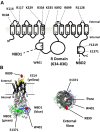
Fig. 1
Fig. 1
Molecular structure of CFTR. a Cartoon of the CFTR protein showing the location and charge of the all amino acids mutated in this study. Note that the extracellular loops are not drawn to scale. b Three-dimensional homology model of CFTR based on the work of Dalton et al. [14], using PyMol software indicating the relative positions of R899, W401 and E1371 residues to other regions of note. Note that the homology model lacks the R domain and that in the right hand diagram, the extracellular surface of CFTR is orientated to face the reader.
Fig. 2
Fig. 2
Arginine residue 889 in extracellular loop 4 of CFTR is essential for [Cl]o sensing. a Representative fWCR current recordings measured between ±100 mV in 20 mV steps from HEK cells transfected with wild type (WT) CFTR and R899Q CFTR, as indicated. The current traces are from the top down: (i) unstimulated in 155.5 mM [Cl]o, (ii) forskolin (FSK)-stimulated in 155.5 mM [Cl]o, (iii) FSK-stimulated in 35.5 mM [Cl]o and (iv) FSK-stimulated in 155.5 mM [Cl]o. Dotted line to the right of the current traces indicates zero current level. b Representative I-V plots for the Vector Control (VEC), WT and R899Q CFTR, for unstimulated in 155.5 mM [Cl]o (black squares); FSK-stimulated in 155.5 mM [Cl]o (black rhombus); FSK-stimulated in 35.5 mM [Cl]o (white circles) and FSK-stimulated in 155.5 mM [Cl]o washoff (white triangles). I-V plots were obtained from whole cell currents in a. All constructs, except the vector control, showed positive E rev shifts (see Table 1) on switching to low [Cl]o consistent with the expression of a Cl-selective conductance. Note the different y-axis scales for the respective I-Vs. c Percentage stimulation of FSK-activated currents by [Cl]o for WT CFTR (n = 24) and for different extracellular loop mutants (see Fig. 1a) (n = 4–9). Percent stimulation was calculated from I-V data at −60 mV from the reversal potential, as described in “Materials and methods.” Data are means ± SEM. **p < 0.01 compared to WT CFTR
Fig. 3
Fig. 3
Gating of CFTR by [Cl]o requires ATP hydrolysis and phosphorylation. a AMP-PNP locks WT CFTR in the open state. Normalised whole cell current density is plotted against time during FSK stimulation (indicated by the dashed line) and the subsequent washoff in the absence (squares, n = 4) and presence (circles, n = 4) of 1 mM AMP-PNP in the pipette solution. b, d Representative fWCR current recordings measured between ±100 mV in 20 mV steps from HEK cells transfected with WT CFTR plus AMP-PNP and E1371Q CFTR, as indicated. The current traces are from the top down: (i) unstimulated in 155.5 mM [Cl]o, (ii) forskolin (FSK)-stimulated in 155.5 mM [Cl]o, (iii) FSK-stimulated in 35.5 mM [Cl]o and (iv) FSK-stimulated in 155.5 mM [Cl]o. Dotted line to the right of the current traces indicates zero current level. c, e Representative I-V plots for the data presented in b and d. f Percentage stimulation of either basal or FSK-activated currents by [Cl]o for WT CFTR (n = 24), the ECL4 mutant R899Q, the hydrolysis-deficient mutant E1371Q and the double-mutant R899Q-E1371Q (see Fig. 1) under different conditions as indicated (n = 4–8). Data are means ± SEM. *p < 0.05, **p < 0.01, ***p < 0.001 between indicated datasets
Fig. 4
Fig. 4
Role of the R domain in [Cl]o sensing by CFTR. a, c Representative fWCR current recordings measured between ±100 mV in 20 mV steps from HEK cells transfected with deltaR-CFTR or deltaR-E1371S CFTR as indicated. Note all experiments used unstimulated conditions. The current traces are from the top down: (i) unstimulated in 155.5 mM [Cl]o, (ii) unstimulated in 35.5 mM [Cl]o and (iii) unstimulated in 155.5 mM [Cl]o. Dotted line to the right of the current traces indicates zero current level. b, d Representative I-V plots for the data presented in a and c. e Percentage current stimulation by [Cl]o for WT CFTR (n = 24) and for DeltaR and DeltaR-E1371Q mutants (see Fig. 1) under unstimulated (No Stim) or after exposure to a combination of forskolin and genistein (FSK/Genistein). (n = 4–10). Data are means ± SEM. *p < 0.05, **p < 0.01 compared to indicated datasets
Fig. 5
Fig. 5
ATP binding to site 1, but not site 2, underlies [Cl]o sensing by CFTR. a, c Representative fWCR current recordings measured between ±100 mV in 20 mV steps from HEK cells transfected with W401G CFTR or Y1219G CFTR, as indicated. The current traces are from the top down: (i) unstimulated in 155.5 mM [Cl]o, (ii) forskolin (FSK)-stimulated in 155.5 mM [Cl]o, (iii) FSK-stimulated in 35.5 mM [Cl]o and (iv) FSK-stimulated in 155.5 mM [Cl]o. Dotted line to the right of the current traces indicates zero current level. b, d Representative I-V plots for the data presented in a and c. e Percentage current stimulation by [Cl]o for WT CFTR (n = 24) and for W401G (NBD1) and Y1219G (NBD2) mutants (see Fig. 1) (n = 7–8). Data are mean ± SEM. **p < 0.01 compared to WT CFTR. f Percentage current stimulation by [Cl]o for WT CFTR at different cytosolic (pipette) ATP concentrations. Data are means ± SEM (n = 5–24). Data points fitted using least squares fit of a log [inhibition] vs response equation, with variable slope function and with no constraints or weighting. The open circle shows the effect of including 50 μM P-ATP with 50 μM ATP on the response to [Cl]o. ***p < 0.001 compared to 100 μM ATP
Fig. 6
Fig. 6
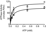
ATPase activity of CFTR is stimulated by a decrease in Cl concentration. Purified WT CFTR was reconstituted into dodecylmaltoside (DDM) micelles in the presence of either 150 mM (squares) or 50 mM Cl (circles) and the ATPase activity measured over a range of ATP concentrations, as described in the “Materials and methods.” Data are means ± SEM for 3 (low Cl) or 4 (high Cl) separate purification/reconstitution experiments. Data points fitted using the Michaelis Menten equation (r 2 = 0.83 (NaCl) and r 2 = 0.8 (NaGlu))

References

    1. Al-Nakkash L, Hu S, Li M, Hwang TC. A common mechanism for cystic fibrosis transmembrane conductance regulator protein activation by genistein and benzimidazolone analogs. J Pharmacol Exp Ther. 2001;296(2):464–472. - PubMed
    1. Aleksandrov L, Aleksandrov AA, Chang XB, Riordan JR. The first nucleotide binding domain of cystic fibrosis transmembrane conductance regulator is a site of stable nucleotide interaction, whereas the second is a site of rapid turnover. J Biol Chem. 2002;277(18):15419–15425. doi: 10.1074/jbc.M111713200. - VSports - DOI - PubMed
    1. Argent BE, Gray MA, Steward MC, Case RM (2012) In: L Johnson (ed) Physiology of the Gastrointestinal Tract. 5th edn. Elsevier, San Diego, pp 1399–1423
    1. Baker JM, Hudson RP, Kanelis V, Choy WY, Thibodeau PH, Thomas PJ, Forman-Kay JD. CFTR regulatory region interacts with NBD1 predominantly via multiple transient helices. Nat Struct Mol Biol. 2007;14(8):738–745. doi: 10.1038/nsmb1278. - DOI - PMC - PubMed
    1. Basso C, Vergani P, Nairn AC, Gadsby DC. Prolonged nonhydrolytic interaction of nucleotide with CFTR’s NH2-terminal nucleotide binding domain and its role in channel gating. J Gen Physiol. 2003;122(3):333–348. doi: 10.1085/jgp.200308798. - DOI - PMC - PubMed

Publication types

V体育ios版 - MeSH terms