<style draggable="gtJzG"><noframes date-time="SmOcabOA"><code dropzone="WbXJv8ZD"></code>
Skip to main page content
U.S. flag

An official website of the United States government

Dot gov

The . gov means it’s official. Federal government websites often end in . gov or . mil. Before sharing sensitive information, make sure you’re on a federal government site VSports app下载. .

Https

The site is secure. The https:// ensures that you are connecting to the official website and that any information you provide is encrypted and transmitted securely V体育官网. .

. 2015 Mar;61(3):883-94.
doi: 10.1002/hep.27489. Epub 2015 Jan 30.

Dysbiosis-induced intestinal inflammation activates tumor necrosis factor receptor I and mediates alcoholic liver disease in mice

Affiliations

Dysbiosis-induced intestinal inflammation activates tumor necrosis factor receptor I and mediates alcoholic liver disease in mice

Peng Chen et al. Hepatology. 2015 Mar.

Abstract

Intestinal barrier dysfunction is an important contributor to alcoholic liver disease (ALD). Translocated microbial products trigger an inflammatory response in the liver and contribute to steatohepatitis. Our aim was to investigate mechanisms of barrier disruption after chronic alcohol feeding VSports手机版. A Lieber-DeCarli model was used to induce intestinal dysbiosis, increased intestinal permeability, and liver disease in mice. Alcohol feeding for 8 weeks induced intestinal inflammation in the jejunum, which is characterized by an increased number of tumor necrosis factor alpha (TNF-α)-producing monocytes and macrophages. These findings were confirmed in duodenal biopsies from patients with chronic alcohol abuse. Intestinal decontamination with nonabsorbable antibiotics restored eubiosis, decreased intestinal inflammation and permeability, and reduced ALD in mice. TNF-receptor I (TNFRI) mutant mice were protected from intestinal barrier dysfunction and ALD. To investigate whether TNFRI on intestinal epithelial cells mediates intestinal barrier dysfunction and ALD, we used TNFRI mutant mice carrying a conditional gain-of-function allele for this receptor. Reactivation of TNFRI on intestinal epithelial cells resulted in increased intestinal permeability and liver disease that is similar to wild-type mice after alcohol feeding, suggesting that enteric TNFRI promotes intestinal barrier dysfunction. Myosin light-chain kinase (MLCK) is a downstream target of TNF-α and was phosphorylated in intestinal epithelial cells after alcohol administration. Using MLCK-deficient mice, we further demonstrate a partial contribution of MLCK to intestinal barrier dysfunction and liver disease after chronic alcohol feeding. .

Conclusion: Dysbiosis-induced intestinal inflammation and TNFRI signaling in intestinal epithelial cells mediate a disruption of the intestinal barrier. Therefore, intestinal TNFRI is a crucial mediator of ALD V体育安卓版. .


"V体育ios版" Conflict of interest statement

None of the authors has a financial, personal or professional conflict of interest to disclose.

Figures

Figure 1
Figure 1. Chronic ethanol administration elevates intestinal TNFα production in mice
C57BL/6 mice were orally fed a control or alcohol diet for 8 weeks. (A) TNFα mRNA level in jejunum (n = 14–19). (B) TNFα mRNA level in isolated lamina propria cells of the jejunum (n = 5–9). (C) Relative amount of TNFα+ inflammatory cells isolated from the jejunum and analyzed by FACS (n = 3–4). *p < 0.05, N.S.: no significance.
Figure 2
Figure 2. Alcohol abuse increases intestinal inflammatory TNFα+ cells in humans
(A) TNFα mRNA expression in duodenal biopsies obtained from healthy controls (n = 15) and patients with chronic alcohol abuse (n = 22). (B and C) Duodenal biopsies obtained from healthy controls (n = 11) and patients with chronic alcohol abuse (n = 8) were stained with CD68 (red) and TNFα (green) by immunofluorescence. Nuclei are stained in blue. (B) Representative intestinal sections are shown (magnification x200). (C) Quantification of CD68/TNFα double positive cells. *p < 0.05.
Figure 3
Figure 3. Intestinal decontamination inhibits alcohol-induced dysbiosis, intestinal inflammation and barrier dysfunction
(A–F) C57BL/6 mice were orally fed a control diet (n = 9), alcohol diet (n = 9) and alcohol diet plus antibiotics (ABX; n = 9). (A) Total cecal bacteria. (B) Jejunal TNFα mRNA level. (C) Quantification of F4/80 TNFα double positive cells in the jejunum as assessed by immunofluorescent staining. (D) Quantification of Lys6C TNFα double positive cells in the jejunum as assessed by immunofluorescent staining. (E) Representative western blot for occludin in the jejunum. (F) Fecal albumin content. (G–H) C57BL/6 mice were orally fed a control or alcohol diet for indicated time points (n = 4–7). (G) Fecal albumin content. Values are presented relative to control-fed animals. (H) Jejunum TNFα mRNA level after 11 days of control and alcohol diet feeding. *p < 0.05, N.S.: no significance.
Figure 4
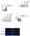
Figure 4. Reactivation of TNFRI on intestinal epithelial cells mediates alcohol-induced intestinal permeability in TNFRI mutant mice
Wild type (WT), TNFRIflxneo/flxneo and VillinCre TNFRIflxneo/flxneo mice were orally fed a control (n = 3–4) and alcohol diet (n = 7–9). (A) Fecal albumin content. (B) Western blot for E. Coli proteins in liver. (C) Representative western blot for occludin in the jejunum. (D) Occludin quantification of western blots. (E) Representative immunofluorescent staining for occludin; nuclei are stained blue. *p < 0.05.
Figure 5
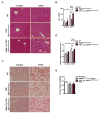
Figure 5. Reactivation of TNFRI on intestinal epithelial cells promotes alcoholic liver disease in TNFRI mutant mice
Wild type (WT), TNFRIflxneo/flxneo and VillinCre TNFRIflxneo/flxneo mice were orally fed a control (n = 3–4) and alcohol diet (n = 7–9). (A) Representative liver sections after hematoxylin-eosin staining. (B) Plasma ALT level. (C) Representative liver sections after oil red O staining. (D) Hepatic triglyceride content. (E) Plasma ethanol concentration. Magnification x200; *p < 0.05, N.S.: no significance.
Figure 6
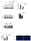
Figure 6. MLCK is involved in TNFα signaling and contributes to alcohol-induced barrier loss
(A) Wild type (WT), TNFRIflxneo/flxneo and VillinCre TNFRIflxneo/flxneo male mice were treated with ethanol or dextrose as control (ctlr) by gavage once (n = 3–4). Representative western blot for phosphorylated MLCK (p-MLCK) in epithelial cells isolated from the jejunum. (B–F) MLCK+/+ and MLCK−/− littermate mice were orally fed a control (n = 4) and alcohol diet (n = 10–14). (B) Fecal albumin content. (C) Western blot for E. Coli proteins in liver (left panel); plasma LPS level (right panel). (D) Representative western blot for occludin in the jejunum. (E) Occludin quantification of western blots. (F) Representative immunofluorescent staining for occludin; nuclei are stained blue. *p < 0.05.
Figure 7
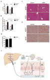
Figure 7. MLCK contributes to alcoholic liver disease
MLCK+/+ and MLCK−/− mice were orally fed a control (n = 4) and alcohol diet (n = 10–14). (A) Plasma ALT level. (B) Hepatic triglyceride content. (C) Representative liver sections after hematoxylin-eosin staining. (D) Representative liver sections after oil red O staining. (E) Plasma ethanol concentration. *p < 0.05. (F) Schematic representation of the proposed model of microbial translocation during alcoholic liver disease: Following the onset of alcoholic dysbiosis, monocytes and macrophages of the intestinal lamina propria are activated and produce TNFα. TNFα binds to TNFRI on enterocytes and increases intestinal permeability. This is in part mediated by the activation of MLCK resulting in disruption of tight junctions. Microbial products cross the mucosal barrier to reach the liver via the portal circulation. Microbial products cause hepatic inflammation and liver disease. The model has been reproduced from with permission from Elsevier and modified with additional permission from Elsevier.

V体育官网 - Comment in

References

    1. Szabo G, Bala S. Alcoholic liver disease and the gut-liver axis. World J Gastroenterol. 2010;16:1321–9. - PMC - PubMed
    1. Nanji AA, Khettry U, Sadrzadeh SM, Yamanaka T. Severity of liver injury in experimental alcoholic liver disease. Correlation with plasma endotoxin, prostaglandin E2, leukotriene B4, and thromboxane B2. Am J Pathol. 1993;142:367–73. - PMC - PubMed
    1. Bjarnason I, Peters TJ, Wise RJ. The leaky gut of alcoholism: possible route of entry for toxic compounds. Lancet. 1984;1:179–82. - PubMed
    1. Hritz I, Mandrekar P, Velayudham A, Catalano D, Dolganiuc A, Kodys K, et al. The critical role of toll-like receptor (TLR) 4 in alcoholic liver disease is independent of the common TLR adapter MyD88. Hepatology. 2008;48:1224–31. - PMC - PubMed
    1. Mathurin P, Deng QG, Keshavarzian A, Choudhary S, Holmes EW, Tsukamoto H. Exacerbation of alcoholic liver injury by enteral endotoxin in rats. Hepatology. 2000;32:1008–17. - PubMed

Publication types

MeSH terms (V体育2025版)

Substances