<b draggable="bmM6w2Q"><bdo draggable="7OaxdS"></bdo></b><area dropzone="RFpga66h"></area> "V体育平台登录" Skip to main page content
U.S. flag

An official website of the United States government

Dot gov

The . gov means it’s official. Federal government websites often end in . gov or . mil VSports app下载. Before sharing sensitive information, make sure you’re on a federal government site. .

Https

The site is secure V体育官网. The https:// ensures that you are connecting to the official website and that any information you provide is encrypted and transmitted securely. .

. 2014 Sep 22;211(10):2047-59.
doi: 10.1084/jem.20131333. Epub 2014 Sep 15.

"VSports手机版" Dysfunctional CD8+ T cells in hepatitis B and C are characterized by a lack of antigen-specific T-bet induction

Affiliations

V体育2025版 - Dysfunctional CD8+ T cells in hepatitis B and C are characterized by a lack of antigen-specific T-bet induction

Peter D Kurktschiev et al. J Exp Med. .

VSports - Abstract

The transcription factor T-bet regulates the production of interferon-γ and cytotoxic molecules in effector CD8 T cells, and its expression correlates with improved control of chronic viral infections. However, the role of T-bet in infections with differential outcome remains poorly defined. Here, we report that high expression of T-bet in virus-specific CD8 T cells during acute hepatitis B virus (HBV) and hepatitis C virus (HCV) infection was associated with spontaneous resolution, whereas T-bet deficiency was more characteristic of chronic evolving infection. T-bet strongly correlated with interferon-γ production and proliferation of virus-specific CD8 T cells, and its induction by antigen and IL-2 stimulation partially restored functionality in previously dysfunctional T-bet-deficient CD8 T cells VSports手机版. However, restoration of a strong interferon-γ response required additional stimulation with IL-12, which selectively induced the phosphorylation of STAT4 in T-bet(+) CD8 T cells. The observation that T-bet expression rendered CD8 T cells responsive to IL-12 suggests a stepwise mechanism of T cell activation in which T-bet facilitates the recruitment of additional transcription factors in the presence of key cytokines. These findings support a critical role of T-bet for viral clearance and suggest T-bet deficiency as an important mechanism behind chronic infection. .


"VSports最新版本" Figures

Figure 1.
Figure 1.
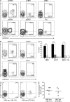
HBV- and HCV-specific CD8 T cells express high amounts of T-bet during acute spontaneously resolving infection. PBMCs were isolated from (A) patients with arHBV, cHBV, rHBV (top), aEBV, pEBV, or resolved Flu infection (bottom) and (B) patients with arHCV (top left), rHCV (top right), acHCV (bottom left), or cHCV infection (bottom right) and analyzed by flow cytometry. The outlined areas indicate the population of pentamer+ CD8 T cells and the numbers indicate the percentage of T-bet+ (above) and T-bet (below) cells among total pentamer+ CD8 T cells. (C) Quantification of ex vivo T-bet expression in virus-specific CD8 T cells of the subjects described in A and B. Bars represent the mean percentage of T-bet+ pentamer+ CD8 T cells among total pentamer+ CD8 T cells of patients with arHBV (n = 19), cHBV (n = 24), rHBV (n = 5), arHCV (n = 7), acHCV (n = 7), cHCV (n = 7), rHCV (n = 7), aEBV (n = 3), pEBV (n = 6), and Flu (n = 6). Error bars indicate the SEM. *, P < 0.05; ***, P < 0.001 (Mann-Whitney-U test). (D) Representative ex-vivo flow cytometry plots showing expression of T-bet in HBV envelope (183–191)–specific (left) and HBV polymerase (573–581)–specific CD8 T cells (right) of patients with arHBV. The numbers indicate the percentage of T-bet+/− and pentamer+/− CD8 T cells among total CD8 T cells. (E) Scatter dot plot showing the percentage of T-bet+ envelope (183–191)–specific (filled circles; n = 5) or polymerase (573–581)-specific (open circles; n = 5) CD8 T cells among total pentamer+ CD8 T cells of patients with arHBV. The data are representative of one experiment due to limited patient material.
Figure 2.
Figure 2.
T-bet expression is lost early in acute chronic-evolving HCV infection. Percentage of T-bet+ pentamer+ CD8 T cells among total pentamer+ CD8 T cells of patients with arHBV (n = 5; left), arHCV (n = 4; middle), and acHCV (n = 4; right) was determined by ex vivo flow cytometry at the indicated time points. Data show the mean percentages and are representative of one experiment due to limited patient material. Error bars indicate SEM.
Figure 3.
Figure 3.
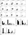
Expression of T-bet is associated with distinct phenotypes of virus-specific CD8 T cells. Ex vivo expression of T-bet, Eomes, PD-1, and CD127 in PBMCs from patients with acute HBV or HCV infection was analyzed by flow cytometry on the earliest available samples obtained within 3 wk of acute symptom onset. The data on cHBV and cHCV patients were obtained at any time point during chronic infection. (A) Contour plots show ex vivo coexpression of T-bet and Eomes in virus-specific CD8 T cells of patients with arHBV, cHBV, arHCV, acHCV, and cHCV infection. The numbers indicate the percentage of T-bet+/− and Eomes+/− CD8 T cells among pentamer+ CD8 T cells. (B) Representative contour plots of T-bet and PD-1 coexpression as described in (A). (C) Representative contour plots of T-bet and CD127 coexpression as described in A. (D) Mean percentage of pentamer+ CD8 T cells with a T-bet+/Eomes, T-bet/Eomes+, and T-bet+/Eomes+ phenotype as determined by flow cytometry. Data were obtained from arHBV (n = 19), cHBV (n = 24), arHCV (n = 7), acHCV (n = 7), and cHCV (n = 7) patients. (E) Mean percentage of pentamer+ CD8 T cells with T-bet+/ PD-1+ and T-bet/ PD-1+ phenotype found in the patients described in D. (F) Mean percentage of T-bet+/ CD127+, T-bet/ CD127+, and T-bet+/CD127 analogous to D. All error bars indicate SEM. ***, P < 0.001 (Mann-Whitney-U test). Data are representative of one experiment due to limited patient material.
Figure 4.
Figure 4.
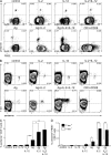
Antigen-specific interferon-γ production correlates with T-bet and can be restored by IL-2+IL-12 co-stimulation. PBMCs of patients with cHBV (n = 11) were cultured for 3 d in culture medium (control) containing either HBV-c18-27 antigen (Ag), c18-27 antigen+IL-2 (Ag+IL-2), c18-27 antigen+IL-12 (Ag+IL-12), IL-2+IL-12, c18-27 antigen+IL-2+IL-12 (Ag+IL-2+IL-12), or CD3+CD28. On day 3 antigen-treated groups were restimulated with antigen and all groups were incubated for 6 h in the presence of Brefeldin A before intracellular flow cytometry was performed. (A) Expression of T-bet and interferon-γ by CD8 T cells. The numbers indicate the percentage of T-bet+/− and interferon-γ+/− CD8 T cells among total CD8 T cells. (B) Mean induction of T-bet by treatment with the respective cytokines. Induction was defined as the percentage of T-bet+ CD8 T cells after stimulation subtracted by the percentage of T-bet+ CD8 T cells in unstimulated controls. Error bars represent the SEM. (C) Mean percentage of T-bet+ interferon-γ+ CD8 T cells after stimulation. Error bars indicate the SEM. Data are representative of one experiment due to limited patient material. ***, P < 0.001 (Mann-Whitney-U test).
Figure 5.
Figure 5.
Induction of T-bet by IL-2 and antigen is associated with antigen-specific proliferation. PBMCs of patients with cHBV (n = 10) were labeled with the proliferation marker PBSE and cultured for 7 d in culture medium in the presence or absence of HBV-c18-27 antigen (Ag), IL-2, or IL-12. Antigen c18-27 was added on day 0, and cytokines were administered on day 4 in the designated groups. Negative controls were cultured in medium without further supplements, while positive controls were stimulated with CD3+CD28. On day 7, cells were stained for flow cytometry. (A) Frequencies of pentamer+ CD8 T cells among total CD8 T cells and their PBSE labeling intensity. (B) Expression of T-bet and the frequency of pentamer+ CD8 T cells after stimulation. (C) Mean percentage of c18-27–specific CD8 T cells among total CD8 T cells. (D) Mean percentage of PBSE/T-bet (white) and mean percentage of PBSE/T-bet+ specific CD8 T cells (black) among total CD8 T cells. Error bars represent the SEM. Data are representative of one experiment due to limited patient material. *, P < 0.05; **, P < 0.01; ***, P < 0.001 (Mann-Whitney-U test).
Figure 6.
Figure 6.
IL-12 selectively induces STAT4 phosphorylation in T-bet+ CD8 T cells. PBMCs of patients with cHBV (n = 7) were cultured for 3 d with either IL-2 or medium as control. On day 3 cells were restimulated for 20 min with either IL-2 or IL-12. Controls were left unstimulated. Cells were then analyzed by flow cytometry. (A) Coexpression of T-bet and pSTAT4 in CD8 T cells of an unstimulated control (left), PBMCs cultured with IL-2 and restimulated with IL-2 (middle), and PBMCs cultured in IL-2 and restimulated with IL-12 (right). Representative plots demonstrate the percentage of T-bet+/− and pSTAT4+/− CD8 T cells among total CD8 T cells. (B) Mean frequency of T-bet pSTAT4+ (white) and mean frequency of T-bet+ pSTAT4+ (black) CD8 T cells among total CD8 T cells after stimulation with the respective cytokines. Error bars represent the SEM. Data are representative of one experiment due to limited patient material. **, P < 0.01 (Mann-Whitney-U test).

Comment in

References

    1. Artillo, S., Pastore G., Alberti A., Milella M., Santantonio T., Fattovich G., Giustina G., Ryff J.C., Chaneac M., Bartolomé J., and Carreño V.. 1998. Double-blind, randomized controlled trial of interleukin-2 treatment of chronic hepatitis B. J. Med. Virol. 54:167–172 10.1002/(SICI)1096-9071(199803)54:3<167::AID-JMV4>3.0.CO;2-3 - DOI - PubMed
    1. Auffermann-Gretzinger, S., Keeffe E.B., and Levy S.. 2001. Impaired dendritic cell maturation in patients with chronic, but not resolved, hepatitis C virus infection. Blood. 97:3171–3176 10.1182/blood.V97.10.3171 - "VSports app下载" DOI - PubMed
    1. Boni, C., Fisicaro P., Valdatta C., Amadei B., Di Vincenzo P., Giuberti T., Laccabue D., Zerbini A., Cavalli A., Missale G., et al. . 2007. Characterization of hepatitis B virus (HBV)-specific T-cell dysfunction in chronic HBV infection. J. Virol. 81:4215–4225 10.1128/JVI.02844-06 - DOI - PMC - PubMed
    1. Bowen, D.G., Zen M., Holz L., Davis T., McCaughan G.W., and Bertolino P.. 2004. The site of primary T cell activation is a determinant of the balance between intrahepatic tolerance and immunity. J. Clin. Invest. 114:701–712 10.1172/JCI200421593 - DOI - PMC - PubMed
    1. Carreño, V., Zeuzem S., Hopf U., Marcellin P., Cooksley W.G., Fevery J., Diago M., Reddy R., Peters M., Rittweger K., et al. . 2000. A phase I/II study of recombinant human interleukin-12 in patients with chronic hepatitis B. J. Hepatol. 32:317–324 10.1016/S0168-8278(00)80078-1 - DOI - PubMed

Publication types

MeSH terms

Substances