Skip to main page content
U.S. flag

An official website of the United States government

Dot gov

The VSports app下载. gov means it’s official. Federal government websites often end in . gov or . mil. Before sharing sensitive information, make sure you’re on a federal government site. .

Https

The site is secure. The https:// ensures that you are connecting to the official website and that any information you provide is encrypted and transmitted securely V体育官网. .

. 2014 Jul 16:12:40.
doi: 10.1186/s12964-014-0040-3.

CD39 is a negative regulator of P2X7-mediated inflammatory cell death in mast cells

CD39 is a negative regulator of P2X7-mediated inflammatory cell death in mast cells

Marcel Kuhny et al. Cell Commun Signal. .

Abstract

Background: Mast cells (MCs) are major contributors to an inflammatory milieu. One of the most potent drivers of inflammation is the cytokine IL-1β, which is produced in the cytoplasm in response to danger signals like LPS. Several controlling mechanisms have been reported which limit the release of IL-1β. Central to this regulation is the NLRP3 inflammasome, activation of which requires a second danger signal with the capacity to subvert the homeostasis of lysosomes and mitochondria VSports手机版. High concentrations of extracellular ATP have the capability to perturb the plasma membrane by activation of P2X7 channels and serve as such a danger signal. In this study we investigate the role of P2X7 channels and the ecto-5'-nucleotidase CD39 in ATP-triggered release of IL-1β from LPS-treated mast cells. .

Results: We report that in MCs CD39 sets an activation threshold for the P2X7-dependent inflammatory cell death and concomitant IL-1β release. Knock-out of CD39 or stimulation with non-hydrolysable ATP led to a lower activation threshold for P2X7-dependent responses. We found that stimulation of LPS-primed MCs with high doses of ATP readily induced inflammatory cell death. Yet, cell death-dependent release of IL-1β yielded only minute amounts of IL-1β V体育安卓版. Intriguingly, stimulation with low ATP concentrations augmented the production of IL-1β in LPS-primed MCs in a P2X7-independent but caspase-1-dependent manner. .

Conclusion: Our study demonstrates that the fine-tuned interplay between ATP and different surface molecules recognizing or modifying ATP can control inflammatory and cell death decisions. V体育ios版.


Figures (VSports最新版本)
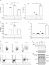
Figure 1
Figure 1
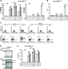
Correlation of IL-1β release and cell death in mast cells. (A) wt BMMCs were primed with 1 μg/ml LPS for 3.5 h and then left untreated or stimulated with the indicated concentrations [mM] of ATP for 1 h. TCL and SN were probed for IL-1β by ELISA (n = 8). (B) Treatment as in (A); SN was probed for IL-6 by ELISA (n = 4). (C) Treatment as in (A); wt BMMCs were stained with Pi and analyzed by FACS (n = 13). (D) Treatment as in (A); cells were stained with FITC-conjugated Annexin V and Pi and analyzed by FACS. The morphology is displayed in the forward and side scatter (FSC/SSC); representative result (n = 12). (E) wt BMMCs were primed with 1 μg/ml LPS for 3.5 h. The cells were then concentrated to 2*106 cells/60 μL and left untreated or stimulated with the indicated concentrations of ATP for 1 h. TCL and SN were then analyzed by immunoblotting with anti-IL-1β (top and middle panel) and anti-p85 (bottom panel, loading control). For details on indicated bands see text. Shown are means and SD of replicates of one representative experiment each. Statistical analysis of n independent experiments by LMM; FDR-corrected p-values: * < 0.05, ** < 0.005, and *** < 0.0005.
Figure 2
Figure 2
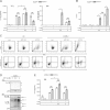
Caspase-1 controls production of IL-1β in mast cells. (A) wt BMMCs were primed with 1 μg/ml LPS for 3.5 h and then left untreated or stimulated with the indicated concentrations [mM] of ATP for 1 h; vehicle (DMSO) or caspase-1 inhibitor (YVAD-CHO) was added 1 h prior to stimulation with ATP. TCL and SN were probed for IL-1β by ELISA (n = 5) (left panel). BMMCs were stained with Pi and analyzed by FACS (n = 3) (right panel). (B) Treatment as in (A); TCL of wt BMMCs was probed for IL- β by ELISA (n = 5). (C) Treatment as in (A); transcripts were analyzed by qPCR (n = 3). Shown are means and SD of replicates of one representative experiment each. Statistical analysis of n independent experiments by LMM; FDR-corrected p-values: * < 0.05, ** < 0.005, and *** < 0.0005.
Figure 3
Figure 3
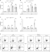
P2X7 is required for ATP-induced IL-1β release and cell death. (A) wt and P2rx7−/− BMMCs were primed with 1 μg/ml LPS for 3.5 h and then left untreated or stimulated with the indicated concentrations [mM] of ATP for 1 h. TCL and SN were probed for IL-1β by ELISA (n = 5). (B) Treatment as in (A); wt and P2rx7−/− BMMCs were stained with Pi and analyzed by FACS (n = 4). (C) Treatment as in (A); cells were stained with FITC-conjugated Annexin V and Pi and analyzed by FACS. The morphology is displayed in the forward and side scatter (FSC/SSC); representative result (n = 4). (D) P2rx7−/− BMMCs were primed with 1 μg/ml LPS for 3.5 h. Cells were then concentrated to 2*106 cells/60 μL and left untreated or stimulated with the indicated concentrations of ATP for 1 h. TCL and SN of wt and P2rx7−/− BMMCs were then analyzed by immunoblotting with anti-IL-1β (top and middle panel) and anti p85 (bottom panel, loading control). For details on indicated bands see text. (E) Treatment as in (A); SN were probed for IL-6 by ELISA (n = 6). Shown are means and SD of replicates of one representative experiment each. Statistical analysis of n independent experiments by LMM; FDR-corrected p-values: * < 0.05, ** < 0.005, and *** < 0.0005.
Figure 4
Figure 4
CD39 is a negative regulator of IL-1β release and cell death. (A) wt and Cd39−/− BMMCs were primed with 1 μg/ml LPS for 3.5 h and then left untreated or stimulated with the indicated concentrations [mM] of ATP for 1 h. TCL and SN were probed for IL-1β by ELISA (n = 5). (B) Treatment as in (A); wt and Cd39−/− BMMCs were stained with Pi and analyzed by FACS (n = 6). (C) Treatment as in (A); cells were stained with FITC-conjugated Annexin V and Pi and analyzed by FACS. The morphology is displayed in the forward and side scatter (FSC/SSC); representative result (n = 4). (D) Cd39−/− BMMCs were primed with 1 μg/ml LPS for 3.5 h. The cells were then concentrated to 2*106 cells/60 μL and left untreated or stimulated with the indicated concentrations of ATP for 1 h. TCL and SN were then analyzed by immunoblotting with anti-IL-1β (top and middle panel) and anti-p85 (bottom panel, loading control). For details on indicated bands see text. (E) Treatment as in (A); SN of wt and Cd39−/− BMMCs were probed for IL-6 by ELISA (n = 6). Shown are means and SD of replicates of one representative experiment each. Statistical analysis of n independent experiments by LMM; FDR-corrected p-values: * < 0.05, ** < 0.005, and *** < 0.0005.
Figure 5
Figure 5
Stimulation with ATPγS aggravates cell death and IL-1β release. (A) wt and P2rx7−/− BMMCs were primed with 1 μg/ml LPS for 3.5 h and then left untreated or stimulated with the indicated concentrations [mM] of ATPγS for 1 h. TCL was probed for IL-1β by ELISA (n = 4). (B) Treatment as in (A); SN of wt and P2rx7−/− BMMCs were probed for IL-6 by ELISA (n = 4). (C) Treatment as in (A); SN of wt and P2rx7−/− BMMCs were probed for IL-1β by ELISA (n = 4). (D) Treatment as in (A); wt and P2rx7−/− BMMCs were stained with Pi and analyzed by FACS (n = 3). (E) Treatment as in (A); cells were stained with FITC-conjugated Annexin V and Pi and analyzed by FACS. The morphology is displayed in the forward and side scatter (FSC/SSC); representative result (n = 3). Shown are means and SD of replicates of one representative experiment each. Statistical analysis of n independent experiments by LMM; FDR-corrected p-values: * < 0.05, ** < 0.005, and *** < 0.0005.

References

    1. Marshall JS. Mast-cell responses to pathogens. Nat Rev Immunol. 2004;4:787–799. doi: 10.1038/nri1460. - DOI - PubMed
    1. Chen GY, Nunez G. Sterile inflammation: sensing and reacting to damage. Nat Rev Immunol. 2010;10:826–837. doi: 10.1038/nri2873. - DOI - PMC - PubMed
    1. Enoksson M, Lyberg K, Möller-Westerberg C, Fallon PG, Nilsson G, Lunderius-Andersson C. Mast cells as sensors of cell injury through IL-33 recognition. J Immunol. 2011;186:2523–2528. doi: 10.4049/jimmunol.1003383. - DOI (VSports) - PubMed
    1. Kawai T, Akira S. The role of pattern-recognition receptors in innate immunity: update on Toll-like receptors. Nat Immunol. 2010;11:373–384. doi: 10.1038/ni.1863. - DOI - PubMed
    1. Lopez-Castejon G, Brough D. Understanding the mechanism of IL-1β secretion. Cytokine Growth Factor Rev. 2011;22:189–195. doi: 10.1016/j.cytogfr.2011.10.001. - VSports手机版 - DOI - PMC - PubMed

V体育官网 - Publication types

MeSH terms (V体育官网)

LinkOut - more resources