Skip to main page content
U.S. flag

An official website of the United States government

Dot gov

The . gov means it’s official. Federal government websites often end in . gov or . mil. Before sharing sensitive information, make sure you’re on a federal government site VSports app下载. .

Https

The site is secure. The https:// ensures that you are connecting to the official website and that any information you provide is encrypted and transmitted securely. V体育官网.

Review
. 2014 Jul 29:5:378.
doi: 10.3389/fmicb.2014.00378. eCollection 2014.

Deciphering the role of DC subsets in MCMV infection to better understand immune protection against viral infections

Affiliations
Review

Deciphering the role of DC subsets in MCMV infection to better understand immune protection against viral infections (V体育ios版)

Yannick O Alexandre et al. Front Microbiol. .

Abstract

Infection of mice with murine cytomegalovirus (MCMV) recapitulates many physiopathological characteristics of human CMV infection and enables studying the interactions between a virus and its natural host. Dendritic cells (DC) are mononuclear phagocytes linking innate and adaptive immunity which are both necessary for MCMV control. DC are critical for the induction of cellular immunity because they are uniquely efficient for the activation of naïve T cells during their first encounter with a pathogen VSports手机版. DC are equipped with a variety of innate immune recognition receptors (I2R2) allowing them to detect pathogens or infections and to engulf molecules, microorganisms or cellular debris. The combinatorial engagement of I2R2 during infections controls DC maturation and shapes their response in terms of cytokine production, activation of natural killer (NK) cells and functional polarization of T cells. Several DC subsets exist which express different arrays of I2R2 and are specialized in distinct functions. The study of MCMV infection helped deciphering the physiological roles of DC subsets and their molecular regulation. It allowed the identification and first in vivo studies of mouse plasmacytoid DC which produce high level of interferons-α/β early after infection. Despite its ability to infect DC and dampen their functions, MCMV induces very robust, efficient and long-lasting CD8 T cell responses. Their priming may rely on the unique ability of uninfected XCR1(+) DC to cross-present engulfed viral antigens and thus to counter MCMV interference with antigen presentation. A balance appears to have been reached during co-evolution, allowing controlled replication of the virus for horizontal spread without pathological consequences for the immunocompetent host. We will discuss the role of the interplay between the virus and DC in setting this balance, and how advancing this knowledge further could help develop better vaccines against other intracellular infectious agents. .

Keywords: NK cells; XCR1+ dendritic cells; cross-presentation; immune evasion; murine cytomegalovirus; plasmacytoid dendritic cells; type I interferons; vaccination V体育安卓版. .


Figures

Figure 1
Figure 1
DC functions are differentially impacted by their infection in vitro vs. the infection of mice in vivo. (A) Impact on DC functions of their infection by MCMV in vitro. In vitro, MCMV-infected DC are “paralyzed” by the virus which prevents them to deliver adequate signals 1 and 2 for antiviral CD8 T cell activation. Specifically, infected DC down-regulate their expression of MHC-I and activating co-stimulation (CD40, CD80, CD86) molecules as a consequence of their expression of viral immune evasion genes. They induce inhibitory co-stimulation molecules (PD-L1 and PD-L2). Hence, infected DC only weakly prime antiviral CD8 T cells. (B) cDC functions in vivo in MCMV-infected mice. cDC are very strongly activated by MCMV infection in vivo, in a way enabling them to induce potent T cell activation in vitro, and consistent with the fact that MCMV infection induces very strong and protective antiviral cellular immune responses in vivo. Specifically, XCR1+ DC and CD11b+ cDC strongly up-regulate MHC and activating co-stimulation molecules in vivo, and can also produce T cell-activating cytokines such as IL-12 and IL-15. (C,D) Proposed explanations to the discrepancy of the impact of MCMV infection on DC in vitro vs. in vivo. (C) Impact of the nature of DC subsets and of their frequency of infection. The DC used in vitro are derived from monocytes (MoDC) and strongly differ from the DC involved in vivo in the induction of anti-viral immune responses in lymphoid tissues (LT-DC). High MOI are used for in vitro infection of DC, leading to a very high proportion of infected cells subjected to the effects of viral immune evasion genes. In contrast, only a very small fraction of XCR1+ cDC and CD11b+ cDC is infected in vivo. (D) Protective functions of cDC in MCMV infected mice are promoted by the inflammatory milieu, in particular by IFN-I and IFN-γ, and by cross-talk with other immune cells.
Figure 2
Figure 2
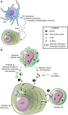
Molecular mechanisms regulating DC subset activation during MCMV infection. (A) Mechanism promoting DC subset maturation in vivo during MCMV infection. High amount of IFN-I are produced in vivo by pDC early after MCMV infection, which drives broad cell-intrinsic responses in all DC, including canonical DC maturation, promoting protective crosstalk with innate and adaptive immune cells. (B) Mechanisms promoting pDC sensing of MCMV infection. Early after completion of the first cycle of virus replication in vivo, pDC sense MCMV nucleotide sequences via endosomal TLRs, which leads to innate cytokine production, in particular IFN-I. How MCMV material ends up in the endosomes of pDC is still not understood. pDC might specifically recognize and engulf materials derived from infected cells and containing viral nucleic acids, either apoptotic bodies or exosomes (❶). pDC may also be able to detect and engulf MCMV particles released by infected cells or even from the viral inoculum (❷). Finally, pDC may directly sense and nibble infected cells (❸).
Figure 3
Figure 3
Requirements for different antigen-presenting cell types during acute vs. latent MCMV infection. (A) Professional cross-presenting XCR1+ cDC are necessary and sufficient for priming of MCMV-specific CD8 T cell during acute infection. XCR1+ cDC are able to take up and process antigenic proteins derived from infected cells, either non-hematopoietic cells as illustrated or the small fraction of infected DC. Once they have processed viral proteins into epitopic peptides, XCR1+ cDC can present them in association with MHC-I molecules (signal 1) to anti-MCMV CD8 T cells. The priming of naïve CD8 T cells for the induction of protective antiviral responses also requires activating co-stimulation signals (signal 2), such as the engagement of CD28 on the lymphocytes by CD80/CD86 expressed by the DC, and activating cytokines (signal 3) such as IL-12 or IFN-I. XCR1+ cDC are also competent for delivery of signals 2 and 3. Upon priming, naïve conventional and inflationary CD8 T cells differentiate into effector conventional and inflationary CD8 T cells, respectively, which control acute viral replication through recognition and killing of infected cells throughout the body. (B) Non-hematopoietic cells are necessary to drive inflationary anti-MCMV CD8 T cell responses during latent infection. After resolution of acute infection, the compartment of antiviral CD8 T cells contracts and gives rise to a low number of memory cells. In latent infection, MCMV can stochastically and transiently reactivate from latently-infected non-hematopoietic cells, causing the expression and presentation of a small number of viral antigens. This drives in turn the reactivation and proliferation of the memory CD8 T cell pool specific for the corresponding viral antigens. These CD8 T cells acquire an effector/effector-memory phenotype and expand continuously over time; a process called “memory inflation.” Even though hematopoietic cells are neither necessary nor sufficient for viral antigen presentation during latent infection, they might contribute to promote memory inflation by delivering other signals to CD8 T cells, such as cytokines or chemokines.
Figure 4
Figure 4
Schematic model of the cross-talk between XCR1+ DC, NK cells, and CD8 T cells during MCMV infection. (A) Comparative kinetics and intensities of innate cytokine, NK and CD8 T cell responses between mice having or lacking an efficient antiviral NK cell response. (B) Potential mechanisms in place early after infection of mice with efficient NK cell responses and promoting accelerated activation of anti-viral CD8 T cells. Early killing of infected cells may provide XCR1+ DC with a faster and increased access to viral antigen for cross-presentation by delivering them immunogenic apoptotic bodies (❶). It may also reduce the amount of viral ligands accessible to pDC and thus decrease systemic IFN-I production to promote its immune activation effects over the immunosuppressive ones (❷). Low IFN-I concentrations may limit the direct negative effects of these cytokines on CD8 T cells (❸), preserve the maintenance of high numbers of XCR1+ DC (❹), and promote an optimal maturation of XCR1+ DC by enabling them to selectively express high levels of activating but not inhibitory co-stimulatory molecules (❺). (C) Potential mechanisms in place later after infection of mice with efficient NK cell responses and promoting accelerated and stronger contraction of anti-viral CD8 T cells. NK cell killing of infected cells could rapidly shortage the supply of viral antigens available for cross-presentation by XCR1+ DC (❻). Cognate engagement of Ly49H by m157 leads to late IL-10 production by the NK cells which limit XCR1+ DC maturation (❼) and directly inhibit CD8 T cell proliferation (❽).

"V体育官网入口" References

    1. Adam C., King S., Allgeier T., Braumuller H., Luking C., Mysliwietz J., et al. (2005). DC-NK cell cross talk as a novel CD4+ T-cell-independent pathway for antitumor CTL induction. Blood 106, 338–344 10.1182/blood-2004-09-3775 - DOI (V体育官网) - PubMed
    1. Andoniou C. E., Van Dommelen S. L., Voigt V., Andrews D. M., Brizard G., Asselin-Paturel C., et al. (2005). Interaction between conventional dendritic cells and natural killer cells is integral to the activation of effective antiviral immunity. Nat. Immunol. 6, 1011–1019 10.1038/ni1244 - DOI - PubMed
    1. Andrews D. M., Andoniou C. E., Granucci F., Ricciardi-Castagnoli P., Degli-Esposti M. A. (2001). Infection of dendritic cells by murine cytomegalovirus induces functional paralysis. Nat. Immunol. 2, 1077–1084 10.1038/ni724 - DOI - PubMed
    1. Andrews D. M., Estcourt M. J., Andoniou C. E., Wikstrom M. E., Khong A., Voigt V., et al. (2010). Innate immunity defines the capacity of antiviral T cells to limit persistent infection. J. Exp. Med. 207, 1333–1343 10.1084/jem.20091193 - DOI - PMC - PubMed
    1. Andrews D. M., Scalzo A. A., Yokoyama W. M., Smyth M. J., Degli-Esposti M. A. (2003). Functional interactions between dendritic cells and NK cells during viral infection. Nat. Immunol. 4, 175–181 10.1038/ni880 - VSports在线直播 - DOI - PubMed

Publication types (VSports app下载)