Skip to main page content
U.S. flag

An official website of the United States government

Dot gov

The . gov means it’s official. Federal government websites often end in . gov or . mil VSports app下载. Before sharing sensitive information, make sure you’re on a federal government site. .

Https

The site is secure. The https:// ensures that you are connecting to the official website and that any information you provide is encrypted and transmitted securely. V体育官网.

. 2014 Aug 1;9(8):e103125.
doi: 10.1371/journal.pone.0103125. eCollection 2014.

Cell wall-anchored nuclease of Streptococcus sanguinis contributes to escape from neutrophil extracellular trap-mediated bacteriocidal activity

Affiliations

Cell wall-anchored nuclease of Streptococcus sanguinis contributes to escape from neutrophil extracellular trap-mediated bacteriocidal activity

"VSports" Chisato Morita et al. PLoS One. .

"VSports" Abstract

Streptococcus sanguinis, a member of the commensal mitis group of streptococci, is a primary colonizer of the tooth surface, and has been implicated in infectious complications including bacteremia and infective endocarditis. During disease progression, S. sanguinis may utilize various cell surface molecules to evade the host immune system to survive in blood. In the present study, we discovered a novel cell surface nuclease with a cell-wall anchor domain, termed SWAN (streptococcal wall-anchored nuclease), and investigated its contribution to bacterial resistance against the bacteriocidal activity of neutrophil extracellular traps (NETs). Recombinant SWAN protein (rSWAN) digested multiple forms of DNA including NET DNA and human RNA, which required both Mg(2+) and Ca(2+) for optimum activity. Furthermore, DNase activity of S. sanguinis was detected around growing colonies on agar plates containing DNA. In-frame deletion of the swan gene mostly reduced that activity VSports手机版. These findings indicated that SWAN is a major nuclease displayed on the surface, which was further confirmed by immuno-detection of SWAN in the cell wall fraction. The sensitivity of S. sanguinis to NET killing was reduced by swan gene deletion. Moreover, heterologous expression of the swan gene rendered a Lactococcus lactis strain more resistant to NET killing. Our results suggest that the SWAN nuclease on the bacterial surface contributes to survival in the potential situation of S. sanguinis encountering NETs during the course of disease progression. .


Conflict of interest statement

Competing Interests: The authors have declared that no competing interests exist.

Figures

Figure 1
Figure 1. DNA digestion around growing colonies of streptococcal species.
The DNase activities of oral streptococcus strains were examined using BHI agar plates containing salmon sperm DNA. The plates were incubated for 72-digested DNA. Halos seen around colonies reflect DNA digestion. The following strains were tested: S. sanguinis, SK36; S. oralis, NCTC 11427T/SK23; S. mutans, MT8148; S. salivarius, HHT; S. parasanguinis, ATCC 903; S. sobrinus, MT10186. Bar, 1 cm.
Figure 2
Figure 2. Chromosomal swan gene locus and SWAN domain organization.
A genetic map around ssa1750 (swan) from Streptococcus sanguinis strain SK36 is shown. Gene designations or gene tag numbers (ssa) are indicated inside the genes (arrows). The domain organization of SWAN is also shown. The positions of the putative signal sequence (SS), the cell wall sorting signal containing a Sortase A-recognizable LPKTG sequence, an RGD sequence, oligosaccharide/nucleotide binding fold (OB-fold), and an MnuA DNase 1-like domain are depicted with start and end positions of the amino acid residue numbers from the N-terminus.
Figure 3
Figure 3. Efficient DNase activity of rSWAN requires Ca2+ and Mg2+.
(A) λDNA (0.3 µg) was incubated with recombinant SWAN (rSWAN, 0.1 µg) with or without CaCl2 (1 mM) and MgCl2 (1 mM) at 37°C for 1 h. Following electrophoresis, DNA was stained with ethidium bromide and visualized under UV light. The sizes of the λDNA Hind III digest markers are indicated on the left. (B) λDNA (0.3 µg) was incubated with rSWAN (0.1 µg) with varying concentrations of either Ca2+ (red circles) or Mg2+ (blue circles) at 37°C for 30 min. Using gel images and Image J software, densitometric analyses were performed to calculate % DNA cleavage. Values shown represent the average ± SE of 3 independent experiments.
Figure 4
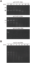
Figure 4. Nuclease activity of rSWAN against variable forms of viral DNA and RNA.
(A) The substrate specificity and preference of rSWAN were examined using φX174RF I DNA (double-stranded, mainly super coiled circular), φX174 RF II DNA (double-stranded, mainly nicked circular), and φX174 DNA (single-stranded, mainly circular). Open arrowhead, open circular form; black arrowhead, super coiled form; gray arrowhead, linear form. (B) Total RNA from human keratinocytes was incubated with rSWAN at 37°C for the indicated time periods.
Figure 5
Figure 5. rSWAN cleaves NET DNA.
Neutrophils were isolated from heparinized human blood and NETs were induced with PMA. NETs were incubated with or without rSWAN (40 µg/ml) at 37°C for 1 h. After fixation and permealization, neutrophil elastase was labeled with rabbit anti-human elastase IgG and an Alexa Fluor 594-conjugated secondary antibody, and DNA was stained with DAPI. Neutrophils without treatment with PMA and rSWAN were utilized as a control. The slides were observed using fluorescent microscopy. Bar, 20 µm.
Figure 6
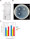
Figure 6. SWAN contributes to escape from bacteriocidal effects of NETs.
(A) Total proteins in the cell wall fractions of S. sanguinis SK36 (WT), the swan deletion mutant (Delswan), and the revertant strain (Wr) were immunoblotted with mouse antiserum against the N-terminal recombinant fragment of SWAN. (B) The DNase activities of WT, Delswan, and Wr on BHI agar plates containing DNA were examined as described in Fig. 1. (C) S. sanguinis strains grown to the late-exponential phase were exposed to NETs at an MOI of 5. After the indicated times, neutrophils were lysed and the lysates were plated on Todd-Hewitt agar plates. The survival rate of S. sanguinis strains is shown as % inoculated colony forming units. Data were pooled from 4 independent experiments performed in triplicate and the values are shown as the mean ± SD. Statistical analysis was performed using one-way ANOVA and Scheffe's test. A confidence interval of p<0.05 was considered to be significant. *p<0.01, **p<0.05.
Figure 7
Figure 7. Heterologous expression of swan in Lactococcus lactis.
(A) Immunoblot analysis for detection of SWAN in cell wall fractions of empty vector-transformed L. lactis (Mock) and swan-expressing (Lswan) strains. (B) The DNase activities of Mock and Lswan were examined by culturing in DNA-containing BHI agar plates for 24 h. (C) Human NETs were exposed to Mock or Lswan at an MOI of 10 for 2 h. The survival rate of recovered bacteria is shown as % inoculated colony forming units. Data were pooled from 3 independent experiments performed in triplicate and the values are shown as the mean ± SD. Statistical analysis was conducted with Student's t-test. *p<0.01.

References

    1. Hamada S, Slade HD (1980) Biology, immunology, and cariogenicity of Streptococcus mutans . Microbiol Rev 44: 331–384. - "VSports手机版" PMC - PubMed
    1. Nobbs AH, Lamont RJ, Jenkinson HF (2009) Streptococcus adherence and colonization. Microbiol Mol Biol Rev 73: 407–450. - PMC - PubMed
    1. Moreillon P, Que YA (2004) Infective endocarditis. Lancet 363: 139–149. - PubMed (VSports手机版)
    1. Strom BL, Abrutyn E, Berlin JA, Kinman JL, Feldman RS, et al. (1998) Dental and cardiac risk factors for infective endocarditis. A population-based, case-control study. Ann Intern Med 129: 761–769. - PubMed (V体育2025版)
    1. Dyson C, Barnes RA, Harrison GA (1999) Infective endocarditis: an epidemiological review of 128 episodes. J Infect 38: 87–93. - "VSports最新版本" PubMed

Publication types (VSports在线直播)

MeSH terms

Substances

LinkOut - more resources