"VSports注册入口" Skip to main page content
U.S. flag

An official website of the United States government

Dot gov

The VSports app下载. gov means it’s official. Federal government websites often end in . gov or . mil. Before sharing sensitive information, make sure you’re on a federal government site. .

Https

The site is secure V体育官网. The https:// ensures that you are connecting to the official website and that any information you provide is encrypted and transmitted securely. .

. 2014 Jul 29;111(3):477-85.
doi: 10.1038/bjc.2014.342. Epub 2014 Jul 10.

Targeting SRPK1 to control VEGF-mediated tumour angiogenesis in metastatic melanoma

Affiliations

Targeting SRPK1 to control VEGF-mediated tumour angiogenesis in metastatic melanoma (V体育官网入口)

M V Gammons et al. Br J Cancer. .

Abstract

Background: Current therapies for metastatic melanoma are targeted either at cancer mutations driving growth (e. g. , vemurafenib) or immune-based therapies (e VSports手机版. g. , ipilimumab). Tumour progression also requires angiogenesis, which is regulated by VEGF-A, itself alternatively spliced to form two families of isoforms, pro- and anti-angiogenic. Metastatic melanoma is associated with a splicing switch to pro-angiogenic VEGF-A, previously shown to be regulated by SRSF1 phosphorylation by SRPK1. Here, we show a novel approach to preventing angiogenesis-targeting splicing factor kinases that are highly expressed in melanomas. .

Methods: We used RT-PCR, western blotting and immunohistochemistry to investigate SRPK1, SRSF1 and VEGF expression in tumour cells, and in vivo xenograft assays to investigate SRPK1 knockdown and inhibition in vivo V体育安卓版. .

Results: In both uveal and cutaneous melanoma cell lines, SRPK1 was highly expressed, and inhibition of SRPK1 by knockdown or with pharmacological inhibitors reduced pro-angiogenic VEGF expression maintaining the production of anti-angiogenic VEGF isoforms. Both pharmacological SRPK1 inhibitors and SRPK1 knockdown reduced growth of human melanomas in vivo, but neither affected cell proliferation in vitro. V体育ios版.

Conclusions: These results suggest that selective blocking of pro-angiogenic isoforms by inhibiting splice-site selection with SRPK1 inhibitors reduces melanoma growth. SRPK1 inhibitors may be used as therapeutic agents. VSports最新版本.


Figures
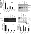
Figure 1
Figure 1
SRPK1, SRSF1 and VEGF expression in cutaneous and uveal melanoma cell lines. (A) SRPK1 mRNA expression was greater in melanoma cell lines compared with primary RPE cells. (B) SRPK1 and SRSF1 protein expression was stronger in metastatic cell lines, A375 and Omm2.5, and this correlated with greater VEGF165 mRNA (C) and panVEGF protein expression (D). VEGFxxxb protein conversely, appeared stronger in primary cell lines Mel270 and 92.1. Experiments were performed in triplicate.
Figure 2
Figure 2
SRPK1 regulates pro-angiogenic VEGF expression in melanoma cells. Serum-starved A375, Omm2.5, Mel270 and 92.1 cells, treated with 5 μM SRPIN340 before IGF-1 administration (100 nM) were stained for (A) SRSF1 (red) or (B) SRPK1 (red) and co-stained for Hoechst (blue). (A) The majority of SRSF1 staining observed was nuclear, but an increase in nuclear intensity following IGF-1 treatment was observed in A375 and Mel270 cells. Pretreatment with SRPIN340 prevented the IGF-1-induced increase in SRSF1 nuclear intensity in Mel270 cells only. #P<0.05. (B) SRPK1 staining was mainly cytoplasmic but nuclear intensity was increased following IGF-1 treatment; SRPIN340 was unable to reverse this response. Scale bar, 10 μm. **P<0.01. (C) Twenty-four hours after inhibitor treatment (1–10 μM) RNA was extracted and cDNA was made for PCR. Treatment with SRPIN340 reduced the expression of VEGF165 relative to GAPDH control in all tested cell lines. Water (W) and sample without reverse transcriptase (–RT) were used as negative controls. (D) Forty-eight hours after treatment, protein was extracted and assayed for total VEGF and VEGFxxxb by ELISA. The expression of pro-angiogenic VEGF (VEGFxxx) was reduced following 10 μM SRPIN340 in A375, Omm2.5 and 92.1 cells (one-way ANOVA, Bonferroni post hoc. *P<0.05). Arrows show non-nuclear-stained cells. NS, not significant. The full colour version of this figure is available at British Journal of Cancer online.
Figure 3
Figure 3
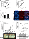
SRPK1 knockdown switches splicing away from pro-angiogenic VEGF. A375 cells were transduced with a lentivirus containing SRPK1 shRNA or scrambled shRNA. (A) Transduction efficiency was determined by GFP expression. (B) Protein was extracted from cells and tested for SRPK1 expression by immunoblotting. There was a significant reduction in SRPK1 expression after lentiviral shRNA SRPK1 transduction relative to tubulin (**P<0.01, one-way ANOVA, Bonferroni post hoc). (C) SRPK1 mRNA expression in A375 melanoma cells was determined by qPCR and expressed relative to internal control (18S ribosomal). SRPK1 was successfully knocked down in SRPK1 shRNA-transduced cells compared with control cells (*P<0.05). (D) Staining for SRSF1 confirmed a knockdown in SRPK1 shRNA cells; cell were imaged and images were processed identically (*P<0.05, Student's t-test). Scale bar, 10 μm. (E) Extracted protein was run on an SDS–PAGE and membranes blotted for SRSF1. SRPK1 knockdown weakly knocks down SRSF1 compared with A375 shRNA scramble relative to α-tubulin expression (17%, n=3). (F) SRSF1 knockdown by siRNA was performed in both control and SRPK1 knockdown cells. SRSF1 siRNA reduced panVEGF without affecting VEGFxxxb expression. (G) RT–PCR with primers spanning VEGF exon 7 and the VEGF 3′-UTR were used to determine expression level. SRPK1 knockdown compared with control cells (scrambled shRNA) showed a significant decrease in VEGF165 relative to GAPDH (P<0.01). (H) Extracted protein was assessed by ELISA. In SRPK1 knockdown cells, pro-angiogenic VEGF was significantly decreased (P<0.05), whereas VEGFxxxb was significantly upregulated (P<0.05).
Figure 4
Figure 4
SRPK1 Knockdown is anti-angiogenic and blocks tumour growth. (A) A375 control shRNA and A375 SRPK1 shRNA cells were seeded and cells were counted every 24 h for 4 days. There was no difference in proliferation rate between the two cell lines as measured by this method. (B) Wells were imaged at time 0 after a 1-mm scratch was performed across the centre of the well, and at 12 and 24 h later. The % coverage of the scratch was calculated. No statistical difference between the coverage of the two cell lines was observed. (C) Cells were then fixed and stained for Ki67, an endogenous proliferation marker. Representative images of staining are shown. Cell counts showed no significant difference in the average number of proliferating cells. Scale bar, 10 μm. (D) Control and SRPK1 KD cells were injected subcutaneously (2 × 10⁶) into the left and right flanks of nude mice, respectively. Tumour growth was measured bi-weekly and tumour excised on day 27. SRPK1 KD significantly reduced tumour growth (P<0.05, *P<0.01, **P<0.001, two-way ANOVA with repeated measures). Images of excised tumours at day 27 are shown below the graph. Scale bar, 1 cm. (E) RNA was extracted from tumour tissue, reverse transcribed and subjected to qPCR using SRPK1 primers and housekeeping gene (18S). SRPK1 levels were expressed as fold increase relative to the smallest tumour and correlated with tumour volume. SRPK1 expression positively correlated with tumour growth, r=0.84 (Pearson's correlation co-efficient). (F) Protein from extracted tissue was assessed for VEGF expression by ELISA. SRPK1 KD tumours expressed reduced panVEGF compared with controls. Although VEGFxxxb expression was unchanged, tubulin confirmed equal loading.
Figure 5
Figure 5
Small molecular weight inhibitors of SRPK1 inhibit melanoma growth. (A) A375 cells were treated with varying doses of SRPIN340 (1–10 μM). Cells were counted every 24 h for 3 days. There was no difference in proliferation rate between the treatments. (B) Wells were imaged at time 0 after a 1-mm scratch was performed across the centre of the well, and at 12 and 24 h later. The % coverage of the scratch was calculated. No statistical difference between the coverage of the cells was observed with treatment. (C) A375 cells were injected subcutaneously (2 × 10⁶) in nude mice. Mice were injected subcutaneously (100 μl) with 2 μg SRPIN340 or 1% DMSO vehicle control daily. Tumour growth was measured bi-weekly and tumour excised on day 27. SRPIN340 significantly reduced tumour growth (P<0.001, one-way ANOVA Bonferroni post hoc). Images of excised tumours at day 35 are shown. Scale bar, 1 cm. (D) Protein from extracted tissue was assessed for VEGF expression. SRPIN340 treatment reduced panVEGF compared with control. (*P<0.05, unpaired t-test) VEGFxxxb expression was detected at very low levels but unchanged following treatment. Probing for actin confirmed equal loading. (E) Tumours were sectioned stained for CD31 (brown) and counterstained with haematoxylin (blue). SRPIN340 significantly reduced MVD as determined by CD31 staining compared with vehicle-treated tumours (P<0.05, Student's unpaired t-test). Scale bar, 50 μm. NS, not significant.

VSports app下载 - References

    1. Amin EM, Oltean S, Hua J, Gammons MV, Hamdollah-Zadeh M, Welsh GI, Cheung MK, Ni L, Kase S, Rennel ES, Symonds KE, Nowak DG, Royer-Pokora B, Saleem MA, Hagiwara M, Schumacher VA, Harper SJ, Hinton DR, Bates DO, Ladomery MR. WT1 mutants reveal SRPK1 to be a downstream angiogenesis target by altering VEGF splicing. Cancer Cell. 2011;20 (6:768–780. - "VSports" PMC - PubMed
    1. Bates DO, Mavrou A, Qiu Y, Carter JG, Hamdollah-Zadeh M, Barratt S, Gammons MV, Millar AB, Salmon AH, Oltean S, Harper SJ. Detection of VEGF-Axxxb isoforms in human tissues. PLoS One. 2013;8 (7:e68399. - PMC - PubMed
    1. Beazley-Long N, Hua J, Jehle T, Hulse RP, Dersch R, Lehrling C, Bevan H, Qiu Y, Lagreze WA, Wynick D, Churchill AJ, Kehoe P, Harper SJ, Bates DO, Donaldson LF. VEGF-A165b is an endogenous neuroprotective splice isoform of vascular endothelial growth factor A in vivo and in vitro. Am J Pathol. 2013;183 (3:918–929. - PMC - PubMed
    1. Bedikian AY, Millward M, Pehamberger H, Conry R, Gore M, Trefzer U, Pavlick AC, DeConti R, Hersh EM, Hersey P, Kirkwood JM, Haluska FG. Bcl-2 antisense (oblimersen sodium) plus dacarbazine in patients with advanced melanoma: the Oblimersen Melanoma Study Group. J Clin Oncol. 2006;24 (29:4738–4745. - PubMed
    1. Boyd SR, Tan D, Bunce C, Gittos A, Neale MH, Hungerford JL, Charnock-Jones S, Cree IA. Vascular endothelial growth factor is elevated in ocular fluids of eyes harbouring uveal melanoma: identification of a potential therapeutic window. Br J Ophthalmol. 2002;86 (4:448–452. - PMC - PubMed

Publication types

MeSH terms