"VSports app下载" Skip to main page content
U.S. flag

An official website of the United States government

Dot gov

The VSports app下载. gov means it’s official. Federal government websites often end in . gov or . mil. Before sharing sensitive information, make sure you’re on a federal government site. .

Https

The site is secure. The https:// ensures that you are connecting to the official website and that any information you provide is encrypted and transmitted securely. V体育官网.

. 2014 Jul;42(13):8343-55.
doi: 10.1093/nar/gku561. Epub 2014 Jul 3.

"V体育安卓版" Long non-coding RNA INXS is a critical mediator of BCL-XS induced apoptosis

Affiliations

Long non-coding RNA INXS is a critical mediator of BCL-XS induced apoptosis (V体育安卓版)

Carlos DeOcesano-Pereira et al. Nucleic Acids Res. 2014 Jul.

Retraction in

Abstract

BCL-X mRNA alternative splicing generates pro-apoptotic BCL-XS or anti-apoptotic BCL-XL gene products and the mechanism that regulates splice shifting is incompletely understood. We identified and characterized a long non-coding RNA (lncRNA) named INXS, transcribed from the opposite genomic strand of BCL-X, that was 5- to 9-fold less abundant in tumor cell lines from kidney, liver, breast and prostate and in kidney tumor tissues compared with non-tumors. INXS is an unspliced 1903 nt-long RNA, is transcribed by RNA polymerase II, 5'-capped, nuclear enriched and binds Sam68 splicing-modulator. Three apoptosis-inducing agents increased INXS lncRNA endogenous expression in the 786-O kidney tumor cell line, increased BCL-XS/BCL-XL mRNA ratio and activated caspases 3, 7 and 9. These effects were abrogated in the presence of INXS knockdown. Similarly, ectopic INXS overexpression caused a shift in splicing toward BCL-XS and activation of caspases, thus leading to apoptosis. BCL-XS protein accumulation was detected upon INXS overexpression VSports手机版. In a mouse xenograft model, intra-tumor injections of an INXS-expressing plasmid caused a marked reduction in tumor weight, and an increase in BCL-XS isoform, as determined in the excised tumors. We revealed an endogenous lncRNA that induces apoptosis, suggesting that INXS is a possible target to be explored in cancer therapies. .


Figures

Figure 1.
Figure 1.
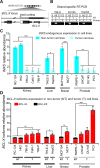
Identification and characterization of INXS as an intronic antisense lncRNA downregulated in tumor cell lines. (A) Structure of the BCL-X gene locus on chromosome 20 with the BLC-X protein-coding mRNA being transcribed from the minus genomic strand and the antisense unspliced INXS lncRNA transcribed from the intronic region on the opposite strand. Gray boxes in INXS indicate transcript portions extended by RACE-PCR and sequencing. Small blue arrows next to INXS indicate PCR primers positions. (B) Antisense transcription [AS] of INXS lncRNA was detected by strand-specific RT-PCR in 786-O, DU145 and HepG2 cell lines. Sense transcription [S] was not detected in this locus region. C1 and C2 are negative controls. (C) INXS expression levels across a panel of tumor (light blue) and non-tumor (dark blue) cell lines from kidney, liver, breast and prostate. Absolute quantification of INXS was obtained for each cell line and the relative abundance is shown with respect to the absolute amount measured in the CaKi-1 cell line (175 molecules per ng of total RNA). (D) BCL-XS (black) and BCL-XL (red) mRNA isoform levels across a panel of tumor (T) and non-tumor (NT) cell lines from kidney, liver, breast and prostate. Expression levels are relative to the expression of the BCL-XS isoform in the CaKi-1 cell line. The data are the mean ± SD of three independent experiments. *(P <0.05), **(P<0.01) and ***(P <0.001).
Figure 2.
Figure 2.
INXS knockdown in the non-tumor kidney cell line RC-124 reduces the BCL-XS/BCL-XL ratio. (A) INXS knockdown in the RC-124 cell line using two distinct INXS-knockdown ASOs (INXS-ASO-1 and -2) reduces by 60% the endogenous levels of INXS compared to a control scrambled oligonucleotide transfection. (B) The levels of BCL-XS (black) and BCL-XL (red) mRNA isoforms in the same cells were measured by RT-qPCR. (C) BCL-XS/BCL-XL mRNA ratio in these cells was significantly reduced from 0.25 to 0.15 upon transfection with INXS-ASO-1 and -2. The data are the mean ± SD of three independent experiments. *(P <0.05), and **(P<0.01).
Figure 3.
Figure 3.
Characterization of INXS biogenesis, stability and cellular localization. (A) RNAPII inhibition with α-amanitin decreases the levels of INXS. Known RNAPII-transcribed (ACTB, MYC) and RNAPIII-transcribed genes (pre-tRNATyr, 7SK) were assayed as controls. (B) The presence of a 5′-end cap modification in INXS was determined by digestion with terminator 5′-phosphate-dependent exonuclease (5′Exo) in combination with tobacco acid pyrophosphatase (TAP), as indicated. TUBA1C tubulin gene was assayed as a control. (C) INXS decay rate in cells treated with actinomycin D, a transcription inhibitor. MYC was measured in parallel as a positive control of the decay assay. (D) Relative distribution of INXS in the nuclear and cytoplasmic fractions (N/C ratio). The nuclear-enriched MALAT1 lincRNA and the 45S rRNA were used as nuclear fraction controls and the 18S rRNA as a cytoplasmic fraction control. As an additional control, western blot of the protein extracts from the fractions was performed, detecting GAPDH only in the cytoplasmic fraction, and histone H3 only in the nuclear fraction. Further controls for cell fractionation are described in the Supplementary Data. *(P <0.05), **(P <0.01) and ***(P <0.001).
Figure 4.
Figure 4.
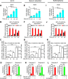
INXS endogenous expression is increased by apoptosis-inducing agents and is associated with increased BCL-XS mRNA and caspases activation. (A, E, I) Induced expression of INXS lncRNA in 786-O kidney tumor cells by exposure (A) to 40 J/m2 UV-C light for 40 s at time zero, (E) to serum reduction for 24 h or (I) to sulforaphane anti-cancer drug (SFN) for up to 24 h. (B, F, J) Levels of BCL-XS (black) and BCL-XL (red) mRNA isoforms in the same cells. (C, G, K) BCL-XS/BCL-XL mRNA ratio in the cells. (D, H, L) Activation of caspase 3 and caspase 9 in 786-O cells at 24 h after the exposure to (D) 40 J/m2 UV-C, (H) 0.5% serum or (L) SFN. (See Supplementary Figure S4 for other caspases.) *(P <0.05), **(P <0.01) and ***(P <0.001).
Figure 5.
Figure 5.
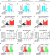
INXS knockdown abrogates the effect of apoptosis-inducing agents. (A, E, I) Abrogated induction of INXS expression in the presence of two distinct INXS-knockdown ASOs (INXS-ASO-1 or -2), with cells exposed (A) to 40 J/m2 UV-C light for 40 s at time zero and incubated for 24 h, (E) to serum reduction (0.5 % serum) for 24 h or (I) to sulforaphane anti-cancer drug (SFN) for 24 h. (B, F, J) The increase in BCL- XS (black) and the decrease in BCL-XL (red) mRNA isoforms induced by the apoptotic agents are abrogated in the presence of two distinct INXS-knockdown ASOs (INXS-ASO-1 or -2). (C, G, K) The increase in BCL-XS/BCL-XL mRNA ratio is significantly abrogated in the INXS-knockdown treated cells. (D, H, L) Abrogated activation of caspase 3 and caspase 9 in the presence of two distinct INXS-knockdown ASOs (INXS-ASO-1 or -2). (See Supplementary Figure S4 for other caspases.) In all panels, the data are the mean ± SD of three independent experiments. *(P <0.05), **(P <0.01) and ***(P <0.001).
Figure 6.
Figure 6.
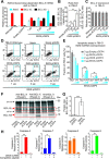
Overexpression of INXS induces BCL-XS mRNA and protein and promotes apoptosis. (A) The levels of BCL-XS (black) and BCL-XL (red) mRNA isoforms were measured by RT-qPCR in 786-O kidney tumor cells 24 h after transient transfection with increasing amounts of pCEP4-INXS plasmid (INXS lncRNA levels = blue bars). All expression levels are shown as relative abundance with respect to the endogenous INXS in wild-type cells. (See Supplementary Figure S6A and F for similar effects on the MCF7 and PC3 cell lines.) (B) The increase in BCL-XS/BCL-XL ratio is dependent on the extent of INXS overexpression. (C) Total BCL-X mRNA does not change upon INXS overexpression. (D) Augmented apoptosis upon transfection of 786-O cells with increasing amounts of pCEP-INXS plasmid, as detected by flow cytometry using double labeling with Annexin V FITC (AV FITC, x-axis) and propidium iodide (PI, y-axis). The percentage of cells that were labeled with AV FITC is shown in the quadrants marked with blue broken lines. (See Supplementary Figure S6D and I for similar effects on the MCF7 and PC3 cell lines.) (E) The results from (D) are shown as the fraction of labeled cells relative to the total. (F) Western blot detects the BCL-XS protein isoform upon INXS overexpression. Antibody anti-BCL-X was used for immunoprecipitation of 786-O cell lysates, and the IP fraction was analyzed by western blot with the same antibody. Three independent replicate transfection experiments are shown. (G) Densitometric intensity ratio between BCL-XS and BCL-XL signals from the data on panel F (the background intensity signal was used as a proxy for BCL-XS in the controls). (H) Caspases 3, 7 and 9 are activated upon INXS overexpression in 786-O cell line, while caspase 8 is not affected. (See Supplementary Figure S9 for detection of active caspase 3 by immunofluorescence microscopy.) In all panels except in D and F, the data are the mean ± SD of three independent experiments. **(P <0.01) and ***(P <0.001).
Figure 7.
Figure 7.
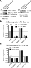
INXS interacts with the Sam68 splicing-modulator complex. (A) Native RIP (RNA-binding protein immunoprecipitation) assay with anti-Sam68 antibody was performed, followed by RT-PCR with primers for the indicated genes. A negative control, from RNA-IP with immunoglobulin G (IgG) was included. For INXS lncRNA, a strand-specific primer was used for RT. For the positive controls (BCL-XL and BCL-XS mRNAs) and the negative control (BCL-2 mRNA), oligo-dT primer was used for RT. The protein fraction from the native RIP assay with anti-Sam68 antibody was analyzed by western blot, which was developed with the same antibody. A negative control sample, from RIP with IgG, was included. (B) INXS overexpression was performed in the 786-O kidney tumor cell line with 3 μg of INXS-expressing plasmid for 24 h and the alternative splicing isoforms of two Sam68 target mRNAs that have been identified in the literature, namely, CCDN1-v1 and SRSF1-v1 and -v2, were measured by RT-qPCR. (C) INXS knockdown was performed in the RC-124 kidney non-tumor cell line as in Figure 2, and the levels of CCDN1-v1 and SRSF1-v1 and -v2 were measured by RT-qPCR. The data are the mean ± SD of three independent experiments.
Figure 8.
Figure 8.
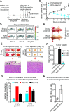
Overexpression of INXS induces tumor regression in vivo. (A) Schematic representation showing the subcutaneous inoculation of mice with 786-O human kidney tumor cells (open arrow, on day –63), followed by a waiting period until all the tumors had implanted and grown to reach the approximate same volume of 250 mm3 (on day zero), when the injection of animals began (vertical arrows). Intra-tumor injections of INXS-plasmid or empty-plasmid were performed every third day over a period of 15 days. (B) Tumor volume was monitored by caliper on the days of injection in six different animals from each of two groups, which were injected either with INXS-plasmid (blue) or control empty-plasmid (black). The data are the mean ± SD of measurements from the six animals of each injection group. (C) An EGF-labeled dye marker was detected in the tumor by in vivo scanning with a near-infrared optical imaging system. Only one representative mouse is shown for each group, namely, INXS-plasmid or control empty-plasmid. (D) Pictures of the scanned animals, taken on day 15; note the difference in size between the tumors of the two animals, as indicated by arrows. (E) On day 15, all six animals from each of the two groups were euthanized, and their tumors were excised and photographed. Subsequently, tumors were formalin-fixed, paraffin-embedded, stained and observed by light microscopy. Arrows point to tumor peripheral vascularization. (F) Average weights of the excised tumors for each of the two injection groups, INXS-plasmid (blue) or control empty-plasmid (black). The data are the mean ± SD of measurements from the six animals of each injection group. An additional animal whose tumor was injected with only transfection solution is shown (vehicle, white bar). (G) INXS lncRNA (blue), BCL-XL mRNA (red) and BCL-XS mRNA (black) expression levels measured by RT-qPCR in the excised xenograft tumors. (H) BCL-X mRNA isoforms ratio in the excised xenograft tumors. *(P <0.05), **(P <0.01) and ***(P <0.001).
Figure 9.
Figure 9.
Proposed model of action of INXS lncRNA. BCL-X pre-mRNA undergoes either a BCL-XL anti-apoptotic or BCL-XS pro-apoptotic alternative processing (4). We propose that the control of BCL-X pre-mRNA alternative splicing, between pathway [1] toward BCL-XL anti-apoptotic isoform and pathway [2] toward BCL-XS pro-apoptotic isoform, depends on INXS endogenous lncRNA. Apoptosis inducing agents, such as UV-C light exposure, serum starvation or anti-cancer drugs, lead to an increased expression of endogenous INXS. Sam68 increases the level of the pro-apoptotic BCL-XS isoform, while its absence leads to the accumulation of BCL-XL (12). We propose that the augmented levels of INXS would favor the positioning of Sam68 splicing-modulator and of possible additional splice factors (SF) of the splicing machinery near the distal donor 5′ splice site on the pre-mRNA (5′ss, dotted blue line) and favor the splicing predominantly through pathway 2, thus leading to BCL-XS protein synthesis and to apoptosis.

References

    1. Cory S., Adams J.M. The Bcl2 family: regulators of the cellular life-or-death switch. Nat. Rev. Cancer. 2002;2:647–656. - PubMed
    1. Kelly P.N., Strasser A. The role of Bcl-2 and its pro-survival relatives in tumourigenesis and cancer therapy. Cell Death Diff. 2011;18:1414–1424. - PMC - PubMed
    1. Tait S.W., Green D.R. Mitochondria and cell death: outer membrane permeabilization and beyond. Nat. Rev. Mol. Cell Biol. 2010;11:621–632. - PubMed (V体育官网入口)
    1. Boise L.H., Gonzalez-Garcia M., Postema C.E., Ding L., Lindsten T., Turka L.A., Mao X., Nunez G., Thompson C.B. bcl-x, a bcl-2-related gene that functions as a dominant regulator of apoptotic cell death. Cell. 1993;74:597–608. - PubMed
    1. Plati J., Bucur O., Khosravi-Far R. Apoptotic cell signaling in cancer progression and therapy. Integrative Biol. 2011;3:279–296. - PMC - PubMed

Publication types

"V体育2025版" MeSH terms

Associated data