Skip to main page content
U.S. flag

An official website of the United States government

Dot gov

The . gov means it’s official. Federal government websites often end in VSports app下载. gov or . mil. Before sharing sensitive information, make sure you’re on a federal government site. .

Https

The site is secure V体育官网. The https:// ensures that you are connecting to the official website and that any information you provide is encrypted and transmitted securely. .

Review
. 2014 May 6;9 Suppl 1(Suppl 1):51-63.
doi: 10.2147/IJN.S26592. eCollection 2014.

Endocytosis and exocytosis of nanoparticles in mammalian cells

Affiliations
Review

Endocytosis and exocytosis of nanoparticles in mammalian cells

"V体育平台登录" Nuri Oh et al. Int J Nanomedicine. .

"V体育2025版" Abstract

Engineered nanoparticles that can be injected into the human body hold tremendous potential to detect and treat complex diseases. Understanding of the endocytosis and exocytosis mechanisms of nanoparticles is essential for safe and efficient therapeutic application. In particular, exocytosis is of significance in the removal of nanoparticles with drugs and contrast agents from the body, while endocytosis is of great importance for the targeting of nanoparticles in disease sites VSports手机版. Here, we review the recent research on the endocytosis and exocytosis of functionalized nanoparticles based on various sizes, shapes, and surface chemistries. We believe that this review contributes to the design of safe nanoparticles that can efficiently enter and leave human cells and tissues. .

Keywords: cancer cell; drug delivery; endocytosis; exocytosis; macrophage; nanoparticle; toxicity V体育安卓版. .


"V体育安卓版" Figures

Figure 1
Figure 1
Schematic of endocytosis and exocytosis patterns of nanoparticles. Nanoparticles enter the cell via four types of pathway: clathrin/caveolar-mediated endocytosis, phagocytosis, macropinocytosis, and pinocytosis. Nanoparticles exit the cell via three types of pathway: lysosome secretion, vesicle-related secretion, and non-vesicle-related secretion. Abbreviation: MVBs, multivesicular bodies.
Figure 2
Figure 2
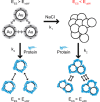
Scheme of aggregation or agglomeration mechanism. The stabilizing electrostatic forces (EES) on the surface of bare nanoparticles are neutralized by NaCl ions in the biological solution, causing the van der Waals forces (EvdW) to drive formation of aggregation or agglomeration. The protein coating of nanoparticles can reduce the aggregation or agglomeration. Note: Reproduced with permission from Albanese A, Chan WC. Effect of gold nanoparticle aggregation on cell uptake and toxicity. ACS Nano. 2011;5:5478–5489. Copyright © 2011 American Chemical Society.
Figure 3
Figure 3
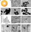
(A) Schematic representation of gold nanoparticles. (B) Transmission electron microscope images of citrate-coated gold nanoparticles with various sizes. (C) Transmission electron microscope images of the gold nanoparticles entrapped in cellular vesicles. Graph showing the number of the gold nanoparticles per vesicle diameter. Note: Reproduced with permission from Chithrani BD, Ghazani AA, Chan WC. Determining the size and shape dependence of gold nanoparticle uptake into mammalian cells. Nano Lett. 2006;6:662–668. Copyright © 2007 American Chemical Society.
Figure 4
Figure 4
Kinetics of cellular uptake of negatively (–COOH) (A) and positively (–NH2) (B) charged polymeric nanoparticles in macrophages and monocytes (THP-1). Confocal fluorescence images (right) were taken after 2 hours’ incubation with the indicated nanoparticles. Cell membrane was stained with red dyes and the nanoparticles were tagged with green dyes. Note: Reproduced with permission from Lunov O, Syrovets T, Loos C, et al. Differential uptake of functionalized polystyrene nanoparticles by human macrophages and a monocytic cell line. ACS Nano. 2011;5:1657–1669. Copyright © 2011 American Chemical Society. Abbreviation: MFI, mean fluorescence index.
Figure 5
Figure 5
(A) Schematic depicting of transferrin-coated gold nanoparticles. (B) Kinetics of exocytosis patterns of the nanoparticles with different sizes. (C) Different stages of exocytosis patterns: (a) Movement of the vesicles containing nanoparticles toward the cell membrane; (b) Docking of one of the vesicles at the cell membrane; (c) Excretion of nanoparticles; (d) Cluster of nanoparticles after exocytosis. Note: Reproduced with permission from Chithrani BD, Chan WC. Elucidating the mechanism of cellular uptake and removal of protein-coated gold nanoparticles of different sizes and shapes. Nano Lett. 2007;7:1542–1550. Copyright © 2007 American Chemical Society.
Figure 6
Figure 6
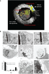
(A) A scanning electron microscopy image of a macrophage showing endocytosis of porous silicon particles incorporated with iron oxide nanoparticles, intracellular partitioning of the particles, endosomal escape of the particles, and exocytosis of the incorporated iron oxide nanoparticles. (B) Exocytosis of iron oxide nanoparticles. Upper column shows transmission electron microscopy images of iron oxide nanoparticles released from the porous silicon carrier in a macrophage. Middle column shows transmission electron microscopy images of the released iron oxide nanoparticles located in the intracellular region of the macrophage 6 days after uptake of the porous silicon particles. Bottom column shows that graph displayed time-dependent iron content in the supernatant and transmission electron microscopy images showed the internalized iron oxide nanoparticles were exocytosed by membrane vesicles. Note: Reproduced with permission from Serda RE, Mack A, van de Ven AL, et al. Logic-embedded vectors for intracellular partitioning, endosomal escape, and exocytosis of nanoparticles. Small. 2010;6:2691–2700. Copyright © 2010 John Wiley & Sons, Inc.
Figure 7
Figure 7
(A) Schematic diagram showing the endocytosis and exocytosis processes of D-penicillamine-coated quantum dots; a: Clathrin-mediated endocytosis; b: Macropinocytosis. (B) Interaction of D-penicillamine-coated quantum dots (green) with plasma membrane of a HeLa cell before internalization. The plasma membrane was stained with the red membrane dye. Scale bar: 10 μm. (C) Kinetics of exocytosis of D-penicillamine-coated quantum dots after removing the nanoparticles in the media. Note: Reproduced with permission from Jiang X, Röcker C, Hafner M, Brandholt S, Dörlich RM, Nienhaus GU. Endo- and exocytosis of zwitterionic quantum dot nanoparticles by live HeLa cells. ACS Nano. 2010;4:6787–6797. Copyright © 2010 American Chemical Society. Abbreviations: QDs, quantum dots; min, minutes.

References

    1. Ferrari M. Cancer nanotechnology: opportunities and challenges. Nat Rev Cancer. 2005;5(3):161–171. - PubMed
    1. Ruoslahti E, Bhatia SN, Sailor MJ. Targeting of drugs and nanoparticles to tumors. J Cell Biol. 2010;188(6):759–768. - PMC - PubMed
    1. Jain RK, Stylianopoulos T. Delivering nanomedicine to solid tumors. Nat Rev Clin Oncol. 2010;7(11):653–664. - PMC - PubMed
    1. Choi HS, Liu W, Misra P, et al. Renal clearance of quantum dots. Nat Biotechnol. 2007;25(10):1165–1170. - PMC - PubMed
    1. Park JH, Gu L, von Maltzahn G, Ruoslahti E, Bhatia SN, Sailor MJ. Biodegradable luminescent porous silicon nanoparticles for in vivo applications. Nat Mat. 2009;8(4):331–336. - "V体育2025版" PMC - PubMed

Publication types (V体育ios版)