Skip to main page content
U.S. flag

An official website of the United States government

Dot gov

The . gov means it’s official. Federal government websites often end in . gov or . mil. Before sharing sensitive information, make sure you’re on a federal government site. VSports app下载.

Https

The site is secure V体育官网. The https:// ensures that you are connecting to the official website and that any information you provide is encrypted and transmitted securely. .

Clinical Trial
. 2014 Jul;2(7):632-42.
doi: 10.1158/2326-6066.CIR-14-0053. Epub 2014 Apr 21.

Bevacizumab plus ipilimumab in patients with metastatic melanoma

Affiliations
Clinical Trial

Bevacizumab plus ipilimumab in patients with metastatic melanoma

F Stephen Hodi et al. Cancer Immunol Res. 2014 Jul.

Erratum in

  • Cancer Immunol Res. 2014 Sep;2(9):923

"V体育ios版" Abstract

Ipilimumab improves survival in advanced melanoma and can induce immune-mediated tumor vasculopathy. Besides promoting angiogenesis, vascular endothelial growth factor (VEGF) suppresses dendritic cell maturation and modulates lymphocyte endothelial trafficking. This study investigated the combination of CTLA4 blockade with ipilimumab and VEGF inhibition with bevacizumab. Patients with metastatic melanoma were treated in four dosing cohorts of ipilimumab (3 or 10 mg/kg) with four doses at 3-week intervals and then every 12 weeks, and bevacizumab (7. 5 or 15 mg/kg) every 3 weeks. Forty-six patients were treated. Inflammatory events included giant cell arteritis (n = 1), hepatitis (n = 2), and uveitis (n = 2) VSports手机版. On-treatment tumor biopsies revealed activated vessel endothelium with extensive CD8(+) and macrophage cell infiltration. Peripheral blood analyses demonstrated increases in CCR7(+/-)/CD45RO(+) cells and anti-galectin antibodies. Best overall response included 8 partial responses, 22 instances of stable disease, and a disease-control rate of 67. 4%. Median survival was 25. 1 months. Bevacizumab influences changes in tumor vasculature and immune responses with ipilimumab administration. The combination of bevacizumab and ipilimumab can be safely administered and reveals VEGF-A blockade influences on inflammation, lymphocyte trafficking, and immune regulation. These findings provide a basis for further investigating the dual roles of angiogenic factors in blood vessel formation and immune regulation, as well as future combinations of antiangiogenesis agents and immune checkpoint blockade. .

PubMed Disclaimer (V体育官网入口)

Figures

Figure 1
Figure 1
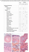
High-grade treatment-related adverse events. A. Grade 4 events. B. Grade 3 Events. Eleven study patients had treatment-related, grade 3 events [23.9%, (95% exact CI: 13% to 39%)]. Hematoxylin and Eosin stained, formalin-fixed, paraffin embedded tissue sections showing: (C-D) Temporal artery, cut in cross section, with transmural acute and chronic inflammation at 200x (C) and 400x (D) final magnification. (E-F) Skin with chronic inflammation intermixed with eosinophils within the mid-dermis at 400x (E) and 1000x (F) final magnification. (G-H) Core of liver with acute and chronic inflammation including prominent eosinophils, at 400x (G) and 1000x (H) final magnification.
Figure 2
Figure 2
Intratumoral blood vessel changes from treatment with bevacizumab plus ipilimumab in subcutaneous melanoma deposits. A. Characterization of tumor-associated vasculature before and while on therapy. Endothelial cells lining small vessels within melanomas of ipilimumab plus bevacizumab-treated patients were rounded and columnar as assessed by H&E and CD31 staining, in contrast to pre-treatment samples and on-treatment samples from patients receiving ipilimumab alone (column to extreme right; v = vessel). Scale bar = 50 microns. Endothelial cells in tumor deposits of patients receiving ipilimumab plus bevacizumab were also associated with increased expression of E-selectin, and adhesion and diapedesis of CD8+ T cells. Enlarged central panels highlight the focally occlusive appearance of this endothelial activation (top H&E, bottom CD31, [inset, E-selectin]). Base membrane of vessels approximated by dotted lines.
Figure 3
Figure 3
Histologic changes in tumor deposits resulting from treatment with bevacizumab plus ipilimumab. A. Phenotypic characterization of immune-cell infiltrates in biopsies from responders before and after initiation of therapy. Tumors after initiation (ON) of ipilimumab-bevacizumab therapy were characterized as compared to pre-treatment samples (PRE). Significant infiltration by CD3+CD8+ T cells and CD163+ macrophages with minimal change in Foxp3+ component were observed. The enlarged panels (center) emphasize the tumor-infiltrating architecture of the immune response (skeletal muscle is seen in the upper left corner). In contrast, patients treated only with ipilimumab demonstrated a lesser degree of immune-cell infiltration while on therapy. The two ipilimumab-bevacizumab specimens are subcutaneous tissues, and the ipilimumab alone specimen was from the oropharyngeal submucosa.
Figure 4
Figure 4
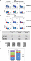
Cellular and humoral immune responses in the peripheral blood are altered by the addition of bevacizumab to ipilimumab. All samples were obtained pre-treatment and at week 12 at the completion of ipilimumab or ipilimumab-bevacizumab induction. A. Example of changes as a function of treatment in CD4+CCR7+CD45RO+ and CD4+CCR7CD45RO+ T-cell populations to ipilimumab plus bevacizumab treatment, compared to changes with ipilimumab treatment alone. B. Example of changes as a function of treatment in CD8+CCR7+CD45RO+ and CD8+CCR7CD45RO+ T-cell populations to ipilimumab plus bevacizumab treatment, compared to the responses to ipilimumab treatment alone. C. Numbers of melanoma patients that have at least 50% enhancement in CD4+/CD8+ CCR7+CD45RO+ and CD4+/CD8+ CCR7CD45RO+ T-cell populations following treatment with ipilimumab (3mg/kg), or ipilimumab (3mg/kg) plus bevacizumab, or ipilimumab (10mg/kg) plus bevacizumab. * indicates P<0.05 between ipilimumab and ipilimumab plus bevacizumab. ** indicates P<0.01. D. Representative immunoblots of antibody responses in four bevacizumab plus ipilimumab-treated patients to a total of zero, one, two, or three galectins. Arrows indicate increased antibody levels in post-treatment sera samples. Galectin-1, -3 and -9 proteins were mixed together and equally loaded and separated by gel electrophoresis. Following transfer onto a membrane, strips were incubated with equally diluted pre-treatment and post-treatment sera. Density analysis using the NIH ImageJ software confirmed the increases in densities of the indicated bands after subtracting background taken from nearby areas of each band. As in most of the cases, density change was seen in only one or two of the 3 galectins, protein(s) without density change serving as loading controls. E. Antibody responses to galectins in patients treated with bevacizumab plus ipilimumab and ipilimumab alone. Anti-galectin-1, -galectin-3, and -galectin-9 antibodies were detected in pre-treatment and post-treatment patient sera by immunoblot analyses. Percentages of patients with increased levels of antibodies to a total of zero, one, two, or three galectins in bevacizumab plus ipilimumab-treated patients (n=45) and ipilimumab alone patients (n=18). Density of each band was measured using the NIH ImmageJ software and the antibody fold-change was calculated using the formula: Fold-change = (DensityPost – DensityPre)/DensityPre. Antibody levels were considered as increased when the fold-change ≥ 0.5.
Figure 5
Figure 5
Activity in treated patients by cohort according to RECIST criteria. Arrows indicate alive at the time of analysis. Crosses indicate death. Black bars indicate discontinuation of treatment other than due to progressive disease. Five patients came off trial due to toxicity requiring systemic steroids. One patient withdrew consent after week 12 without dose-limiting toxicity. PD= progressive disease. SD = stable disease. PR = partial response. CR = complete response.
Figure 6
Figure 6
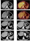
Example of pre-treatment (A and B) and 8-week post-treatment (C and D) PET CT images. An early metabolic response is noted in a dominant left hepatic metastasis. E. Axial contrast-enhanced CT image obtained at baseline demonstrates a solitary heterogeneous hypodense lesion in segment II of the liver consistent with a metastatic deposit. F. Axial contrast-enhanced CT obtained 4 months after treatment demonstrates slight increase in the lesions with interval decrease in density (from 40 HU to 19 HU, approximately 50%) consistent with treatment effect. The increase in size represents pseudo-progression. G and H. Follow-up CT scans obtained 6 and 36 months after the start of treatment demonstrate gradual decrease in size of the metastatic deposit.

References

    1. Hodi FS, O'Day SJ, McDermott DF, Weber RW, Sosman JA, Haanen JB, et al. Improved Survival with Ipilimumab in Patients with Metastatic Melanoma. N Engl J Med. 2010;363:711–23. - PMC - PubMed
    1. Robert C, Thomas L, Bondarenko I, O'Day S, Weber JW, Garbe C, et al. Ipilimumab plus dacarbazine for previously untreated metastatic melanoma. N Engl J Med. 2011;364:2517–26. - PubMed
    1. Ohm JE, Carbone DP. VEGF as a mediator of tumor-associated immunodeficiency. Immunol Res. 2001;23:263–72. - PubMed
    1. Oyama T, Ran S, Ishida T, Nadaf S, Kerr L, Carbone DP, Gabrilovich DI. Vascular endothelial growth factor affects dendritic cell maturation through the inhibition of nuclear factor-kappa B activation in hemopoietic progenitor cells. J Immunol. 1998;160:1224–32. - PubMed
    1. Kandalaft LE, Motz GT, Busch J, Coukos G. Angiogenesis and the tumor vasculature as antitumor immune modulators: the role of vascular endothelial growth factor and endothelin. Curr Top Microbiol Immunol. 2011;344:129–48. - PubMed

V体育安卓版 - Publication types

MeSH terms