Skip to main page content
U.S. flag

An official website of the United States government

Dot gov

The . gov means it’s official. Federal government websites often end in . gov or . mil VSports app下载. Before sharing sensitive information, make sure you’re on a federal government site. .

Https

The site is secure V体育官网. The https:// ensures that you are connecting to the official website and that any information you provide is encrypted and transmitted securely. .

. 2014 Aug;66(8):2222-33.
doi: 10.1002/art.38639.

Fibrin accumulation secondary to loss of plasmin-mediated fibrinolysis drives inflammatory osteoporosis in mice

Affiliations

Fibrin accumulation secondary to loss of plasmin-mediated fibrinolysis drives inflammatory osteoporosis in mice

Heather A Cole et al. Arthritis Rheumatol. 2014 Aug.

Abstract

Objective: Osteoporosis is a skeletal disorder characterized by low bone mass and increased bone fragility associated with aging, menopause, smoking, obesity, or diabetes. Persistent inflammation has been identified as an instigating factor in progressive bone loss. In addition to the role of fibrin in coagulation, inordinate fibrin deposition within a tissue matrix results in increased local inflammation. Given that fibrin accumulation is a hallmark of osteoporosis-related comorbidities, we undertook this study to test the hypothesis that persistent fibrin deposition causes inflammatory osteoporosis VSports手机版. .

Methods: Multiple imaging modalities, bone integrity metrics, and histologic analyses were employed to evaluate skeletal derangements in relation to fibrin deposition, circulating fibrinogen levels, and systemic markers of inflammation in mice that were plasminogen deficient and in plasminogen-deficient mice that were concomitantly either fibrinogen deficient or carrying a mutant form of fibrinogen lacking the αM β2 binding motif V体育安卓版. .

Results: Mice generated with a genetic deficit in the key fibrinolytic protease, plasmin, uniformly developed severe osteoporosis. Furthermore, the development of osteoporosis was fibrin(ogen) dependent, and the derangements in the bone remodeling unit were mechanistically tied to fibrin(ogen)-mediated activation of osteoclasts via activation of the leukocyte integrin receptor αM β2 on monocytes and secondary stimulation of osteoblasts by RANKL. Notably, the genetic elimination of fibrin(ogen) or the expression of a mutant form of fibrinogen retaining clotting function but lacking the αM β2 binding motif prevented the degenerative skeletal phenotypes, resulting in normal local and systemic cytokine levels. V体育ios版.

Conclusion: Taken together, these data reveal for the first time that fibrin promotes inflammation-driven systemic osteoporosis, which suggests a novel association between hemostasis, inflammation, and bone biology VSports最新版本. .


Figures (VSports注册入口)

Figure 1
Figure 1
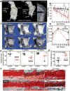
Plasminogen deficiency imposes early degeneration and biomechanical incompetency of the axial skeleton. A, Lateral radiographs of 20-week-old control and plasminogen-deficient (PLG−/−) mice show substantial kyphosis of the axial spine. Bars = 5 mm. B, Serial measurements of kyphotic indices indicate that following proper axial development through 5 weeks, PLG−/− mice (triangles) show progressive degeneration compared to control mice (squares) (P=0.0003 by two-way repeated-measures analysis of variance [ANOVA]). C, Three-dimensional renderings of micro–computed tomography (micro-CT) data show evidence of rapid bone loss in the L5 vertebrae (asterisk) of PLG−/− mice. Bars =1 mm. D, Shown are micro-CT–derived bone fraction volumes corresponding to images in C. Triangles represent PLG−/− mice; squares represent control mice. ** = P<0.01 versus control mice, by Student’s t-test. E, Vertebral body compression ratios of T11, T12, and L1 determined from lateral radiographs demonstrate biomechanical incompetence of the anterior cortex of the vertebral bodies at age 20 weeks in PLG−/− mice. *= P<0.05; ***= P<0.001 versus control mice, by two-way ANOVA with Bonferroni adjustment for multiple comparisons. F, Shown is premature ossification (asterisks) of the articular end plate of L5/L6 observed in PLG−/− mice at age 10 weeks and in both control and PLG−/− mice at age 20 weeks (Safranin O staining). Arrow indicates cartilage. Bars =100 μm. G, Shown is the percentage area of end plate ossification of L5/L6 determined by histologic examination at age 20 weeks (n = 8 wild-type mice; n = 11 PLG−/− mice). P<0.001 versus control mice, by Student’s t-test. Bars in B, D, E, and G show the mean ±SD. Symbols in E and G represent individual mice. Color figure can be viewed in the online issue, which is available at http://onlinelibrary.wiley.com/doi/10.1002/art.38639/abstract.
Figure 2
Figure 2
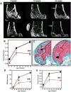
Plasminogen-deficient (PLG−/−) mice develop early degeneration and reduced fracture resistance of the appendicular skeleton. Triangles represent PLG−/− mice; squares represent control mice. A, Two-dimensional images rendered from micro–computed tomography (micro-CT) data reveal cortical thinning in PLG−/− mice compared to control mice (arrows) occurring at age 20 weeks despite relatively normal bone architecture in younger mice. Bars = 1 mm. B, Shown are tabulated cortical measurements (Student’s t-test was performed for each time point using data collected from separate mice at the time that they were killed). C, Safranin O staining of the distal femur at age 20 weeks demonstrates loss of trabecular bone in the distal femoral epiphysis in PLG−/− mice compared to control mice (arrowheads) (see Supplementary Table 1, available on the Arthritis & Rheumatology web site at http://onlinelibrary.wiley.com/doi/10.1002/art.38639/abstract). Bars = 200 μm. D and E, Biomechanical analyses of stiffness (D) and fracture resistance by peak force (E) indicate that observed progressive loss of bone results in significantly reduced biomechanical strength (means, standard deviations, and sample sizes, along with additional biomechanical and micro-CT data, are presented in Supplementary Tables 1 and 2, available on the Arthritis & Rheumatology web site at http://onlinelibrary.wiley.com/doi/10.1002/art.38639/abstract). Bars show the mean ±SD (n = 8 wild-type mice; n=11 PLG−/− mice). *=P<0.05; **=P<0.01 versus control mice, by Student’s t-test. Color figure can be viewed in the online issue, which is available at http://onlinelibrary.wiley.com/doi/10.1002/art.38639/abstract.
Figure 3
Figure 3
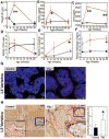
Exuberant osteoclast (OC) activity is associated with increased circulating levels of osteoprotegerin (OPG), interleukin-6 (IL-6), and fibrinogen and with accumulation of fibrin(ogen) in bone. Triangles represent plasminogen-deficient (PLG−/−) mice; squares represent control mice. A–D, Plasma levels of tartrate-resistant acid phosphatase 5b (TRAP-5b) (A), soluble RANKL (sRANKL) (B), and OPG (C) and the resulting sRANKL:OPG ratios (D) were determined by enzyme-linked immunosorbent assay (ELISA). Increased osteoclast activity was observed without a change in RANKL:OPG ratio. E and F, Plasma IL-6 levels determined by flow cytometry (E) and fibrinogen levels determined by ELISA (F) indicate a progressive increase in inflammation and acute-phase reactants at ages 5, 10, and 20 weeks (measurements for all data were collected from separate mice at the time that they were killed; Student’s t-test was performed at each time point). Bars show the mean ±SD (n=4 at each time point). *=P<0.05; ** = P<0.01 versus control mice. G, Immunolocalization of fibrinogen demonstrates abundant fibrin within the L5 vertebrae of 20-week-old PLG−/− mice (red signal), but not in control mice. Bars =100 μm. H, Left, The number of osteoclasts per unit bone surface in L5 vertebrae at age 8 weeks (time point of maximal TRAP-5b concentration) was determined by TRAP staining. Representative images from each group are shown. Insets are higher-magnification views of boxed areas. Original magnification ×200; ×400 in insets. Bars =500 μm. Right, Quantification of osteoclasts/bone surface area illustrates increased numbers of osteoclasts in PLG−/− mice (n=4) compared to control mice (n = 4) at age 8 weeks. Bars show the mean ±SD. *=P<0.01 versus control mice.
Figure 4
Figure 4
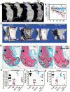
Targeting fibrin prevents bone loss in plasminogen-deficient (PLG−/−) mice. A, Lateral radiographs at age 20 weeks demonstrate that the degree of kyphosis seen in PLG−/− mice is reduced in mice deficient in both plasminogen and fibrinogen (PLG−/−FBG−/− mice [or Plg−/−Fib−/− mice]) as well as in plasminogen-deficient mice expressing a mutant fibrinogen unable to bind αMβ2 (PLG−/−FBGg390–396A mice [or Plg−/−Fib7390–396A mice]). Bars = 5 mm. B, By age 20 weeks there is complete prevention of kyphosis in PLG−/−FBG−/− mice (green symbols) and partial prevention in PLG−/−FBGg390–396A mice (blue symbols). Wild-type (WT) mice are represented by black symbols; PLG−/− mice are represented by red symbols. Bars show the mean ± SD. C, Three-dimensional reconstruction of micro-computed tomography data from L5 vertebrae in 20-week-old mice shows decreased bone loss in PLG−/−FBG−/− and PLG−/−FBGg390–396A mice compared to PLG−/− mice. Bars = 1 mm. D, Similar reductions in cortical bone loss (black arrows) and trabecular bone loss (yellow arrows) are observed in Safranin O-stained sections of distal femurs from 20-week-old PLG−/−FBG−/− and PLG−/−FBGg390–396A mice. Bars = 200 /μm. E-G, These changes in bone architecture are reflected by enhanced bone fraction volume (E) and biomechanical properties contributing to fracture resistance (peak force [F] and stiffness [G]). Symbols represent individual mice (n = 10 WT mice; n = 8 PLG−/− mice; n = 5 PLG−/−FBG−/− mice; n = 5 PLG−/−FBGg390–396A mice). Bars show the mean ± SD. ** = P < 0.01 versus control, by one-way analysis of variance. Color figure can be viewed in the online issue, which is available at http://onlinelibrary.wiley.com/doi/10.1002/art.38639/abstract.
Figure 5
Figure 5
Fibrin(ogen) (Fib) stimulates osteoclastogenesis by engaging αMβ2 and promoting osteoblast RANKL expression through an alternative mechanism. A, Isolated splenic monocytes incubated with 0.5 μM fibrinogen show increased osteoclastogenesis as indicated by tartrate-resistant acid phosphatase (TRAP) staining of multinucleated cells. Bars =1 mm. B, Shown is the percent of TRAP-stained area. Symbols represent individual mice. *= P<0.01 versus control, by one-way analysis of variance (ANOVA) followed by Dunn’s multiple comparison test. C, The observed stimulatory behavior of fibrinogen is inhibited in a dose-dependent manner by addition of a γ390–396A inhibitor. D, Fibrin(ogen) stimulates a 2-fold increase in cell surface expression of RANKL by MC3T3 cells (an osteoblast precursor cell line). Blue line indicates normal RANKL production; green line indicates RANKL production in the presence of 1 μM fibrinogen for 18 hours; red line indicates RANKL production in the presence of 1 uM fibrinogen for 36 hours. E, Shown is the fold increase in cell surface expression of RANKL. *= P<0.01 versus 0 hours, by one-way ANOVA followed by Dunn’s multiple comparison test. Bars show the mean ±SD. Color figure can be viewed in the online issue, which is available at http://onlinelibrary.wiley.com/doi/10.1002/art.38639/abstract.

References

    1. Raisz LG. Pathogenesis of osteoporosis: concepts, conflicts, and prospects. J Clin Invest. 2005;115:3318–25. - PMC - PubMed
    1. Canalis E, Giustina A, Bilezikian JP. Mechanisms of anabolic therapies for osteoporosis. N Engl J Med. 2007;357:905–16. - PubMed
    1. Mundy GR. Osteoporosis and inflammation. Nutr Rev. 2007;65:S147–51. - PubMed
    1. Daniell HW. Osteoporosis and smoking. JAMA. 1972;221:509. - PubMed
    1. Hofbauer LC, Brueck CC, Singh SK, Dobnig H. Osteoporosis in patients with diabetes mellitus. J Bone Miner Res. 2007;22:1317–28. - PubMed

Publication types