Skip to main page content
U.S. flag

An official website of the United States government

Dot gov

The . gov means it’s official. Federal government websites often end in VSports app下载. gov or . mil. Before sharing sensitive information, make sure you’re on a federal government site. .

Https

The site is secure V体育官网. The https:// ensures that you are connecting to the official website and that any information you provide is encrypted and transmitted securely. .

. 2014 Apr 15;192(8):3837-46.
doi: 10.4049/jimmunol.1103516. Epub 2014 Mar 12.

TNFR1/phox interaction and TNFR1 mitochondrial translocation Thwart silica-induced pulmonary fibrosis

Affiliations

TNFR1/phox interaction and TNFR1 mitochondrial translocation Thwart silica-induced pulmonary fibrosis

Fabrizio Fazzi et al. J Immunol. .

V体育ios版 - Abstract

Macrophages play a fundamental role in innate immunity and the pathogenesis of silicosis. Phagocytosis of silica particles is associated with the generation of reactive oxygen species (ROS), secretion of cytokines, such as TNF, and cell death that contribute to silica-induced lung disease VSports手机版. In macrophages, ROS production is executed primarily by activation of the NADPH oxidase (Phox) and by generation of mitochondrial ROS (mtROS); however, the relative contribution is unclear, and the effects on macrophage function and fate are unknown. In this study, we used primary human and mouse macrophages (C57BL/6, BALB/c, and p47(phox-/-)) and macrophage cell lines (RAW 264. 7 and IC21) to investigate the contribution of Phox and mtROS to silica-induced lung injury. We demonstrate that reduced p47(phox) expression in IC21 macrophages is linked to enhanced mtROS generation, cardiolipin oxidation, and accumulation of cardiolipin hydrolysis products, culminating in cell death. mtROS production is also observed in p47(phox-/-) macrophages, and p47(phox-/-) mice exhibit increased inflammation and fibrosis in the lung following silica exposure. Silica induces interaction between TNFR1 and Phox in RAW 264. 7 macrophages. Moreover, TNFR1 expression in mitochondria decreased mtROS production and increased RAW 264. 7 macrophage survival to silica. These results identify TNFR1/Phox interaction as a key event in the pathogenesis of silicosis that prevents mtROS formation and reduces macrophage apoptosis. .


Figures
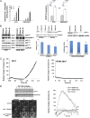
FIGURE 1.
FIGURE 1.
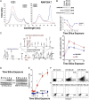
Mitochondria are a significant source of ROS in silica-exposed IC21 macrophages. (A) Following exposure to silica (left panel) or PMA (right panel), mitochondrial superoxide anion production increased in IC21 and RAW 264.7 macrophages, but the production was significantly greater in IC21 macrophages. Superoxide anion production was measured by cytochrome c (nmol/μg protein) reduction in response to silica (20 μg/cm2) (left panel) or as changes in dihydroethidium fluorescence in response to PMA exposure (right panel). Basal level of cytochrome c reduction in untreated cells (5.3 nmol/μg protein in IC21 macrophages; 3.8 nmol/μg protein in RAW 264.7 macrophages) was subtracted from values obtained from silica-stimulated cells. Data are representative of duplicate samples obtained on three occasions. *p < 0.001 versus control, #p < 0.001 versus RAW 264.7 cell line at same time point. (B) In response to silica, p47phox protein decreased in IC21, but not in RAW 264.7, macrophages. Western blot analysis of NADPH oxidase proteins in IC21 and RAW 264.7 macrophages revealed reduced p47phox expression in IC21 macrophages in response to silica (20 μg/cm2) (left panel). Macrophages isolated from the bone marrow of C57BL/6 mice, but not from BALB/C mice (middle panel), as well as monocyte-derived human macrophages (right panel), reacted to silica with significant reductions in p47phox expression. *p < 0.05 versus control. (C) Following exposure to silica, mitochondrial superoxide anion production increased in both macrophage cell lines, but the production was greater in IC21 macrophages. Graphs illustrate the time course of MitoSOX emission by IC21 and RAW 264.7 cells (n = 5 tests/cell line, with 10 stage positions/test and six cells/stage position). (D) IC21 macrophages were infected with p47phox-specific shRNA lentiviral particles also coding for puromycin resistance. Reduced p47phox expression was confirmed by Western blot (upper left panel), and shRNA p47phox IC21 cells (lane 2) were selected and expanded, under puromycin, for further experimentation. Photomicrographs (lower left panel, using original magnification ×10) and graph (right panel) illustrate the time-course of MitoSOX emission by IC21 and shRNA p47phox IC21 macrophages, alone or in response to silica (20 μg/cm2) Data are representative of five different experiments with each experiment monitoring 10 stage positions that included six cells per stage position.
FIGURE 2.
FIGURE 2.
mtROS production is associated with mitochondrial injury, CL oxidation, and apoptosis in silica-exposed IC21 macrophages. (A) Mitochondrial membrane potential decreased in IC21, but not RAW 264.7, macrophages following silica exposure. Silica (20 μg/cm2)-exposed IC21 and RAW 264.7 macrophages were incubated with JC-1 and analyzed by FACS. In response to silica, ∼600-nm emission is low in IC21 macrophages, indicating that JC-1 cannot be processed to give an additional red fluorescent signal as a consequence of lost mitochondrial membrane potential. RAW 264.7 macrophages with normal mitochondrial membrane potential have preserved ∼600-nm fluorescent signal. Data are from a representative test repeated three times. (B) Silica inhibits the enzymatic activity of mitochondrial Complex I. Mitochondria were isolated from IC21 and RAW 264.7 macrophages exposed to silica (20 μg/cm2) for the indicated times, and Respiratory Complex I (39 kD subunit) abundance was determined by Western blot while using Complex IV expression as a loading control (upper panel); enzymatic activity (lower panel) was measured spectrophotometrically. In IC21 macrophages, but not in RAW 264.7 macrophages, silica induced a time-dependent decrease in the Complex I 39-kDa unit that was associated with progressive inhibition of the enzymatic activity of the complex. (C) Silica exposure induced time-dependent oxidation of CL in both macrophage cell lines, but the effects were significantly greater (p < 0.001) in IC21 macrophages, as determined by Amplex Red analysis. Silica exposure resulted in oxidation of primarily C18:2-containing molecular species of CL at m/z 1452, which corresponded to CL-OH and CL-OOH, with the dominant products of C18:1/C8:2-O/C18:1/C18:2-OO at m/z 1500, as determined by electrospray ionization MS analysis. (D) Cytochrome c was detected in cytosolic fractions of IC21 macrophages but not RAW 264.7 macrophages. Cells were exposed to 20 μg/cm2 silica, and cytosolic fractions were analyzed by Western blot. (E) Silica exposure increased caspase 3 activity in IC21 macrophages. Caspase 3 activity was measured by colorimetric assay of cell extracts isolated from macrophages exposed to 20 μg/cm2 silica. (F) In response to silica, the number of cells with increased annexin V binding and propidium iodide uptake was greater in IC21 macrophages compared with RAW 264.7 macrophages. FACS colocalization of annexin V binding and propidium iodine uptake on silica (20 μg/cm2)-exposed macrophage populations. Each panel depicts data gathered from 10,000 individual cells. Horizontal axis depicts fluorescein-labeled annexin V, and the vertical axis shows binding of propidium iodide fluorescence. Data are representative of five independent tests. *p < 0.001 versus RAW264.7 macrophage cell line.
FIGURE 3.
FIGURE 3.
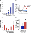
Silica induces TNF production in RAW 264.7, but not in IC21, macrophages. (A) TNF protein (pg/ml) released in cell-free supernatants of RAW 264.7 macrophage exposed to 10–100 μg/cm2 silica for 2 h. (B) Time-course of TNF release by RAW 264.7 macrophages during silica exposure (50 μg/cm2). (C) IC21 macrophages were exposed to 10–100 μg/cm2 silica for 2 h. (D) IC21 macrophages exposed to 4 mg/ml LPS for 4 h can produce TNF. *p < 0.001 versus control-treated cells, #p < 0.05 versus LPS-treated RAW 264.7 cells.
FIGURE 4.
FIGURE 4.
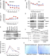
Silica alters TNFR1 expression and cell distribution and induces TNFR1/Phox interaction in silica-exposed RAW 264.7 macrophages. TNFR1 protein measured by ELISA in whole-cell lysates (A and B) or culture supernatants (C) extracted from RAW 264.7 (A and C) or IC21 (B) macrophages following silica (20 μg/cm2) exposure (0–8 h). ▪, silica-exposed cells; ▴, control-treated cells. (D) In RAW 264.7 macrophages, silica induces expression of RFK, which coimmunoprecipitates with TRADD and p22phox. TNFR1 immunoprecipitates (IP) from IC21 or RAW 264.7 macrophages (5 × 10⁸ cells), exposed to silica (20 μg/cm2) for 0–4 h, were immunoblotted with Abs specific to TRADD, RFK, and p22phox. Results are representative of three independent exposures. (E) TNFR1 is expressed in mitochondria from silica-exposed-RAW 264.7 macrophages but not IC21 macrophages. Mitochondria were isolated from 1 × 10⁹ RAW 264.7 and IC21 macrophages following silica exposure, and TNFR1 and tBid localization were analyzed by Western blotting using Abs against TNFR1 or tBid. Cytochrome c was used as a mitochondrial marker and loading control. Twice the concentration of proteins (100 μg) loaded in lanes from IC21 samples. Gel is representative of four experiments. (F) Silica exposure induces TNFR1 endocytosis in caveolin-containing rafts. Following silica (20 μg/cm2) exposure for 0–6 h, RAW 264.7 macrophages (5 × 10⁸ cells) were lysed and subjected to a 45% sucrose gradient, as described in Materials and Methods. Gels from each gradient were stained with Abs against TNFR1 or caveolin. (G) TNFR1 protein measured by ELISA in sucrose gradients isolated from RAW 264.7 macrophages 4 h after silica exposure. (H) Proteins from specific sucrose density gradients were resolved by two-dimensional gel electrophoresis and subjected to MALDI-TOF analysis to identify proteins that were differentially expressed in the gradients following silica exposure (Supplemental Table I).
FIGURE 5.
FIGURE 5.
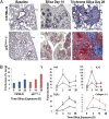
p47phox deficiency enhances silica-induced lung injury. (A) Lung photomicrographs (original magnifications ×4 [middle panel] and ×10 [right panel]) obtained from C57BL/6 (strain-matched controls, left panel, original magnification ×4) or p47phox−/− mice 0–28 d after intratracheal crystalline silica (0.2 g/kg) administration. In C57BL/6 mice, silica predominantly induced lesions that encased the terminal bronchiolar–alveolar duct region, and the nodule-like lesions show increased staining with trichrome blue, consistent with localized collagen deposition 28 d after silica exposure. In contrast, p47phox−/− mice react to silica with diffuse parenchymal accumulation of foamy macrophages and multinucleated giant cells and exhibit diffuse parenchymal deposition of collagen. (B) Quantitative analysis of trichrome staining demonstrates that both mouse strains accumulate significant amounts of collagen in their lungs compared with control (*p < 0.05), but the accumulation is statistically significantly greater (p < 0.05) in the lungs of p47phox−/− mice. Enhanced expression of inflammatory cytokines (TNF, IL-6), and fibrotic-related (TGFβ and collagen type 1, α 1) transcripts were measured by real-time PCR in the lungs as a function of time following intratracheal exposure to crystalline silica (0.2 mg/kg). Data are mean (± SE) fold increase normalized to RPL32 (Mm02528467_g1) gene expression as internal control (n = 5 mice/time point). Squares represent data from p47phox−/− and triangles represent data from C57BL/6 mice. *p < 0.001 versus C57BL/6 mouse strain.

References

    1. Di Giuseppe M., Gambelli F., Hoyle G. W., Lungarella G., Studer S. M., Richards T., Yousem S., McCurry K., Dauber J., Kaminski N., et al. 2009. Systemic inhibition of NF-kappaB activation protects from silicosis. PLoS ONE 4: e5689. - PMC - PubMed
    1. Dostert C., Pétrilli V., Van Bruggen R., Steele C., Mossman B. T., Tschopp J. 2008. Innate immune activation through Nalp3 inflammasome sensing of asbestos and silica. Science 320: 674–677 - PMC - PubMed
    1. Cassel S. L., Eisenbarth S. C., Iyer S. S., Sadler J. J., Colegio O. R., Tephly L. A., Carter A. B., Rothman P. B., Flavell R. A., Sutterwala F. S. 2008. The Nalp3 inflammasome is essential for the development of silicosis. Proc. Natl. Acad. Sci. USA 105: 9035–9040 - PMC (VSports注册入口) - PubMed
    1. Hornung V., Bauernfeind F., Halle A., Samstad E. O., Kono H., Rock K. L., Fitzgerald K. A., Latz E. 2008. Silica crystals and aluminum salts activate the NALP3 inflammasome through phagosomal destabilization. Nat. Immunol. 9: 847–856 - PMC - PubMed
    1. Costantini L. M., Gilberti R. M., Knecht D. A. 2011. The phagocytosis and toxicity of amorphous silica. PLoS ONE 6: e14647. - PMC - PubMed

VSports在线直播 - Publication types

MeSH terms

Substances

LinkOut - more resources