Skip to main page content
U.S. flag

An official website of the United States government

Dot gov

The . gov means it’s official. Federal government websites often end in . gov or . mil VSports app下载. Before sharing sensitive information, make sure you’re on a federal government site. .

Https

The site is secure V体育官网. The https:// ensures that you are connecting to the official website and that any information you provide is encrypted and transmitted securely. .

. 2014 Jul;63(7):2440-53.
doi: 10.2337/db13-1810. Epub 2014 Mar 7.

"VSports" Role of transcription factor acetylation in diabetic kidney disease

Affiliations

"V体育ios版" Role of transcription factor acetylation in diabetic kidney disease

Ruijie Liu et al. Diabetes. 2014 Jul.

Abstract

Nuclear factor (NF)-κB and signal transducer and activator of transcription 3 (STAT3) play a critical role in diabetic nephropathy (DN). Sirtuin-1 (SIRT1) regulates transcriptional activation of target genes through protein deacetylation. Here, we determined the roles of Sirt1 and the effect of NF-κB (p65) and STAT3 acetylation in DN. We found that acetylation of p65 and STAT3 was increased in both mouse and human diabetic kidneys. In human podocytes, advanced glycation end products (AGEs) induced p65 and STAT3 acetylation and overexpression of acetylation-incompetent mutants of p65 and STAT3 abrogated AGE-induced expression of NF-κB and STAT3 target genes VSports手机版. Inhibition of AGE formation in db/db mice by pyridoxamine treatment attenuated proteinuria and podocyte injury, restored SIRT1 expression, and reduced p65 and STAT3 acetylation. Diabetic db/db mice with conditional deletion of SIRT1 in podocytes developed more proteinuria, kidney injury, and acetylation of p65 and STAT3 compared with db/db mice without SIRT1 deletion. Treatment of db/db mice with a bromodomain and extraterminal (BET)-specific bromodomain inhibitor (MS417) which blocks acetylation-mediated association of p65 and STAT3 with BET proteins, attenuated proteinuria, and kidney injury. Our findings strongly support a critical role for p65 and STAT3 acetylation in DN. Targeting protein acetylation could be a potential new therapy for DN. .


"VSports注册入口" Figures

Figure 1
Figure 1
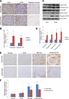
Acetylation of p65 and STAT3 is increased in diabetic kidneys. A: Kidney sections from db/db and db/m mice were stained for acetyl-p65 and acetyl-STAT3 using specific antibodies. The representative pictures of six mice in each group are shown. B: Immunostaining was quantified as described in Research Design and Methods. *P < 0.01 compared with db/m mice (n = 6). COD, corrected optical density. C: Western blot analyses of glomerular lysates from db/db and db/m mice were performed for acetyl-, phosphor-, and total p65 and STAT3. The representative blots of three independent experiments are shown. D: Western blots from all experiments were quantified by densitometry analysis, as described in Research Design and Methods. The ratios of acetyl-protein or phosphor-protein to total protein were calculated for p65 and STAT3. The fold changes relative to db/m mice are shown. *P < 0.01 compared with db/m mice (n = 6). E: Kidney sections from nephrectomized samples of patients with normal kidneys, minimal change disease (MCD), and mild and advanced DN were stained for acetyl-STAT3 and NF-κB. The representative pictures are shown (n = 5). F: The quantitation data of immunostaining in kidney biopsies from patients with normal kidneys, MCD, mild DN, and advanced DN are shown. *P < 0.05 compared with normal and MCD (n = 5).
Figure 2
Figure 2
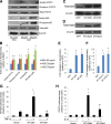
AGE induced acetylation of p65 and STAT3, which is required for their transcriptional activation. A: Human podocytes were cultured with AGE or BSA at the indicated doses for 24 h. Cell lysates were subject to Western blot analysis for acetyl-, phosphor-, and total p65 and STAT3. The representative blots of three independent experiments are shown. B: The Western blots from all experiments were quantified by densitometry analysis. The ratios of acetyl-protein or phosphor-protein to total protein were calculated for p65 and STAT3. The fold changes relative to BSA-treated cells are shown. *P < 0.01 compared with BSA-treated cells (n = 3). C and D: Podocytes were infected with wild-type (WT) p65 or STAT3 constructs or p65 and STAT3 constructs with mutated acetyl residues (MT) for 3 days. Western blots were performed to confirm the expression of p65 and STAT3. E and F: The Western blots from these experiments were quantified by densitometry analysis. The ratios of p65/β-actin (E) and STAT3/β-actin (F) were calculated; the fold changes relative to cells infected with green fluorescent protein are shown. *P < 0.01 compared with GFP-infected cells (n = 3). G and H: Podocytes were infected with WT or MT p65 and STAT3 and then transfected with p65 and Stat3 reporter constructs for 3 days. The cells were then further stimulated with either tumor necrosis factor (TNF)-α (10 ng/mL) or interleukin (IL)-6 (10 ng/mL) for an additional 24 h before lysis of cells for measurement of luciferase activity. *P < 0.01 compared with WT-p65 or WT-STAT3 without TNF-α or IL-6 stimulation; #P < 0.05 compared with WT-p65 or WT-STAT3 with TNF-α or IL-6 stimulation (n = 3).
Figure 3
Figure 3
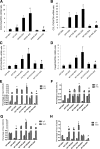
Acetylation of p65 and STAT3 is required for AGE-induced transcriptional activation of their target genes. Podocytes were infected with p65 or STAT3 constructs with mutated acetyl residues for 3 days then stimulated with AGE or BSA at the indicated doses for an additional 24 h. Total RNA was isolated from these cells for real-time PCR analysis of NF-κB target genes (CXCL5 [A] and CCL17 [B]) and STAT3 target genes (SOC3 [C] and Casp9 [D]). For chromatin immunoprecipitation (ChiP) assay for NF-κB and STAT3 target genes, podocytes were infected with p65 or STAT3 constructs with mutated acetyl residues for 3 days then stimulated with AGE or BSA at the indicated doses for an additional 24 h. ChiP assay also was performed in these cells to determine the binding of p65 and STAT3 on the promoter sites of their target genes (CXCL5 [E], CCL17 [F], SOC3 [G], and Casp9 [H]). Immunoprecipitated DNA samples were subjected to PCR analysis and are expressed as a percentage of input. *P < 0.01 compared with corresponding BSA-treated cells; #P < 0.01 compared with AGE-treated WT-p65 or WT-STAT3 cells (n = 4).
Figure 4
Figure 4
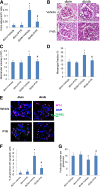
Prevention of AGE formation by PYR attenuates proteinuria and diabetic kidney injury in db/db mice. Eight-week-old female B6 diabetic db/db or nondiabetic db/m mice were randomized to receive either PYR or vehicle for 12 weeks. A: Urinary albumin-to-creatinine ratio was measured as described in Research Design and Methods (P < 0.01; n = 6). B: The representative pictures of the kidney histology of these mice are shown after periodic acid Schiff (PAS) staining. Morphometric analysis was performed in these kidney sections with PAS staining for the calculation of glomerular volume (C) and mesangial/glomerular fraction area (D). Kidney sections also were used for transferase dUTP nick end labeling to determine the rate of apoptosis in podocytes (E and F) and costained for wild-type 1 (WT-1) to determine the number of podocytes per glomerulus (E and G). The representative pictures are shown in E. *P < 0.01 compared with db/m mice treated with vehicle; #P < 0.05 compared with db/db mice treated with vehicle (n = 6).
Figure 5
Figure 5
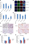
Treatment of db/db mice with PYR restores SIRT1 expression and reduces acetylation of p65 and STAT3 in the kidney of db/db mice. A: mRNA levels of SIRT1 were assessed by real-time PCR. B: Immunofluorescence staining for SIRT1 and synaptopodin was performed in kidney sections of these mice. C: SIRT1 staining was quantified by counting the number of cells stained positively for SIRT1 and colocalized with synaptopodin per glomerulus. D: Synaptopodin staining was quantified by calculating the percentage of glomerular area with positive staining for synaptopodin. E: Immunostaining for acetyl-p65 in kidney sections of these mice. F: Immunostaining of acetyl-STAT3 in kidney sections of these mice. The quantification data of immunostaining are shown for acetyl-p65 (G) and acetyl-STAT3 (H). *P < 0.01 compared with db/m mice treated with vehicle; #P < 0.05 compared with db/db mice treated with vehicle (n = 6). COD, corrected optical density.
Figure 6
Figure 6
Knockout of SIRT1 in podocytes aggravates proteinuria and kidney injury in db/db mice. A: The urine albumin-to-creatinine ratio was measured as described in QUANTIFICATION OF URINE ALBUMIN. *P < 0.01 compared with the corresponding time points of mice in other groups (n = 6). B: Kidney histology of mice with periodic acid Schiff (PAS) staining. The representative images are shown. C: Morphometric analysis was performed in these kidney sections with PAS staining for calculation of mesangial/glomerular fraction area. D: Kidney sections also were used for wild-type 1 staining to determine the number of podocytes per glomerulus. *P < 0.01 compared with Pod-Cre+/−;db/m mice; #P < 0.05 compared with Pod-Cre+/−;db/db mice (n = 6).
Figure 7
Figure 7
Effects of bromodomain inhibitor in DN. MS417, a bromodomain inhibitor, suppressed acetylation of Stat3 and NF-κB in podocytes treated with AGE. Podocytes were incubated with either BSA or AGE-BSA together with DMSO or MS417 (1.0 μmol/L) for 24 h. Western blot analysis was performed in these cells for acetyl, phosphor- and total p65 (A) and STAT3 (C). The representative blots of three independent experiments are shown. The densitometry analyses of these Western blots are shown for p65 (B) and STAT3 (D). The ratios of acetyl-protein or phosphor-protein to total protein were calculated for p65 and STAT3. The fold changes relative to cells treated with BSA + DMSO are shown. *P < 0.001 compared with cells treated with BSA + DMSO; #P < 0.001 compared with cells treated with AGE-DMSO (n = 3). E: Db/m and db/db mice were treated with either MS417 or vehicle from 8 weeks to 20 weeks of age (a total of 12 weeks). The urine albumin-to-creatinine ratio was measured as described in QUANTIFICATION OF URINE ALBUMIN. *P < 0.01 compared with the corresponding time points of mice from other groups (n = 6). F: Kidney histology was analyzed after periodic acid Schiff (PAS) staining in these mice. The representative images are shown. G: Morphometric analysis was performed in these kidney sections with PAS staining for calculation of mesangial/glomerular fraction area. *P < 0.05 compared with vehicle-treated db/m mice; #P < 0.05 compared with vehicle-treated db/db mice (n = 6).
Figure 8
Figure 8
Summary of the role of SIRT1 and acetylation of TFs in DN. AGE suppresses SIRT1 expression, leading to increased acetylation of NF-κB (p65), STAT3, and FOXO4. Acetylated p65 and STAT3 induces the expression of proinflammatory genes in diabetic kidneys. Acetylated FOXO4 induces proapoptotic gene expression, leading to podocyte apoptosis. PYR and bromodomain inhibitors (Brdi) attenuate diabetic kidney injury through the inhibition of AGE formation or by directly inhibiting the acetylation of TFs.

References

    1. Collins AJ, Foley RN, Herzog C, et al. US Renal Data System 2010 annual data report. Am J Kidney Dis 2011;57:A8, e1–526 - PubMed (VSports注册入口)
    1. Vlassara H, Striker LJ, Teichberg S, Fuh H, Li YM, Steffes M. Advanced glycation end products induce glomerular sclerosis and albuminuria in normal rats. Proc Natl Acad Sci U S A 1994;91:11704–11708 - PMC - PubMed
    1. Bohlender JM, Franke S, Stein G, Wolf G. Advanced glycation end products and the kidney. Am J Physiol Renal Physiol 2005;289:F645–F659 - PubMed (V体育平台登录)
    1. Zheng F, Cai W, Mitsuhashi T, Vlassara H. Lysozyme enhances renal excretion of advanced glycation endproducts in vivo and suppresses adverse age-mediated cellular effects in vitro: a potential AGE sequestration therapy for diabetic nephropathy? Mol Med 2001;7:737–747 - PMC - PubMed
    1. Zheng F, Zeng YJ, Plati AR, et al. Combined AGE inhibition and ACEi decreases the progression of established diabetic nephropathy in B6 db/db mice. Kidney Int 2006;70:507–514 - PubMed

"V体育安卓版" MeSH terms

Substances