Skip to main page content
U.S. flag

An official website of the United States government

Dot gov

The . gov means it’s official. Federal government websites often end in . gov or . mil. Before sharing sensitive information, make sure you’re on a federal government site. VSports app下载.

Https

The site is secure. The https:// ensures that you are connecting to the official website and that any information you provide is encrypted and transmitted securely. V体育官网.

Comparative Study
. 2014 Apr 15;189(8):909-31.
doi: 10.1164/rccm.201308-1458OC.

Z α1-antitrypsin confers a proinflammatory phenotype that contributes to chronic obstructive pulmonary disease

Affiliations
Comparative Study

Z α1-antitrypsin confers a proinflammatory phenotype that contributes to chronic obstructive pulmonary disease

Samuel Alam et al. Am J Respir Crit Care Med. .

V体育安卓版 - Abstract

Rationale: Severe α1-antitrypsin deficiency caused by the Z variant (Glu342Lys; ZZ-AT) is a well-known genetic cause for emphysema VSports手机版. Although severe lack of antiproteinase protection is the critical etiologic factor for ZZ-AT-associated chronic obstructive pulmonary disease (COPD), some reports have suggested enhanced lung inflammation as a factor in ZZ-AT homozygotes. .

Objectives: To provide molecular characterization of inflammation in ZZ-AT. V体育安卓版.

Methods: Inflammatory cell and cytokine profile (nuclear factor-κB, IL-6, tumor necrosis factor-α), intracellular polymerization of Z-AT, and endoplasmic reticulum (ER) stress markers (protein kinase RNA-like ER kinase, activator transcription factor 4) were assessed in transgenic mice and transfected cells in response to cigarette smoke, and in explanted lungs from ZZ and MM individuals with severe COPD V体育ios版. .

Measurements and main results: Compared with M-AT, transgenic Z-AT mice lungs exposed to cigarette smoke had higher levels of pulmonary cytokines, neutrophils, and macrophages and an exaggerated ER stress. Similarly, the ER overload response was greater in lungs from ZZ-AT homozygotes with COPD, and was particularly found in pulmonary epithelial cells. Cigarette smoke increased intracellular Z-AT polymers, ER overload response, and proinflammatory cytokine release in Z-AT-expressing pulmonary epithelial cells, which could be prevented with an inhibitor of polymerization, an antioxidant, and an inhibitor of protein kinase RNA-like ER kinase. VSports最新版本.

Conclusions: We show here that aggregation of intracellular mutant Z-AT invokes a specific deleterious cellular inflammatory phenotype in COPD. Oxidant-induced intracellular polymerization of Z-AT in epithelial cells causes ER stress, and promotes excess cytokine and cellular inflammation. This pathway is likely to contribute to the development of COPD in ZZ-AT homozygotes, and therefore merits further investigation V体育平台登录. .


Figures

Figure 1.
Figure 1.
Acute cigarette smoke (CS) exposure up-regulates inflammatory mediator mRNA in vivo in Z variant of antitrypsin (Z-AT) mice lungs. Acute CS exposure significantly up-regulated expression of tumor necrosis factor (TNF) mRNA (P = 0.006 and P < 0.001, respectively) (A) and IL-6 mRNA (P = 0.003 and P < 0.001, respectively) (B) in lungs of normal variant AT (M-AT) and Z-AT mice compared with their respective non–CS-exposed controls. CS-exposed Z-AT mice lungs had significantly elevated TNF-α and IL-6 mRNA compared with CS-exposed M-AT mice (P = 0.003 and P < 0.001, respectively). At 24 hours JE mRNA was unaffected by CS exposure in Z-AT or M-AT mice lungs (C). Negative control was mastermix (final reaction volume compensated with water) and positive control was LPS (20 ng) treated J774.1 cells. Band density was determined using the ImageJ program and expressed relative to murine GAPDH mRNA. Results presented are from analysis of bronchoalveolar lavage fluid and lung homogenates (lungs) from n = 8 mice per group, and of three independent experiments (n = 3). *Non–CS-exposed versus CS exposed, **M-AT mice versus Z-AT mice. Values are expressed as mean ± SEM. GAPDH = glyceraldehyde phosphate dehydrogenase.
Figure 2.
Figure 2.
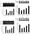
Z variant of antitrypsin (Z-AT) mice have an enhanced endoplasmic reticulum (ER) stress following acute cigarette smoke (CS) exposure. Acute CS exposure significantly increased ER stress markers; protein kinase RNA–like ER kinase (PERK) (A and B) and activator transcription factor (ATF) 4 (C and D) in Z-AT mice compared with normal variant AT (M-AT). (A and B) Baseline expression of PERK mRNA was higher in unstimulated Z-AT controls compared with M-AT controls (P = 0.026). CS exposure significantly up-regulated PERK mRNA in both M-AT and Z-AT mice lungs compared with non–CS exposed M-AT and Z-AT controls (P = 0.004 and P = 0.044, respectively). (A) CS-exposed Z-AT mice had significantly greater PERK mRNA than CS-exposed M-AT mice (P = 0.010). (B) There was significant up-regulation of PERK protein product (140 kD) in non–CS-exposed and CS-exposed Z-AT mice compared with their respective M-AT mice (P = 0.015 and P = 0.012, respectively). (C and D) Baseline expression of ATF4 mRNA was higher in Z-AT mice lungs (P = 0.006) (C). CS exposure induced significant up-regulation of ATF4 mRNA in Z-AT mice lungs compared with the non–CS-exposed Z-AT mice lungs (P = 0.038). ATF4 mRNA was not affected by CS exposure in M-AT mice lungs compared with non–CS-exposed M-AT mice lungs (P = 0.345). (D) There was significant up-regulation of ATF4 protein product (38 kD) in non–CS-exposed and CS-exposed Z-AT mice compared with their respective M-AT mice (P = 0.013 and P = 0.021, respectively). (E and F) Baseline expression of ATF6 mRNA and protein was unaffected in control M-AT and Z-AT mice. CS exposure significantly up-regulated expression of ATF6 mRNA and protein in both M-AT and Z-AT compared with their respective non–CS-exposed controls; P < 0.001 for all. There was no difference in the induction of ATF6 between CS-M and CS-Z mice (E and F). Thapsigargin (0.5 µg/ml) was used to induce ER stress and thus provide a positive control for ATF4, PERK, and ATF6 (39). Band density was determined using the ImageJ program and expressed relative to murine GAPDH mRNA (A, C, and E) or murine GAPDH protein (B, D, and F). Results are presented for n = 8 mice per group and of three independent experiments (n = 3). Values are expressed as mean ± SEM. *Non–CS-exposed versus CS exposed, **M-AT mice versus Z-AT mice. GAPDH = glyceraldehyde phosphate dehydrogenase.
Figure 2.
Figure 2.
Z variant of antitrypsin (Z-AT) mice have an enhanced endoplasmic reticulum (ER) stress following acute cigarette smoke (CS) exposure. Acute CS exposure significantly increased ER stress markers; protein kinase RNA–like ER kinase (PERK) (A and B) and activator transcription factor (ATF) 4 (C and D) in Z-AT mice compared with normal variant AT (M-AT). (A and B) Baseline expression of PERK mRNA was higher in unstimulated Z-AT controls compared with M-AT controls (P = 0.026). CS exposure significantly up-regulated PERK mRNA in both M-AT and Z-AT mice lungs compared with non–CS exposed M-AT and Z-AT controls (P = 0.004 and P = 0.044, respectively). (A) CS-exposed Z-AT mice had significantly greater PERK mRNA than CS-exposed M-AT mice (P = 0.010). (B) There was significant up-regulation of PERK protein product (140 kD) in non–CS-exposed and CS-exposed Z-AT mice compared with their respective M-AT mice (P = 0.015 and P = 0.012, respectively). (C and D) Baseline expression of ATF4 mRNA was higher in Z-AT mice lungs (P = 0.006) (C). CS exposure induced significant up-regulation of ATF4 mRNA in Z-AT mice lungs compared with the non–CS-exposed Z-AT mice lungs (P = 0.038). ATF4 mRNA was not affected by CS exposure in M-AT mice lungs compared with non–CS-exposed M-AT mice lungs (P = 0.345). (D) There was significant up-regulation of ATF4 protein product (38 kD) in non–CS-exposed and CS-exposed Z-AT mice compared with their respective M-AT mice (P = 0.013 and P = 0.021, respectively). (E and F) Baseline expression of ATF6 mRNA and protein was unaffected in control M-AT and Z-AT mice. CS exposure significantly up-regulated expression of ATF6 mRNA and protein in both M-AT and Z-AT compared with their respective non–CS-exposed controls; P < 0.001 for all. There was no difference in the induction of ATF6 between CS-M and CS-Z mice (E and F). Thapsigargin (0.5 µg/ml) was used to induce ER stress and thus provide a positive control for ATF4, PERK, and ATF6 (39). Band density was determined using the ImageJ program and expressed relative to murine GAPDH mRNA (A, C, and E) or murine GAPDH protein (B, D, and F). Results are presented for n = 8 mice per group and of three independent experiments (n = 3). Values are expressed as mean ± SEM. *Non–CS-exposed versus CS exposed, **M-AT mice versus Z-AT mice. GAPDH = glyceraldehyde phosphate dehydrogenase.
Figure 3.
Figure 3.
Expression of endoplasmic reticulum (ER) stress markers in emphysematous lungs. (A) Immunolocalization of protein kinase RNA–like ER kinase (PERK) and activator transcription factor (ATF) 4 in frozen lung sections from MM-AT and ZZ-AT emphysematous lung tissue and controls. PERK (arrows, top panels) and ATF4 (arrows, middle panels) are localized to the cytoplasm of respiratory epithelial cells with staining significantly more intense and frequent in ZZ-AT as compared with MM-AT and control lungs. Representative images (n = 3–4). Original magnification ×40. (B–D) Immunohistochemistry localization of macrophage phenotypes in MM-AT and ZZ-AT chronic obstructive pulmonary disease (COPD). (B) Sections of lung stained with the monocyte marker CD14 demonstrate a significantly increased number of monocytes in ZZ-AT when compared with MM-AT (arrows, top panels) (P < 0.001) (graph). (C) Sections of lung stained for matured and activated macrophage marker the panmacrophage marker CD68 show significantly increased number of macrophages in ZZ-AT when compared with MM-AT (arrows, top) (P < 0.001) (graph). (D) Sections of lung stained for MAC387, a marker of blood-derived macrophages, showing localization to cells present within the parenchymal wall and within the lumen of vessels. Significantly more cells were positive for MAC387 in ZZ-AT when compared with MM-AT (arrows, top) (P < 0.001) (graph). (B–D) Although MM-AT COPD lung sections had significantly more monocytes, macrophages, and recently blood-derived macrophages compared with normals (P < 0.001 for all) ZZ-AT COPD demonstrated significantly increased numbers of all of these macrophages compared with normals or MM-AT COPD lungs (P < 0.001 for all). Note the presence of cells within the alveolar wall and alveolar spaces (arrows). Images are representative of n = 10 in each group and are at a magnification of ×400. Values are expressed as mean (± SEM). *Normal versus MM-AT or ZZ-AT and **MM-AT versus ZZ-AT.
Figure 3.
Figure 3.
Expression of endoplasmic reticulum (ER) stress markers in emphysematous lungs. (A) Immunolocalization of protein kinase RNA–like ER kinase (PERK) and activator transcription factor (ATF) 4 in frozen lung sections from MM-AT and ZZ-AT emphysematous lung tissue and controls. PERK (arrows, top panels) and ATF4 (arrows, middle panels) are localized to the cytoplasm of respiratory epithelial cells with staining significantly more intense and frequent in ZZ-AT as compared with MM-AT and control lungs. Representative images (n = 3–4). Original magnification ×40. (B–D) Immunohistochemistry localization of macrophage phenotypes in MM-AT and ZZ-AT chronic obstructive pulmonary disease (COPD). (B) Sections of lung stained with the monocyte marker CD14 demonstrate a significantly increased number of monocytes in ZZ-AT when compared with MM-AT (arrows, top panels) (P < 0.001) (graph). (C) Sections of lung stained for matured and activated macrophage marker the panmacrophage marker CD68 show significantly increased number of macrophages in ZZ-AT when compared with MM-AT (arrows, top) (P < 0.001) (graph). (D) Sections of lung stained for MAC387, a marker of blood-derived macrophages, showing localization to cells present within the parenchymal wall and within the lumen of vessels. Significantly more cells were positive for MAC387 in ZZ-AT when compared with MM-AT (arrows, top) (P < 0.001) (graph). (B–D) Although MM-AT COPD lung sections had significantly more monocytes, macrophages, and recently blood-derived macrophages compared with normals (P < 0.001 for all) ZZ-AT COPD demonstrated significantly increased numbers of all of these macrophages compared with normals or MM-AT COPD lungs (P < 0.001 for all). Note the presence of cells within the alveolar wall and alveolar spaces (arrows). Images are representative of n = 10 in each group and are at a magnification of ×400. Values are expressed as mean (± SEM). *Normal versus MM-AT or ZZ-AT and **MM-AT versus ZZ-AT.
Figure 4.
Figure 4.
Cigarette smoke (CS) extract induces nuclear factor (NF)-κB activity in Z variant of antitrypsin (Z-AT) cells. CS extract (12.5%) exposed A549–Z-AT cells had significantly greater NF-κB activity than A549–normal variant AT (M-AT) cells (Z-AT cells vs. M-AT cells at 24 h; P < 0.001). Positive control for NF-κB was Jurkat cells. Results presented are from three independent experiments (n = 3). Values are expressed as mean ± SEM. (A) *CS extract M-AT cells versus CS extract Z-AT cells.
Figure 5.
Figure 5.
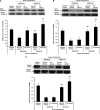
Cigarette smoke (CS) extract induces the endoplasmic reticulum (ER) stress in Z variant of antitrypsin (Z-AT) cells. Representative Western blot analysis of whole cell extract on 12% sodium dodecyl sulfate–polyacrylamide gel electrophoresis. (A) A polyclonal anti–protein kinase RNA–like ER kinase (PERK) antibody detected significant up-regulated PERK protein (125 kD) in CS extract A549–Z-AT cells compared with their respective non–CS extract Z-AT cells (P < 0.001). (B–D) Polyclonal antibodies against activator transcription factor (ATF) 4 protein (38 kD), regulator of G-protein signaling protein 16 protein (RGS16) (predicted at 23 kD and detected at 29 kD) and the ER chaperone calnexin (predicted at 90 kD and detected at 75 kD), respectively, in CS extract A549–Z-AT cells compared with their respective non–CS extract Z-AT cells (P < 0.001, P = 0.001, and P < 0.001, respectively). (E) A polyclonal anti-ATF6 antibody detected ATF6 protein (85 kD) in CS extract exposed both A549–normal variant AT (M-AT) and A549–Z-AT cells compared with their respective non–CS extract A549–Z-AT cells (P < 0.001 for both). There was no difference between non–CS extract exposed A549–M-AT and A549–Z-AT controls (P = 0.795) or CS extract exposed A549–M-AT and A549–Z-AT cells (P = 0.912). Positive control, the ER stress–inducing control agent thapsigargin, treated HeLa cell RNA for ATF4, PERK, and ATF6. Untreated MCF-7 cells constitutively express RGS16 and calnexin (24) (n = 3). Band density was determined using ImageJ program and expressed relative to human GAPDH protein. Values are expressed as mean ± SEM. *Non –CS extract versus CS extract and **M-AT cells versus Z-AT cells. GAPDH = glyceraldehyde phosphate dehydrogenase.
Figure 5.
Figure 5.
Cigarette smoke (CS) extract induces the endoplasmic reticulum (ER) stress in Z variant of antitrypsin (Z-AT) cells. Representative Western blot analysis of whole cell extract on 12% sodium dodecyl sulfate–polyacrylamide gel electrophoresis. (A) A polyclonal anti–protein kinase RNA–like ER kinase (PERK) antibody detected significant up-regulated PERK protein (125 kD) in CS extract A549–Z-AT cells compared with their respective non–CS extract Z-AT cells (P < 0.001). (B–D) Polyclonal antibodies against activator transcription factor (ATF) 4 protein (38 kD), regulator of G-protein signaling protein 16 protein (RGS16) (predicted at 23 kD and detected at 29 kD) and the ER chaperone calnexin (predicted at 90 kD and detected at 75 kD), respectively, in CS extract A549–Z-AT cells compared with their respective non–CS extract Z-AT cells (P < 0.001, P = 0.001, and P < 0.001, respectively). (E) A polyclonal anti-ATF6 antibody detected ATF6 protein (85 kD) in CS extract exposed both A549–normal variant AT (M-AT) and A549–Z-AT cells compared with their respective non–CS extract A549–Z-AT cells (P < 0.001 for both). There was no difference between non–CS extract exposed A549–M-AT and A549–Z-AT controls (P = 0.795) or CS extract exposed A549–M-AT and A549–Z-AT cells (P = 0.912). Positive control, the ER stress–inducing control agent thapsigargin, treated HeLa cell RNA for ATF4, PERK, and ATF6. Untreated MCF-7 cells constitutively express RGS16 and calnexin (24) (n = 3). Band density was determined using ImageJ program and expressed relative to human GAPDH protein. Values are expressed as mean ± SEM. *Non –CS extract versus CS extract and **M-AT cells versus Z-AT cells. GAPDH = glyceraldehyde phosphate dehydrogenase.
Figure 6.
Figure 6.
Cigarette smoke (CS) extract induces formation of intracellular polymeric Z variant of antitrypsin (Z-AT) in Z-AT cells. (Top) Representative Western blot on 7.5% nondenaturing polyacrylamide gel electrophoresis using antihuman AT. Monomeric AT was detected in non–CS extract or CS extract–exposed A549–normal variant AT (M-AT) cell supernatant, and in the supernatant of non–CS extract A549–Z-AT cells. Monomeric Z-AT and polymeric Z-AT were detected in inclusion bodies of non–CS extract A549–Z-AT cells and in the supernatant and inclusion bodies of CS extract A549–Z-AT cells. (Bottom) Representative Western blot using a monoclonal antioxidized AT antibody. This detected monomeric oxidized AT in CS extract A549–M-AT cell supernatant, and both oxidized AT and oxidized-polymeric Z-AT in the supernatant and inclusion bodies of CS extract A549–Z-AT cells. (Top and bottom) Protein loading was equalized for 150 ng of total AT per lane. Oxidized AT and oxidized-polymeric AT were prepared by oxidizing plasma purified native AT or polymer AT, respectively, using N-chlorosuccinamide oxidizing agent.
Figure 7.
Figure 7.
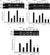
The effect of inhibition of polymerization and an antioxidant. (A–D) Cigarette smoke (CS) extract significantly up-regulated expression of human protein kinase RNA–like endoplasmic reticulum (ER) kinase (PERK), activator transcription factor (ATF) 4, regulator of G-protein signaling protein 16 (RGS16), and calnexin mRNA in A549–Z variant of antitrypsin (Z-AT) cells (P < 0.001 for all). Inhibitor of polymerization (4M, 20 μg) and N-acetylcysteine (NAC, 10 mM) independently significantly inhibited CS extract–induced human PERK, ATF4, RGS16, and calnexin mRNA (P < 0.001 for all). (E) CS extract–induced up-regulation of ATF6 mRNA in A549–Z-AT cells was unaffected by 4M (P = 0.925). However, the antioxidant NAC significantly reduced CS extract–induced ATF6 expression in A549–Z-AT cells (P < 0.001). Positive control, the ER stress–inducing control agent thapsigargin treated HeLa cell RNA for PERK, ATF4, and ATF6. Untreated MCF-7 cells constitutively express RGS16 and calnexin. Band density was determined using ImageJ program and expressed relative to human GAPDH mRNA. n = 3. Values are expressed as mean ± SEM. *Non–CS extract normal variant AT (M-AT) cells versus non–CS extract Z-AT cells, **non–CS extract Z-AT cells versus CS extract Z-AT cells, and ***CS extract Z-AT cells versus CS extract 4M or + N-acetylcysteine. GAPDH = glyceraldehyde phosphate dehydrogenase.
Figure 7.
Figure 7.
The effect of inhibition of polymerization and an antioxidant. (A–D) Cigarette smoke (CS) extract significantly up-regulated expression of human protein kinase RNA–like endoplasmic reticulum (ER) kinase (PERK), activator transcription factor (ATF) 4, regulator of G-protein signaling protein 16 (RGS16), and calnexin mRNA in A549–Z variant of antitrypsin (Z-AT) cells (P < 0.001 for all). Inhibitor of polymerization (4M, 20 μg) and N-acetylcysteine (NAC, 10 mM) independently significantly inhibited CS extract–induced human PERK, ATF4, RGS16, and calnexin mRNA (P < 0.001 for all). (E) CS extract–induced up-regulation of ATF6 mRNA in A549–Z-AT cells was unaffected by 4M (P = 0.925). However, the antioxidant NAC significantly reduced CS extract–induced ATF6 expression in A549–Z-AT cells (P < 0.001). Positive control, the ER stress–inducing control agent thapsigargin treated HeLa cell RNA for PERK, ATF4, and ATF6. Untreated MCF-7 cells constitutively express RGS16 and calnexin. Band density was determined using ImageJ program and expressed relative to human GAPDH mRNA. n = 3. Values are expressed as mean ± SEM. *Non–CS extract normal variant AT (M-AT) cells versus non–CS extract Z-AT cells, **non–CS extract Z-AT cells versus CS extract Z-AT cells, and ***CS extract Z-AT cells versus CS extract 4M or + N-acetylcysteine. GAPDH = glyceraldehyde phosphate dehydrogenase.
Figure 8.
Figure 8.
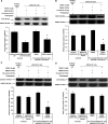
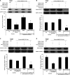
The effect of protein kinase RNA–like endoplasmic reticulum (ER) kinase (PERK) inhibitor I on ER stress in Z variant of antitrypsin (Z-AT) cells. (A–C) Pretreatment of A549–Z-AT cells with the PERK inhibitor I (2 μg) prevented cigarette smoke (CS) extract–induced up-regulated expression of (A) activator transcription factor (ATF) 4 protein (38 kD) (P = 0.021), which was comparable with non–CS extract A549–Z-AT cells (P = 0.439), (B) regulator of G-protein signaling protein 16 protein (RGS16) (29 kD) (P < 0.001), and (C) calnexin protein (75 kD) (P < 0.001). (D) Pretreatment of A549–Z-AT cells with the PERK inhibitor I did not prevent CS extract–induced up-regulated expression of ATF6 protein (85 kD) (P = 0. 935). Vehicle DMSO (0.03%) had no effect compared with CS extract. Band density was determined using the ImageJ program and expressed relative to human GAPDH protein. Positive control, the ER stress–inducing control agent thapsigargin treated HeLa cell RNA for PERK, ATF4, and ATF6. Untreated MCF-7 cells constitutively express RGS16 and calnexin. n = 3. Values are expressed as mean ± SEM. *CS extract versus PERK inhibitor I + CS extract. DMSO = dimethyl sulfoxide; GAPDH = glyceraldehyde phosphate dehydrogenase.
Figure 9.
Figure 9.
N-Acetylcysteine (NAC) inhibited cigarette smoke (CS) extract induced endoplasmic reticulum (ER) overload response in primary normal human bronchial epithelial cells transfected with human Z variant of antitrypsin (Z-AT) gene. (A–D) In primary normal human bronchial epithelial (primary NHBE) cells transfected with human Z-AT (primary NHBE–Z-AT cells) CS extract significantly induced up-regulation of (A) protein kinase RNA–like ER kinase (PERK) protein (125 kD), (B) activator transcription factor (ATF) 4 protein (38 kD), (C) regulator of G-protein signaling protein 16 protein (RGS16) (29 kD), and (D) calnexin protein (75 kD) (P < 0.001, P < 0.001, P = 0.042, and P = 0.036, respectively). Inhibitor of polymerization (4M, 20 μg) and NAC (10 mM) independently significantly inhibited CS extract–induced human PERK, ATF4, RGS16, and calnexin proteins (P < 0.001 for all). (E) CS extract induced up-regulation of ATF6 protein (85 kD) in primary NHBE–Z-AT cells was unaffected by 4M (P = 0.789). However, the antioxidant NAC significantly reduced CS extract–induced ATF6 expression in NHBE–Z-AT cells (P < 0.001). Band density was determined using the ImageJ program and expressed relative to human GAPDH protein. Positive control, the ER stress inducing control agent thapsigargin treated HeLa cell RNA for ATF4, PERK, and ATF6. Untreated MCF-7 cells constitutively express RGS16 and calnexin. n = 3. Values are expressed as media. *Non–CS extract primary NHBE cells versus non–CS extract primary NHBE–Z-AT cells, **non–CS extract primary NHBE–Z-AT cells versus CS extract primary NHBE–Z-AT cells, and ***CS extract primary NHBE–Z-AT cells versus CS extract primary NHBE–Z-AT cells + N-acetylcysteine or 4M. GAPDH = glyceraldehyde phosphate dehydrogenase.
Figure 9.
Figure 9.
N-Acetylcysteine (NAC) inhibited cigarette smoke (CS) extract induced endoplasmic reticulum (ER) overload response in primary normal human bronchial epithelial cells transfected with human Z variant of antitrypsin (Z-AT) gene. (A–D) In primary normal human bronchial epithelial (primary NHBE) cells transfected with human Z-AT (primary NHBE–Z-AT cells) CS extract significantly induced up-regulation of (A) protein kinase RNA–like ER kinase (PERK) protein (125 kD), (B) activator transcription factor (ATF) 4 protein (38 kD), (C) regulator of G-protein signaling protein 16 protein (RGS16) (29 kD), and (D) calnexin protein (75 kD) (P < 0.001, P < 0.001, P = 0.042, and P = 0.036, respectively). Inhibitor of polymerization (4M, 20 μg) and NAC (10 mM) independently significantly inhibited CS extract–induced human PERK, ATF4, RGS16, and calnexin proteins (P < 0.001 for all). (E) CS extract induced up-regulation of ATF6 protein (85 kD) in primary NHBE–Z-AT cells was unaffected by 4M (P = 0.789). However, the antioxidant NAC significantly reduced CS extract–induced ATF6 expression in NHBE–Z-AT cells (P < 0.001). Band density was determined using the ImageJ program and expressed relative to human GAPDH protein. Positive control, the ER stress inducing control agent thapsigargin treated HeLa cell RNA for ATF4, PERK, and ATF6. Untreated MCF-7 cells constitutively express RGS16 and calnexin. n = 3. Values are expressed as media. *Non–CS extract primary NHBE cells versus non–CS extract primary NHBE–Z-AT cells, **non–CS extract primary NHBE–Z-AT cells versus CS extract primary NHBE–Z-AT cells, and ***CS extract primary NHBE–Z-AT cells versus CS extract primary NHBE–Z-AT cells + N-acetylcysteine or 4M. GAPDH = glyceraldehyde phosphate dehydrogenase.
Figure 10.
Figure 10.
The effect of protein kinase RNA–like endoplasmic reticulum (ER) kinase (PERK) inhibitor I on ER overload response in primary normal human bronchial epithelial (NHBE)–Z variant of antitrypsin (Z-AT) cells. (A–C) Pretreatment of primary NHBE–Z-AT cells with the PERK inhibitor I (2 μg) prevented cigarette smoke (CS)–extract induced up-regulated expression of (A) activator transcription factor (ATF) 4 protein (38 kD) (P = 0.007), which was comparable with non–CS extract NHBE–Z-AT cells (P = 0.617), (B) regulator of G-protein signaling protein 16 (RGS16) (29 kD) (P < 0.001), and (C) calnexin protein (75 kD) (P < 0.001). (D) Pretreatment of NHBE–Z-AT cells with the PERK inhibitor I did not prevent CS extract–induced up-regulated expression of ATF6 protein (85 kD) (P = 0. 916). Band density was determined using the ImageJ program and expressed relative to human GAPDH protein. Positive control, the ER stress–inducing control agent thapsigargin treated HeLa cell RNA for ATF4, PERK, and ATF6. Untreated MCF-7 cells constitutively express RGS16 and calnexin. n = 3. Values are expressed as mean ± SEM. *CS extract versus PERK inhibitor I + CS extract. DMSO = dimethyl sulfoxide; GAPDH = glyceraldehyde phosphate dehydrogenase.
Figure 11.
Figure 11.
Schematic diagram showing the mechanisms contributing to the development of lung disease of ZZ-AT homozygotes. The left side of the diagram details our new findings; the right side (separated by the dashed line) shows what is already established in the study of α1-antitrypsin (AT) deficiency. Severe deficiency of AT is the main predisposing factor to emphysema. In addition, there is baseline intracellular polymerization and aggregation in alveolar epithelial cells, which activates endoplasmic reticulum (ER) stress. Oxidants from cigarette smoke (CS) and inflammatory cells potentiate this process by accelerating the formation of oxidized-polymeric Z-AT, which in turn activates protein kinase RNA–like ER kinase (PERK)-dependent nuclear factor (NF)-κB production of inflammatory mediators contributing to lung damage. Confirmation of this pathway is provided by the interruption of this process by (1) targeting the structural differences in Z-AT by directly preventing polymerization (with 4M), (2) by inhibiting oxidant-mediated acceleration of polymerization with an antioxidant (N-acetylcysteine), and (3) inhibiting PERK with the PERK inhibitor I (GSK2606414). CS-induced ER overload response markers; PERK, activator transcription factor (ATF) 4, and regulator of G-protein signaling protein 16 (RGS16), and the ER chaperone calnexin could be inhibited below the expression level of non–CS exposed Z-AT cells independently by both the inhibitor of Z-AT polymerization, 4M or an antioxidant, N-acetylcysteine (NAC). Diagram also shows the previously reported effect of oxidants on inducing polymerization of extracellular Z-AT in plasma and lung, further resulting in reduced inhibitory activity of Z-AT and an effect on neutrophil chemotaxis (17, 20).

V体育2025版 - Comment in

References

    1. Stoller JK, Aboussouan LS. A review of α1-antitrypsin deficiency. Am J Respir Crit Care Med. 2012;185:246–259. - PubMed
    1. Eriksson S. Studies in α 1-antitrypsin deficiency. Acta Med Scand Suppl. 1965;432:1–85. - PubMed
    1. Blanco I, de Serres FJ, Fernandez-Bustillo E, Lara B, Miravitlles M. Estimated numbers and prevalence of PI*S and PI*Z alleles of alpha1-antitrypsin deficiency in European countries. Eur Respir J. 2006;27:77–84. - PubMed
    1. de Serres FJ, Blanco I, Fernández-Bustillo E. Genetic epidemiology of alpha-1 antitrypsin deficiency in North America and Australia/New Zealand: Australia, Canada, New Zealand and the United States of America. Clin Genet. 2003;64:382–397. - PubMed
    1. Lomas DA, Evans DL, Finch JT, Carrell RW. The mechanism of Z alpha 1-antitrypsin accumulation in the liver. Nature. 1992;357:605–607. - PubMed

Publication types

MeSH terms