Skip to main page content (VSports在线直播)
U.S. flag

An official website of the United States government

Dot gov

The . gov means it’s official. Federal government websites often end in . gov or VSports app下载. mil. Before sharing sensitive information, make sure you’re on a federal government site. .

Https

The site is secure. The https:// ensures that you are connecting to the official website and that any information you provide is encrypted and transmitted securely V体育官网. .

. 2014 Apr 1;111(13):4952-7.
doi: 10.1073/pnas.1319963111. Epub 2014 Feb 28.

Multiple cells-of-origin of mutant K-Ras-induced mouse lung adenocarcinoma

Affiliations

VSports最新版本 - Multiple cells-of-origin of mutant K-Ras-induced mouse lung adenocarcinoma

Kate D Sutherland et al. Proc Natl Acad Sci U S A. .

Abstract

Much controversy surrounds the cell-of-origin of mutant K-Ras (K-RasG12D)-induced lung adenocarcinoma. To shed light on this issue, we have used technology that enables us to conditionally target K-RasG12D expression in Surfactant Protein C (SPC)(+) alveolar type 2 cells and in Clara cell antigen 10 (CC10)(+) Clara cells by use of cell-type-restricted recombinant Adeno-Cre viruses. Experiments were performed both in the presence and absence of the tumor suppressor gene p53, enabling us to assess what effect the cell-of-origin and the introduced genetic lesions have on the phenotypic characteristics of the resulting adenocarcinomas. We conclude that both SPC-expressing alveolar type 2 cells and CC10-expressing Clara cells have the ability to initiate malignant transformation following the introduction of these genetic alterations. The lungs of K-Ras(lox-Stop-lox-G12D/+) and K-Ras(lox-Stop-lox-G12D/+);tumor suppressor gene Trp53(F/F) mice infected with Adeno5-SPC-Cre and Adeno5-CC10-Cre viruses displayed differences in their tumor spectrum, indicating distinct cellular routes of tumor initiation. Moreover, using a multicolor Cre reporter line, we demonstrate that the resulting tumors arise from a clonal expansion of switched cells. Taken together, these results indicate that there are multiple cellular paths to K-RasG12D-induced adenocarcinoma and that the initiating cell influences the histopathological phenotype of the tumors that arise VSports手机版. .

Keywords: BASCs; NSCLC; bronchioalveolar stem cells; non small cell lung cancer. V体育安卓版.


Conflict of interest statement

The authors declare no conflict of interest.

Figures
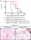
Fig. 1.
Fig. 1.
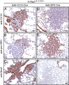
Using a cell-type–restricted adenoviral infection approach to identify the cell-of-origin of K-RasG12D–induced lung adenocarcinomas. (A) Kaplan Meier survival curves of K-RasLSL-G12D/+ mice (solid lines) and K-RasLSL-G12D/+;Trp53F/F mice (dashed lines) infected with Ad5–SPC–Cre virus (red curve) or Ad5–CC10–Cre virus (blue curve). The median survival of K-RasLSL-G12D/+ and K-RasLSL-G12D/+;Trp53F/F mice infected with Ad5–SPC–Cre virus is 360 d (n = 5) and 162 d (n = 8), respectively. The median survival of K-RasLSL-G12D/+ and K-RasLSL-G12D/+;Trp53F/F mice infected with Ad5–CC10–Cre virus is 308 d (n = 8) and 177 d (n = 9), respectively. (B) A representative H&E section from a K-RasLSL-G12D/+ animal examined 32 wk following Ad5–SPC–Cre infection. The majority of lesions arise in the alveolar compartment at the periphery of the lung. (C) A representative H&E section from a K-RasLSL-G12D/+ animal 32 wk following Ad5–CC10–Cre infection. The majority of lesions observed are associated and/or in close proximity to the BADJ. (Scale bar in B and C, 500 μM.)
Fig. 2.
Fig. 2.
Distinct cells-of-origin of K-RasG12D–induced lung adenocarcinoma. (A) Quantification of lesions observed in the lungs of K-RasLSL-G12D/+ following infection with Ad5–SPC–Cre virus (black bars; n = 5; 323–485 d) and Ad5–CC10–Cre virus (white bars; n = 8; 220–491 d). (B) Quantification of lesions observed in the lungs of K-RasLSL-G12D/+;Trp53F/F mice following infection with Ad5–SPC–Cre virus (black bars; n = 8; 146–210 d) and Ad5–CC10–Cre virus (white bars; n = 9; 159–197 d). The bar graphs represent the average number of lesions per lung: AAH/adenomas, papillary hyperplasia/papilloma, adenocarcinoma, papillary carcinoma, mixed carcinoma:adenocarcinoma/papillary carcinoma, per mouse examined. Data represent means ± SEM.
Fig. 3.
Fig. 3.
Tumor initiation and progression occurs in distinct cell types and locations following K-RasG12D activation. (A–C) Microphotographs of H&E-stained sections from K-RasLSL-G12D/+ mice following Ad5–SPC–Cre infection showing (A) alveolar hyperplasia, (B) AAH, and (C) adenocarcinoma. (D–F) H&E stained sections from K-RasLSL-G12D/+ mice following Ad5–CC10–Cre infection showing (D) focal hyperplasia of bronchiolar epithelial cells residing at the terminal bronchiole (arrow), (E) papillary hyperplasia of bronchiolar epithelial cells at the terminal bronchiole, and (F) papillary carcinoma in the lumen of a dilated bronchiole. (Scale bar in A, B, D, and E, 20 μM; C and F, 50 μM.)
Fig. 4.
Fig. 4.
Immunohistochemical heterogeneity of papilloma and AAH at the terminal bronchiole and alveoli. (A–C) Sections (4 μm) of papilloma observed in K-RasLSL-G12D/+ mice following Ad5–CC10–Cre infection at the terminal bronchiole, (A) showing positive staining for the Clara cell marker, CC10. (B) A number of hyperplasic cells (CC10+) at the terminal bronchiole stain positive for the AT2 cell marker, pro-SPC. (C) Sox2-positive staining of cells lining bronchiolar and hyperplasic cells at the terminal bronchiole. (D–F) Representative images of AAH and adenoma observed in K-RasLSL-G12D/+ mice following Ad5–SPC–Cre infection. (D) Adenomas are negative for the Clara cell marker, CC10. (E) AAH stain positive for the AT2 marker, pro-SPC, but are negative for Sox2 (F). (Scale bar in A–F, 50 μM.)
Fig. 5.
Fig. 5.
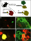
Clonal outgrowth of CC10+ and SPC+ cells following K-RasG12D activation. (A) Schematic representation of the genetic strategy to trace CC10- and SPC-expressing cells in K-RasG12D lung lesions. Regarding the lesions observed in K-RasLSL-G12D/+ mice following Ad5–CC10–Cre infection, (a) do the lesions in the alveoli arise from CC10-expressing cells present in the alveoli, or (b) do the lesions in the alveoli arise from transformed cells present at the BADJ? (B and C) Confocal imaging of multiple areas of alveolar hyperplasia (B) and AAH/adenomas (C) present in the lungs of K-RasLSL-G12D/+;R26R-Confetti animals following Ad5–SPC–Cre infection. Each lesion is marked uniformly by a single Confetti color. (D and E) Confocal imaging of CC10-stained lung tissue sections from a K-RasLSL-G12D/+;R26R-Confetti mouse following Ad5–CC10–Cre infection. (D) Single Confetti+ switched cells resident on the bronchiolar wall stained positive with anti-CC10 immunofluorescence staining. This immunofluorescence staining pattern indicates that not all K-RasG12D–expressing (Confetti+) switched cells are capable of further expansion/proliferation. (E) Areas of clonal hyperplasia (red Confetti color) located at the terminal bronchiole contain cells that coexpress the Clara cell marker, CC10 (yellow). (Scale bar in B–D, 100 μM; E, 500 μM.)
Fig. 6.
Fig. 6.
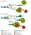
Schematic representation of the consequences of K-RasG12D activation and Trp53 loss in distinct lung epithelial cells. (Upper) Ad5–SPC–Cre virus directs Cre-recombinase activity to SPC-expressing AT2 cells (yellow). Activation of K-RasG12D in these cells (green halo) results in adenoma and adenocarcinoma formation in the alveoli. Cells resident in the bronchiolar lining and BADJ are normal. The additional loss of Trp53 in Ad5–SPC–Cre-infected K-RasLSL-G12D/+;Trp53F/F animals results in moderate to poorly differentiated adenocarcinomas. Well- to moderately differentiated areas of the tumors stain positive for are Nkx2-1, whereas Hmga2-positive tumor cells (orange) are more poorly differentiated. (Lower) In a wild-type lung, Clara cells (blue) lining the bronchiolar and CC10+SPC+ BASC cells (brown) resident at the BADJ are targeted by the Ad5–CC10–Cre virus. Infection of K-RasLSL-G12D/+ mice with Ad5–CC10–Cre results in papillary hyperplasia at the BADJ that involve not only CC10+SPC+ BASCs but also Clara cells. Moreover, some hyperplasic cells lose expression of CC10, but gain expression of the AT2 cell marker, SPC, resulting in SPC+ adenomas in Ad5–CC10–Cre-infected K-RasLSL-G12D/+ animals, and these are generally associated with the BADJ. The additional loss of Trp53 in K-RasLSL-G12D/+;Trp53F/F mice results in adenocarcinomas with pleomorphism and atypia. These tumors express Hmga2 (orange cells) and have the propensity to invade (red arrow).

Comment in

  • Diverse cells at the origin of lung adenocarcinoma.
    Rowbotham SP, Kim CF. Rowbotham SP, et al. Proc Natl Acad Sci U S A. 2014 Apr 1;111(13):4745-6. doi: 10.1073/pnas.1401955111. Epub 2014 Mar 20. Proc Natl Acad Sci U S A. 2014. PMID: 24707043 Free PMC article. No abstract available.

V体育官网入口 - References

    1. Mao C, et al. KRAS mutations and resistance to EGFR-TKIs treatment in patients with non-small cell lung cancer: A meta-analysis of 22 studies. Lung Cancer. 2010;69(3):272–278. - "VSports手机版" PubMed
    1. Giangreco A, Reynolds SD, Stripp BR. Terminal bronchioles harbor a unique airway stem cell population that localizes to the bronchoalveolar duct junction. Am J Pathol. 2002;161(1):173–182. - PMC - PubMed
    1. Dovey JS, Zacharek SJ, Kim CF, Lees JA. Bmi1 is critical for lung tumorigenesis and bronchioalveolar stem cell expansion. Proc Natl Acad Sci USA. 2008;105(33):11857–11862. - PMC - PubMed
    1. Kim CF, et al. Identification of bronchioalveolar stem cells in normal lung and lung cancer. Cell. 2005;121(6):823–835. - PubMed
    1. Ventura JJ, et al. p38alpha MAP kinase is essential in lung stem and progenitor cell proliferation and differentiation. Nat Genet. 2007;39(6):750–758. - PubMed (VSports)

Publication types

MeSH terms