"V体育ios版" Skip to main page content
U.S. flag

An official website of the United States government

Dot gov

The VSports app下载. gov means it’s official. Federal government websites often end in . gov or . mil. Before sharing sensitive information, make sure you’re on a federal government site. .

Https

The site is secure. The https:// ensures that you are connecting to the official website and that any information you provide is encrypted and transmitted securely V体育官网. .

. 2014 Aug 1;210(3):473-82.
doi: 10.1093/infdis/jiu091. Epub 2014 Feb 12.

Increased neutrophil extracellular trap-mediated Staphylococcus aureus clearance through inhibition of nuclease activity by clindamycin and immunoglobulin

Affiliations

Increased neutrophil extracellular trap-mediated Staphylococcus aureus clearance through inhibition of nuclease activity by clindamycin and immunoglobulin

Katrin Schilcher et al. J Infect Dis. .

Abstract

The Gram-positive human pathogen Staphylococcus aureus causes a variety of human diseases such as skin infections, pneumonia, and endocarditis. The micrococcal nuclease Nuc1 is one of the major S VSports手机版. aureus virulence factors and allows the bacterium to avoid neutrophil extracellular trap (NET)-mediated killing. We found that addition of the protein synthesis inhibitor clindamycin to S. aureus LAC cultures decreased nuc1 transcription and subsequently blunted nuclease activity in a molecular beacon-based fluorescence assay. We also observed reduced NET degradation through Nuc1 inhibition translating into increased NET-mediated clearance. Similarly, pooled human immunoglobulin specifically inhibited nuclease activity in a concentration-dependent manner. Inhibition of nuclease activity by clindamycin and immunoglobulin enhanced S. aureus clearance and should be considered in the treatment of S. aureus infections. .

Keywords: NET; Staphylococcus aureus; clindamycin; immunoglobulin; molecular beacon; nuclease. V体育安卓版.


Figures

Figure 1.
Figure 1.
Quantification of Nuc1 activity in Staphylococcus aureus supernatants in the presence or absence of subclindamycin. A, S. aureus cultures grown to postexponential phase in the presence of subclindamycin resulted in a reduction of nuclease activity. DNA cleavage events were measured as increase in fluorescence (AU, arbitrary units). Reduced nuclease activity of LAC wild-type (wt) supernatants in the presence of subclindamycin is indicated by a lower fluorescence signal compared with control without subclindamycin (P < .01 at last measurement time point). Data were pooled from 3 independent experiments, each performed in triplicate (means ± standard errors of the mean) and statistically analyzed with Student t test. B, Representative agarose gel of bacterial culture supernatants of LAC wt and LAC Δnuc1 in the presence or absence of subclindamycin after incubation with calf thymus DNA. Nuclease activity resulted in a smear of degraded DNA; undegraded DNA appeared as a high molecular weight band. Addition of subclindamycin to the LAC wt cultures resulted in less degraded eukaryotic DNA. Abbreviations: ku, Kunitz units; MNase, micrococcal nuclease; CLI, clindamycin.
Figure 2.
Figure 2.
Dose-dependent inhibition of Staphylococcus aureus Nuc1 activity by immunoglobulin (IG). Supernatants of LAC wild-type (wt) and LAC Δnuc1 were preincubated with various amounts of immunoglobulin for 30 minutes at 37°C. A, Nuclease activity in the presence or absence of immunoglobulin was quantified with the molecular beacon–based reporter assay. Immunoglobulin dose-dependent inhibition of Nuc1 is shown from ≥3 independent experiments (means ± standard errors of the mean). Nuclease activity was significantly reduced after preincubation with 25 mg/mL (P < .01) and 10 mg/mL (P < .05) immunoglobulin, compared with control without immunoglobulin. No significant reduction in nuclease activity was observed after preincubation with 2.5 mg/mL immunoglobulin. Statistical analysis using Student t test was performed at the last measurement time point. B, Representative agarose gel of calf thymus DNA after incubation with bacterial culture supernatants of LAC wt and LAC Δnuc1 strains incubated with or without immunoglobulin. In the presence of high concentrations of immunoglobulin (25–10 mg/mL), less DNA degradation was observed, suggesting nuclease inhibition. Abbreviations: AU, arbitrary units; ku, Kunitz units; MNase, micrococcal nuclease.
Figure 3.
Figure 3.
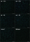
Inhibitory effect of subclindamycin and immunoglobulin (IG) on neutrophil extracellular trap (NET) degradation, which was used as an indicator of nuclease activity. Neutrophils were stimulated with phorbol 12-myristate 13-acetate for NET induction, followed by coincubation with bacterial supernatants. Reduced nuclease activity translated into reduced NET degradation in the presence of subclindamycin or preincubation with immunoglobulin (25 mg/mL). Supernatants of LAC Δnuc1 did not show NET degradation. Micrococcal nuclease (MNase; 4 Kunitz units/mL) was used as a positive control for efficient NET degradation. Representative images are shown. Nuclei were stained with DAPI (blue), and histone-DNA complexes were stained with AlexaFluor 488–labeled antibody (green). Scale bars represent 100 µm. Abbreviations: CLI, clindamycin; wt, wild type.
Figure 4.
Figure 4.
Reduced nuclease activity and enhanced bacterial clearance by neutrophils in the presence of subclindamycin and immunoglobulin. A, In extracellular neutrophil killing experiments, LAC wild-type (wt) showed significantly reduced growth when either treated with subclindamycin or preincubated with 25 mg/mL immunoglobulin, compared with bacteria grown without subclindamycin or immunoglobulin (control). LAC Δnuc1 was not affected by addition of subclindamycin or immunoglobulin. B, In total neutrophil killing experiments, LAC wt and LAC Δnuc1 grown with subclindamycin or preincubated with immunoglobulin showed significantly reduced growth compared with bacteria without subclindamycin or immunoglobulin (control). No significant difference in relative growth was observed comparing LAC wt and LAC Δnuc1. Data are presented as relative bacterial growth compared with LAC wt in the presence of neutrophils and absence of clindamycin. Pooled data from 2 independent blood donors (n = 12 in A upper graph; n = 9 in A lower graph and B upper and lower graph means ± standard errors of the mean), analyzed with the Wilcoxon signed rank test (**P < .01). Abbreviations: IG, immunoglobulin; CLI, clindamycin; NS, not significant.
Figure 5.
Figure 5.
Down-regulation of Nuc1 transcription in response to subclindamycin. Relative transcript levels of nuc1 derived from LAC wild-type (wt) cultures grown to postexponential phase with and without subclindamycin were determined by means of quantitative reverse-transcription polymerase chain reaction. Fold change ratios were calculated by normalizing complementary DNA levels of nuc1 against one of the reference genes (gyrB or rpoD). Data are presented as median fold change of nuc1 gene expression, and error bars indicate interquartile ranges for each data set. Experiments were performed with ≥4 independent samples, using the Mann–Whitney U test; **P < .01. Abbreviation: CLI, clindamycin.
Figure 6.
Figure 6.
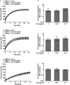
Effect of clindamycin on nuclease activity and nuc1 transcription in clindamycin-resistant methicillin-resistant Staphylococcus aureus (MRSA) isolates. A, Clindamycin-resistant S. aureus cultures (MRSA 1, 2, and 3) grown to postexponential phase with 0.03 and 0.6 µg/mL clindamycin did not result in significant alteration of nuclease activity. DNA cleavage events were measured as increase in fluorescence (AU, arbitrary units). Data were pooled from 3 independent experiments, each performed in triplicate (means ± standard errors of the mean). B, Relative transcript levels of nuc1 derived from MRSA 1, 2, and 3 grown without clindamycin and 0.03 and 0.6 µg/mL clindamycin were determined by means of quantitative reverse-transcription polymerase chain reaction. Fold change ratios were calculated by normalizing complementary DNA levels of nuc1 against one of the reference genes (gyrB or rpoD). Data are presented as median fold change of nuc1 gene expression, and error bars indicate interquartile range for each data set. Experiments were performed with ≥3 independent samples. Abbreviation: CLI, clindamycin.

References

    1. Lowy FD. Staphylococcus aureus infections. N Engl J Med. 1998;339:520–32. - PubMed
    1. Chambers HF, Deleo FR. Waves of resistance: Staphylococcus aureus in the antibiotic era. Nat Rev Microbiol. 2009;7:629–41. - PMC - PubMed
    1. Miller LG, Perdreau-Remington F, Rieg G, et al. Necrotizing fasciitis caused by community-associated methicillin-resistant Staphylococcus aureus in Los Angeles. N Engl J Med. 2005;352:1445–53. - PubMed
    1. Francis JS, Doherty MC, Lopatin U, et al. Severe community-onset pneumonia in healthy adults caused by methicillin-resistant Staphylococcus aureus carrying the Panton-Valentine leukocidin genes. Clin Infect Dis. 2005;40:100–7. - PubMed (V体育官网入口)
    1. Liu C, Bayer A, Cosgrove SE, et al. Clinical practice guidelines by the infectious diseases society of america for the treatment of methicillin-resistant Staphylococcus aureus infections in adults and children. Clin Infect Dis. 2011;52:e18–55. - PubMed

Publication types

MeSH terms