Skip to main page content
U.S. flag

An official website of the United States government

Dot gov

The . gov means it’s official. Federal government websites often end in . gov or . mil. Before sharing sensitive information, make sure you’re on a federal government site VSports app下载. .

Https

The site is secure. The https:// ensures that you are connecting to the official website and that any information you provide is encrypted and transmitted securely. V体育官网.

. 2014 Jan 23;5(1):e1013.
doi: 10.1038/cddis.2013.552.

Discrepant NOXA (PMAIP1) transcript and NOXA protein levels: a potential Achilles' heel in mantle cell lymphoma

Affiliations

Discrepant NOXA (PMAIP1) transcript and NOXA protein levels: a potential Achilles' heel in mantle cell lymphoma

"V体育平台登录" M A Dengler et al. Cell Death Dis. .

Abstract (VSports)

Mantle cell lymphoma (MCL) is an aggressive lymphoid neoplasm with transient response to conventional chemotherapy. We here investigated the role of the Bcl-2 homology domain 3-only protein NOXA for life-death decision in MCL. Surprisingly, NOXA (PMAIP1) mRNA and NOXA protein levels were extremely discrepant in MCL cells: NOXA mRNA was found to be highly expressed whereas NOXA protein levels were low. Chronic active B-cell receptor signaling and to a minor degree cyclin D1 overexpression contributed to high NOXA mRNA expression levels in MCL cells. The phoshatidyl-inositol-3 kinase/AKT/mammalian target of rapamycin pathway was identified as the major downstream signaling pathway involved in the maintenance of NOXA gene expression. Interestingly, MCL cells adapt to this constitutive pro-apoptotic signal by extensive ubiquitination and rapid proteasomal degradation of NOXA protein (T½∼15-30 min). In addition to the proteasome inhibitor Bortezomib, we identified the neddylation inhibitor MLN4924 and the fatty acid synthase inhibitor Orlistat as potent inducers of NOXA protein expression leading to apoptosis in MCL VSports手机版. All inhibitors targeted NOXA protein turnover. In contrast to Bortezomib, MLN4924 and Orlistat interfered with the ubiquitination process of NOXA protein thereby offering new strategies to kill Bortezomib-resistant MCL cells. Our data, therefore, highlight a critical role of NOXA in the balance between life and death in MCL. The discrepancy between NOXA transcript and protein levels is essential for sensitivity of MCL to ubiquitin-proteasome system inhibitors and could therefore provide a druggable Achilles' heel of MCL cells. .


V体育2025版 - Figures

Figure 1
Figure 1
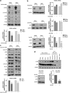
MCL cells express high levels of NOXA (PMAIP1) mRNA but low NOXA protein. (a) Analysis of NOXA gene expression in MCL cell lines Mino and Rec1 compared with embryonal carcinoma cell lines (NTERA2/D1, 2102EP) lung cancer cell lines (A549, NCI-H23, NCI-H460), ovarian cancer cell lines (OVCAR5, SKOV3, A2780) and PHA-stimulated PBMCs of healthy donors using high-throughput real-time PCR BioMark HD System. NOXA mRNA expression was normalized to TBP. Data represent means±S.D. from three technical replicates. (b) Comparison of NOXA mRNA levels in samples from four MCL patients with the MCL cell lines Mino, Jeko1, Rec1, Jvm2 and Granta519 and PBMCs of healthy donors by conventional real-time PCR. NOXA mRNA expression was normalized to GAPDH. Data of MCL cell lines and PBMCs represent means±S.D. from three experiments. (c) MCL cell lines have relatively low constitutive NOXA protein expression. Comparison of NOXA protein expression levels in MCL cell lines Mino and Rec1 compared with embryonal carcinoma cell lines (NTERA2/D1, 2102EP) lung cancer cell lines (A549, NCI-H23, NCI-H460), ovarian cancer cell lines (OVCAR5, SKOV3, A2780) and PHA-stimulated PBMCs of healthy donors. Upper panel: densitometric evaluation of NOXA protein expression analyzed by western blot. NOXA protein expression was normalized to GAPDH. Data reflect means±S.D. from three experiments. Lower panel: representative western blot of NOXA protein expression. (d) Constitutive NOXA protein expression in MCL cell lines Mino, Jeko1, Rec1, Jvm2 and Granta519 compared with primary MCL samples and PHA-stimulated PBMCs of healthy donors analyzed by western blot
Figure 2
Figure 2
BCR and cyclin D1 mediate the high constitutive NOXA mRNA levels via the PI3/AKT/mTOR pathway. (a) Inhibition of BCR signaling causes reduction of constitutive NOXA mRNA expression in MCL cell lines Mino and Rec1. Effect of RNAi-mediated silencing of CD79A on BCR downstream pathways (upper panel) and constitutive expression of NOXA mRNA (lower panel; *P<0.05). Cells were transfected with control siRNA or siRNA targeting CD79A. After 24 h, cells were harvested for western blot analysis and quantification of NOXA mRNA levels. NOXA mRNA expression was normalized to GAPDH. Data represent means±S.D. from three experiments. (b) Impact of inhibition of BCR downstream pathways on constitutive NOXA expression. Upper panel: inhibition of the MAPK pathway has only minor effects on NOXA mRNA levels. Cell lines Mino and Rec1 were treated with 1 μM of PD0325901 for 6 h then harvested for western blot analysis and quantification of NOXA mRNA. Middle panel: effect of p65 knockdown on constitutive NOXA expression in MCL cell lines Mino and Rec1. Cells were transfected with control siRNA or siRNA targeting p65. After 48 h, cells were harvested for western blot analysis and quantification of NOXA mRNA levels. Lower panel: inhibition of the PI3K/AKT/mTOR pathway significantly reduces NOXA mRNA and protein expression. Cell lines Mino and Rec1 were treated with 1 μM of Bez235 for 6 h then harvested for western blot analysis and quantification of NOXA mRNA levels (***P<0.001). NOXA mRNA expression was normalized to GAPDH. All data represent means±S.D. from three experiments. (c) Knockdown of cyclin D1 attenuates PI3K/AKT/mTOR signaling and reduces constitutive NOXA mRNA levels in MCL cell lines Mino and Rec1. Cells were transfected with control siRNA or siRNA targeting cyclin D1 (CCND1). After 16 h, cells were harvested for western blot (upper panel) analysis and quantification of NOXA mRNA levels (lower panel; *P<0.05, ***P<0.001). NOXA mRNA expression was normalized to GAPDH. Data represent means±S.D. from three experiments. (d) Upper panel: comparison of PI3K/AKT/mTOR signaling in MCL cell lines, primary MCL samples and PBMCs of healthy donors. Phosphorylation of PI3K downstream kinases was measured by western blot. Lower panel: effect of PI3K/AKT/mTOR pathway inhibition on constitutive NOXA mRNA expression in MCL patients. Primary MCL cells were treated with 1 μM of Bez235 for 6 h then harvested for western blot analysis and quantification of NOXA mRNA levels (**P<0.01). NOXA mRNA expression was normalized to GAPDH. Data represent means±S.D. from three technical replicates
Figure 3
Figure 3
Rapid UPS-mediated NOXA protein turnover in MCL cells. (a) Half-life of NOXA protein in MCL cell lines (Mino, Rec1) and PHA-stimulated PBMCs of healthy donors. Cells were exposed to 20 μg/ml cycloheximide and half-life of NOXA was determined by western blot. (b) NOXA is polyubiquitinated and degraded by the proteasome. Cells were cultivated in absence or presence of 1 μM MG132 or 5 μM lactacystin for 8 h then harvest and lysed. Total polyubiquitinated proteins were isolated from cell lysates using agarose-TUBE2 beads. Protein expression and polyubiquitination state of NOXA were analyzed by western blot analysis using an NOXA-specific antibody
Figure 4
Figure 4
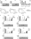
Agents accumulating NOXA protein efficiently induce NOXA-dependent apoptosis in MCL cells. (a) Screening for substances able to induce NOXA protein expression. MCL cell lines Mino and Rec1 were treated with Cisplatin (10 μM), Doxorubicin (100 nM), Bortezomib (10 nM), Orlistat (15 μM) or MLN4924 (0.5 μM) for 16 h and NOXA protein expression was analyzed by western blot. (b) Accumulation of NOXA efficiently induces cell death. MCL cells were treated with Cisplatin (10 μM), Doxorubicin (100 nM), Bortezomib (10 nM), Orlistat (15 μM) or MLN4924 (0.5 μM) for 24 h and cell viability was analyzed by Annexin V staining and flow cytometry. (c) Dose-response curves of MCL cell lines Mino, Jeko1, Rec1, Jvm2 and Granta519 compared with PBMCs and fibroblasts of healthy donors. Cells were treated with increasing concentrations of Bortezomib, Orlistat or MLN4924 and cell viability measured by MTT. (d) NOXA siRNA rescues from Bortezomib, Orlistat and MLN4924 induced apoptosis. Cells were transfected with control siRNA or siRNA targeting NOXA. At 24 h after transfection, cells were treated with Bortezomib (10 nM), Orlistat (15 μM) or MLN4924 (0.5 μM) and further cultivated for 24 h. Cells were then harvested for western blot analysis and quantification of cell death by Annexin V staining and flow cytometry (**P<0.01, ***P<0.001). Data represent means±S.D. from three experiments. (e) Active PI3K/AKT/mTOR signaling is needed for effective accumulation of NOXA protein and induction of cell death upon treatment with Bortezomib, Orlistat or MLN4924. MCL cell lines Mino and Rec1 were pre-treated with 1 μM Bez235 (6 h) before treatment with Bortezomib (10 nM), Orlistat (15 μM) or MLN4924 (0.5 μM). After 16 h, cells were harvested for western blot analysis (lower panel). Quantification of cell death by Annexin V staining and flow cytometry was determined 24 h upon treatment (*P<0.05). Data represent means±S.D. from three experiments
Figure 5
Figure 5
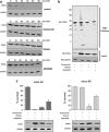
Orlistat and MLN4924 kill Bortezomib-resistant MCL cells by stabilizing NOXA protein in a proteasome-independent manner. (a) Bortezomib, Orlistat and MLN4924 are stabilizing NOXA protein by inhibiting rapid turnover. MCL cell line Mino was treated with Bortezomib (10 nM), Orlistat (15 μM) or MLN4924 (0.5 μM) for 8 h and then exposed to 20 μg/ml cycloheximide and NOXA half-life determined by western blot. (b) Orlistat and MLN4924 stabilize NOXA by inhibiting ubiquitination of NOXA. Cells were cultivated in absence or presence of Bortezomib (10 nM), Orlistat (15 μM) or MLN4924 (0.5 μM) for 8 h then harvest and lysed. Total polyubiquitinated proteins were isolated from cell lysates using agarose-TUBE2 beads. Protein expression and polyubiquitination state of NOXA were analyzed by western blot analysis using an NOXA-specific antibody. (c) Treatment with Orlistat and MLN4924 can kill Bortezomib-resistant MCL cells. Bortetomib-sensitive (Jeko1-BS, left panel) and Bortezomib-resistant (Jeko1-BR) MCL cells were treated for 24 h with Bortezomib (10 nM), Orlistat (15 μM) or MLN4924 (0.5 μM) and cell viability measured by Annexin V staining and flow cytometry (upper panel). Data represent means±S.D. from three experiments. NOXA accumulation was determined after 8 h of treatment by western blot analysis (lower panel)

References

    1. Jares P, Colomer D, Campo E. Genetic and molecular pathogenesis of mantle cell lymphoma: perspectives for new targeted therapeutics. Nat Rev Cancer. 2007;7:750–762. - PubMed
    1. Jares P, Colomer D, Campo E. Molecular pathogenesis of mantle cell lymphoma. J Clin Invest. 2012;122:3416–3423. - PMC (V体育平台登录) - PubMed
    1. Pérez-Galán P, Dreyling M, Wiestner A. Mantle cell lymphoma: biology, pathogenesis, and the molecular basis of treatment in the genomic era. Blood. 2011;117:26–38. - PMC (VSports手机版) - PubMed
    1. Fernàndez V, Hartmann E, Ott G, Campo E, Rosenwald A. Pathogenesis of mantle-cell lymphoma: all oncogenic roads lead to dysregulation of cell cycle and DNA damage response pathways. J Clin Oncol. 2005;23:6364–6369. - PubMed
    1. Young RM, Staudt LM. Targeting pathological B cell receptor signalling in lymphoid malignancies. Nat Rev Drug Discov. 2013;12:229–243. - PMC (VSports) - PubMed

MeSH terms

Substances

LinkOut - more resources (VSports在线直播)