VSports在线直播 - Skip to main page content
U.S. flag

An official website of the United States government

Dot gov

The . gov means it’s official VSports app下载. Federal government websites often end in . gov or . mil. Before sharing sensitive information, make sure you’re on a federal government site. .

Https

The site is secure. The https:// ensures that you are connecting to the official website and that any information you provide is encrypted and transmitted securely. V体育官网.

. 2014 Apr;12(4):504-13.
doi: 10.1158/1541-7786.MCR-13-0489. Epub 2014 Jan 21.

V体育平台登录 - P-cadherin promotes ovarian cancer dissemination through tumor cell aggregation and tumor-peritoneum interactions

Affiliations

P-cadherin promotes ovarian cancer dissemination through tumor cell aggregation and tumor-peritoneum interactions

Akihiro Usui et al. Mol Cancer Res. 2014 Apr.

Abstract

More than 60% of patients who are diagnosed with epithelial ovarian cancer (EOC) present with extensive peritoneal carcinomatosis. EOC cells typically disseminate by shedding into the peritoneal fluid in which they survive as multicellular aggregates and then implant onto peritoneal surfaces. However, the mechanism that facilitates aggregation and implantation of EOC cells is poorly understood VSports手机版. The cell adhesion molecule P-cadherin has been reported to be induced during early progression of EOC and to promote tumor cell migration. In this study, P-cadherin not only promoted migration of EOC cells, but also facilitated the assembly of floating EOC cells into multicellular aggregates and inhibited anoikis in vitro. Furthermore, inhibiting P-cadherin by short hairpin RNAs (shRNA) or a neutralizing antibody prevented EOC cells from attaching to peritoneal mesothelial cells in vitro. In mouse intraperitoneal xenograft models of EOC, inhibition of P-cadherin decreased the aggregation and survival of floating tumor cells in ascites and reduced the number of tumor implants on peritoneal surfaces. These findings indicate that P-cadherin promotes intraperitoneal dissemination of EOC by facilitating tumor cell aggregation and tumor-peritoneum interactions in addition to promoting tumor cell migration. .

Implications: Inhibiting P-cadherin blocks multiple key steps of EOC progression and has therapeutic potential V体育安卓版. .


V体育官网 - Figures

Figure 1
Figure 1
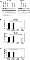
Knockdown of P-cadherin inhibits motility and invasiveness of EOC cells. (A) Western blot of cadherin levels in OVCA429 and SKOV3ip cells stably expressing empty pGIPZ vector, non-targeting shRNA and shRNAs that targeted different sites of CDH3 (sh-PCAD). (B) Tumor cell motility was assayed at 6 h after seeding in uncoated transwell chambers. (C) Tumor cell invasion was assayed at 16 h after seeding in Matrigel-coated transwell chambers. Shown in B and C are average results of 3 independent experiments.
Figure 2
Figure 2
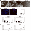
Knockdown of P-cadherin inhibits aggregation of floating EOC cells and increases anoikis. (A-C) Cells of control and sh-PCAD EOC lines were incubated as suspension cultures in polyHEMA-coated plates for 3 days. (A) Morphology of control and sh-PCAD SKOV3ip cells viewed under phase-contrast microscopy. Bar, 100 μm. (B) SKOV3ip cells were stained with Hoechst dye to visualize nuclei and with 7AAD to detect cell death, and viewed under immunofluorescence microscopy. Bar, 50 μm. The percent of 7AAD+ cells was calculated from the number of 7AAD+ cells and total number of cells within a 100x microscopic field. Five random fields were scored per assay. The average total number of cells in one field was 80. (C) Cell death was measured by assaying mono- and oligo- nucleosomes in cell lysates by ELISA. (D) Cell lines were incubated as adherent monolayers on uncoated plates. Cell viability was measured daily by MTT assay. Shown in B, C and D are average results of 3 independent experiments.
Figure 3
Figure 3
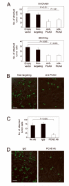
Inhibiting P-cadherin prevents EOC-mesothelial cell interactions. (A) Equivalent numbers of control and sh-PCAD EOC cells that stably expressed GFP were seeded onto confluent monolayers of normal human omental mesothelial cells. At 1 h thereafter, attached EOC cells were viewed under immunofluorescence microscopy and counted in three random 200x microscopic fields per well. Three independent experiments were performed for each assay. (B) Representative examples of attachment assays using control and sh-PCAD OVCA429 cells. Bar, 50 μm. (C) Attachment of OVCA429 control (non-targeting) cells to mesothelial cell monolayers, where mesothelial cell monolayers were pre-incubated with neutralizing P-cadherin Ab, mouse IgG or no Ab prior to seeding of tumor cells. (D) Representative examples of attachment assays where mesothelial cells were pre-incubated with IgG or with P-cadherin Ab. Bar, 50 μm.
Figure 4
Figure 4
Blocking P-cadherin increases anoikis and decreases EOC cell attachment in vivo. (A,B) Female nude mice were injected i.p. with equivalent numbers of GFP-expressing control (non-targeting) SKOV3ip cells together with neutralizing Ab to mouse P-cadherin, control IgG or with no Ab. Mice were sacrificed at 10 days thereafter. (A) Tumor implants that were attached to the peritoneal cavity wall were viewed under a fluorescence stereoscope. Bar, 1 mm. (B) Numbers of tumor implants on the peritoneal cavity wall were counted in five random 25mm2 fields per mouse. (C) GFP-expressing control SKOV3ip cells were pre-incubated with neutralizing Ab to human P-cadherin, control IgG or with no Ab, and then injected i.p. into mice. Mice were sacrificed at 3 days thereafter. Floating cells in the peritoneal cavity were collected and stained with 7AAD. Cell death within the gated population of GFP+ tumor cells was evaluated by flow cytometric analysis of 7AAD staining.
Figure 5
Figure 5
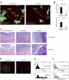
Knockdown of P-cadherin prevents EOC progression in i.p. xenograft models. Female nude mice were inoculated i.p. with equivalent numbers of GFP-expressing control and sh-PCAD SKOV3ip cells and were sacrificed at 3 weeks thereafter (n=5 mice per group). (A) Tumor implants were viewed under a fluorescence stereoscope. (B) I.p. tumor burden was quantified by measuring areas of fluorescence signals in captured images and is expressed as the average percent of area of fluorescence within the abdominal cavity. Fluorescence signals on the peritoneal cavity wall were excluded from analysis. Also shown are volumes of ascites in mice. (C) Representative examples of HE-stained tumor implants on the peritoneal cavity wall and diaphragm. Also shown are omental implants and the adjacent pancreas. Bar, 100 μm. (D) Cells were collected from ascites, stained with Hoechst dye to visualize nuclei (blue) and viewed under immunofluorescence microscopy. GFP-expressing ascitic tumor cells were visualized using a fluorescein filter. Bar, 50 μm. (E,F) Evaluation of cell death within the gated population of GFP+ tumor cells in ascites by flow cytometric analysis of (E) 7AAD staining and (F) staining of active caspase-3.

References

    1. Siegel R, Naishadham D, Jemal A. Cancer statistics, 2013. CA Cancer J Clin. 2013;63:11–30. - PubMed
    1. Naora H, Montell DJ. Ovarian cancer metastasis: Integrating insights from disparate model organisms. Nat Rev Cancer. 2005;5:355–66. - PubMed
    1. Lengyel E. Ovarian cancer development and metastasis. Am J Pathol. 2010;177:1053–64. - PMC - PubMed
    1. Sodek KL, Murphy KJ, Brown TJ, Ringuette MJ. Cell-cell and cell-matrix dynamics in intraperitoneal cancer metastasis. Cancer Metastasis Rev. 2012;31:397–414. - PMC - PubMed
    1. Burleson KM, Casey RC, Skubitz KM, Pambuccian SE, Oegama TR, Skubitz AP. Ovarian carcinoma ascites spheroids adhere to extracellular matrix components and mesothelial cell monolayers. Gynecol Oncol. 2004;93:170–81. - PubMed

Publication types

"V体育官网" MeSH terms

Substances