V体育安卓版 - Skip to main page content
U.S. flag

An official website of the United States government

Dot gov

The . gov means it’s official. Federal government websites often end in . gov or . mil. Before sharing sensitive information, make sure you’re on a federal government site VSports app下载. .

Https

The site is secure. The https:// ensures that you are connecting to the official website and that any information you provide is encrypted and transmitted securely V体育官网. .

. 2014 Feb 13;123(7):1040-50.
doi: 10.1182/blood-2013-08-522698. Epub 2014 Jan 6.

c-Myc inhibition prevents leukemia initiation in mice and impairs the growth of relapsed and induction failure pediatric T-ALL cells

Affiliations

c-Myc inhibition prevents leukemia initiation in mice and impairs the growth of relapsed and induction failure pediatric T-ALL cells

VSports手机版 - Justine E Roderick et al. Blood. .

Abstract

Although prognosis has improved for children with T-cell acute lymphoblastic leukemia (T-ALL), 20% to 30% of patients undergo induction failure (IF) or relapse. Leukemia-initiating cells (LICs) are hypothesized to be resistant to chemotherapy and to mediate relapse. We and others have shown that Notch1 directly regulates c-Myc, a known regulator of quiescence in stem and progenitor populations, leading us to examine whether c-Myc inhibition results in efficient targeting of T-ALL-initiating cells. We demonstrate that c-Myc suppression by small hairpin RNA or pharmacologic approaches prevents leukemia initiation in mice by eliminating LIC activity. Consistent with its anti-LIC activity in mice, treatment with the BET bromodomain BRD4 inhibitor JQ1 reduces C-MYC expression and inhibits the growth of relapsed and IF pediatric T-ALL samples in vitro. These findings demonstrate a critical role for c-Myc in LIC maintenance and provide evidence that MYC inhibition may be an effective therapy for relapsed/IF T-ALL patients. VSports手机版.


Figures

Figure 1
Figure 1
Silencing of c-Myc prolongs survival and reduces LIC frequency in mice transplanted with murine T-ALL cells. (A) Experimental design. Five independent murine Tal1/Lmo2 T-ALL cells were infected with retroviruses encoding an shRNA to c-Myc or Renilla luciferase. Cells were sorted for GFP expression, serially diluted, and transplanted into syngeneic recipients via intraperitoneal injection. Transplanted mice were monitored for evidence of disease. (B) The survival curve for each group of mice was estimated using the Kaplan-Meier method and the difference in overall survival between the 2 groups assessed by the log-rank test (P < .0001). (C) c-Myc silencing reduces LIC frequency in mice transplanted with murine T-ALL cells. A log-log plot and LIC frequency was calculated using ELDA software for ShRenilla (red, 1/7308) and shMyc (black, 1/41 337). A portion of secondary recipients develop leukemia when transplanted with limiting dilutions of shRenilla- and shMyc-infected leukemic cells. (D) Reduced c-Myc protein levels in shMyc-transduced leukemic cells at time of transplant. Protein was isolated from GFP-positive leukemic cells prior to transplant and lysates probed with a c-Myc and Erk1/2 antibodies. (E) Transplanted mice that develop disease do not exhibit reduced Myc expression. RNA was isolated from leukemic cells transduced with the shRen or shMyc retroviruses and c-Myc mRNA levels determined by quantitative real-time PCR. Copy number was normalized to β-actin using the ΔΔCT method. Each point represents c-Myc mRNA levels of leukemic cells isolated from a single mouse.
Figure 2
Figure 2
JQ1 treatment of murine T-ALL cells results in cell-cycle arrest followed by apoptosis. Murine T-ALL cell lines were treated with JQ1 (250 nM) or with the GSI (Compound E, 1 μM), unless otherwise noted. (A) JQ1-induced cell-cycle arrest is evident at 24 hours. Murine T-ALL cell lines were left untreated or treated with JQ1 or Compound E for 24 hours and then stained with propidium iodide followed by flow cytometry. Four mouse T-ALL cell lines were analyzed; 1 representative plot from mouse T-ALL 5109 is shown. (B) JQ1 treatment induces cell-cycle arrest followed by apoptosis. Murine T-ALL cell line 5109 was treated with JQ1 or Compound E for 24, 48, and 96 hours and the percentage of cells in the subG1, G0/G1, and S phases determined. (C) Mouse T-ALL cell lines are more sensitive to treatment with JQ1 than GSI. Four mouse T-ALL cell lines (4673, 5109, 5059, and 6124) were treated with DMSO, JQ1, or the GSI (Compound E) for 96 hours and the total number of viable cells was calculated via trypan blue exclusion assay. The results are averages of 3 independent experiments and error bars represent standard error of the mean (SEM). P < .0001 for all JQ1-treated cell lines, *P < .05, **P < .01, ***P < .001. (D) JQ1 induces apoptosis of mouse T-ALL cells. Four mouse T-ALL cell lines were treated with vehicle, JQ1, or Compound E and the percentage of apoptotic cells determined by Annexin V and 7AAD staining followed by flow cytometry. The results are averages of 3 independent experiments and error bars represent SEM. *P < .05, **P < .01, ***P < .001. (E) Reduced c-Myc mRNA levels in JQ1- and GSI-treated mouse T-ALL cells. RNA was isolated from leukemic cultures treated with vehicle, JQ1, or Compound E for 48 hours and c-Myc expression was analyzed by quantitative real-time PCR. Copy number was normalized to β-actin using the ΔΔCT method. The results are averages of 3 independent experiments and error bars represent SEM. P < .0001 for all JQ1-treated cell lines *P < .05, **P < .01, ***P < .001. (F) c-Myc protein levels were reduced upon treatment with JQ1. Protein was isolated from mouse T-ALL cells treated with DMSO, JQ1, or GSI (Compound E) for 48 hours and c-Myc and β-actin protein levels determined by immunoblotting.
Figure 3
Figure 3
Exogenous c-Myc expression partially rescues cell-cycle arrest and apoptosis of murine T-ALL cells treated with JQ1. Murine T-ALL cell lines were transduced with a MSCV2.2 IRES-GFP retrovirus or with one expressing mouse c-Myc. Murine stem cell virus– or c-Myc–expressing cells were treated with JQ1 (250 nM) or with the GSI (Compound E, 1 μM), unless otherwise noted. (A-B) c-Myc expression rescues JQ1 effects on cell-cycle progression at 24 hours. Murine T-ALL cell lines were left untreated or treated with JQ1 or Compound E for 24 hours and then stained with propidium iodide followed by flow cytometry. Mouse T-ALL cell line 5059 was analyzed; shown are 1 representative histogram overlay of DNA content (A) and collated data for the percentage of cells in S phase (B). The results are averages of 3 independent experiments and error bars represent SEM. *P < .05, **P < .01. (C) Mouse T-ALL cell line 5059 was treated with DMSO, JQ1 or GSI for 10 days and the total number of viable cells was calculated via trypan blue exclusion assay. The results are averages of 3 independent experiments and error bars represent SEM. **P < .01. (D) JQ1 induces apoptosis of mouse T-ALL cells. T-ALL cell lines were treated with vehicle, JQ1, or GSI for 10 days and the percentage of GFP-positive cells were determined by flow cytometry. The results are averages of 3 independent experiments and error bars represent SEM. *P < .05, **P < .001.
Figure 4
Figure 4
JQ1 treatment in vivo reduces c-Myc expression and significantly prolongs survival. (A) Experimental design. Tal1/Lmo2 mouse T-ALLs were transplanted (cell dose 105) into syngeneic recipients and vehicle or JQ1 was administered at 50 mg/kg daily starting at the time of transplant and continuing for 3 consecutive weeks. Mice were monitored for disease and euthanized when they became moribund. (B) Survival was estimated using the Kaplan-Meier method and the difference in overall survival between the 2 groups assessed by the log-rank test. (C) c-Myc inhibition targets the LIC in Tal1/Lmo2 T-ALLs. Leukemic cells were diluted and transplanted into syngeneic mice. JQ1 was administered at 50 mg/kg daily starting at the time of transplant and continuing for 3 consecutive weeks. A log-log plot and LIC frequency was calculated using ELDA software for vehicle, (red, 1/17 970) and JQ1 (black, 1/199 871). A fraction of secondary recipients develop leukemia when transplanted with limiting dilutions of leukemic cells and treated with either vehicle or JQ1. (D) Leukemic mice remain responsive to JQ1 treatment. When disease was evident, a single dose of JQ1 was readministered and RNA isolated from mouse leukemic tissues. c-Myc expression was analyzed by quantitative real-time PCR. Copy number was normalized to β-actin using the ΔΔCT method. Each bar represents a single mouse and error bars represent SEM. **P < .01, ***P < .001, ****P < .0001.
Figure 5
Figure 5
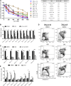
JQ1 treatment impairs growth and induces apoptosis of human T-ALL cell lines. (A) JQ1 treatment inhibits human T-ALL cell line growth. Human T-ALL cell lines were cultured with vehicle or increasing concentrations of JQ1 (100-1000 nM) for 5 days and growth and metabolism assayed by MTS. The absorbance levels were normalized to the vehicle control and the GI50 of each cell line was calculated using Graph Pad Prism 5 software. The results are averages of 3 to 5 independent experiments and error bars represent SEM. (B) JQ1 treatment reduces C-MYC expression in human T-ALL lines. Human T-ALL lines were cultured with DMSO or JQ1 (1 μM) for 24 hours and C-MYC expression levels quantified by quantitative PCR. C-MYC expression was normalized to β-ACTIN and calculated using the ΔΔCT method. The results are averages of 3 independent experiments and error bars represent SEM. ***P < .001 for all JQ1-treated cell lines. (C-D) JQ1 induces apoptosis of human T-ALL lines. (C) KOPTK1 and Jurkat cell lines were treated with DMSO or JQ1 (1 μM) for 96 hours and the apoptotic cells detected by Annexin V and 7AAD staining followed by flow cytometry. A representation fluorescence-activated cell sorter plot is shown. (D) Collated data of Annexin V–positive cells from 7 human T-ALL cell lines treated with vehicle or JQ1 (1 μM) for 96 hours. The results are averages of 3 to 5 independent experiments and error bars represent SEM. *P < .05, **P < .01, ***P < .001.
Figure 6
Figure 6
JQ1 treatment inhibits the growth of pediatric T-ALL cells in vitro. (A-B) Treatment of pediatric T-ALL cells are sensitive to GSI or JQ1 treatment. (A) Diagnostic, relapsed, or IF pediatric T-ALL cells were cultured with vehicle, GSI (1 μM), or increasing concentrations of JQ1 (100-1000 nM) for 5 days and growth and metabolism assayed by MTS. The absorbance levels were normalized to the vehicle control and the GI50 of each cell line was calculated using Graph Pad Prism 5 software. The results are averages of 3 to 5 independent experiments and error bars represent SEM. (B) Absorbance values determined by MTS assay are plotted for each patient sample treated with DMSO, GSI (DBZ, 1 μM), or JQ1 (1 μM). The results are averages of 3 to 5 independent experiments and error bars represent SEM. P < .0001 for all patient samples treated with JQ1; *P < .05, **P < .01, ***P < .001. (C) JQ1 treatment reduces C-MYC expression in diagnostic, relapsed, or IF patient T-ALL samples. Primary leukemic cells from patients were cultured with DMSO, GSI (DBZ, 1 μM), or JQ1 (1 μM) for 24 hours and C-MYC expression levels were measured using quantitative real-time PCR. C-MYC expression was normalized to β-ACTIN and calculated using the ΔΔCT method. The results are averages of 3 independent experiments and error bars represent SEM. P < .001 for all samples treated with JQ1; *P < .05, ***P < .001. (D-E) JQ1 treatment of some diagnostic and relapsed/IF T-ALL samples results in apoptosis. (D) Patient samples TALL-x-13 (relapse) and TALL-x-9 (diagnostic) were treated with DMSO, GSI (DBZ, 1 μM), or JQ1 (1 μM) for 96 hours and apoptotic cells detected by staining with Annexin V and 7AAD followed by flow cytometry. A representative fluorescence-activated cell sorter plot is shown. (E) Collated data of Annexin V–positive cells from relapsed/IF patients treated with DMSO, GSI (DBZ, 1 μM), or JQ1 (1 μM) for 96 hours. The results are averages of 3 independent experiments and error bars represent SEM. *P < .05, ***P < .001.

References

    1. Weng AP, Ferrando AA, Lee W, et al. Activating mutations of NOTCH1 in human T cell acute lymphoblastic leukemia. Science. 2004;306(5694):269–271. - PubMed
    1. O’Neil J, Grim J, Strack P, et al. FBW7 mutations in leukemic cells mediate NOTCH pathway activation and resistance to gamma-secretase inhibitors. J Exp Med. 2007;204(8):1813–1824. - PMC - PubMed
    1. Malyukova A, Dohda T, von der Lehr N, et al. The tumor suppressor gene hCDC4 is frequently mutated in human T-cell acute lymphoblastic leukemia with functional consequences for Notch signaling. Cancer Res. 2007;67(12):5611–5616. - PubMed
    1. Thompson BJ, Buonamici S, Sulis ML, et al. The SCFFBW7 ubiquitin ligase complex as a tumor suppressor in T cell leukemia. J Exp Med. 2007;204(8):1825–1835. - VSports最新版本 - PMC - PubMed
    1. Maser RS, Choudhury B, Campbell PJ, et al. Chromosomally unstable mouse tumours have genomic alterations similar to diverse human cancers. Nature. 2007;447(7147):966–971. - PMC - PubMed

Publication types

MeSH terms (V体育官网)