"V体育ios版" Skip to main page content
U.S. flag

An official website of the United States government

Dot gov

The . gov means it’s official. Federal government websites often end in . gov or VSports app下载. mil. Before sharing sensitive information, make sure you’re on a federal government site. .

Https

The site is secure. The https:// ensures that you are connecting to the official website and that any information you provide is encrypted and transmitted securely V体育官网. .

. 2014;43(3):255-66.
doi: 10.3109/08820139.2013.864667. Epub 2013 Dec 30.

"V体育官网入口" Pro-inflammatory effects of uric acid in the gastrointestinal tract

Affiliations

Pro-inflammatory effects of uric acid in the gastrointestinal tract

John K Crane et al. Immunol Invest. 2014.

V体育ios版 - Abstract

Uric acid can be generated in the gastrointestinal (GI) tract from the breakdown of nucleotides ingested in the diet or from purines released from host cells as a result of pathogen-induced cell damage. Xanthine oxidase (XO) is the enzyme that converts hypoxanthine or xanthine into uric acid, a reaction that also generates hydrogen peroxide. It has been assumed that the product of XO responsible for the pro-inflammatory effects of this enzyme is hydrogen peroxide. Recent literature on uric acid, however, has indicated that uric acid itself may have biological effects. We tested whether uric acid itself has detectable pro-inflammatory effects using an in vivo model using ligated rabbit intestinal segments ("loops") as well as in vitro assays using cultured cells. Addition of exogenous uric acid increased the influx of heterophils into rabbit intestinal loops, as measured by myeloperoxidase activity. In addition, white blood cells adhered avidly to uric acid crystals, forming large aggregates of cells. Uric acid acts as a leukocyte chemoattractant in the GI tract. The role of uric acid in enteric infections and in non-infectious disorders of the GI tract deserves more attention VSports手机版. .


Figures (VSports在线直播)
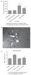
Figure 1
Figure 1
Formation of uric acid in vivo in rabbit intestinal loops in response to EPEC infection. Panel A. Uric acid levels were measured in the fluid that accumulates in the intestinal loops following infection with rabbit EPEC strain E22 at 20 h after infection, with and without 1 μM EHNA, an adenosine deaminase (ADA) inhibitor. Panel B, microscopic evidence for uric acid crystal formation in the unfiltered loop fluid of a rabbit infected with E22 with 35 U/mL ADA. The large group of birefringent crystals was 470 μm in size in the vertical dimension. Panel C, pro-inflammatory effects of exogenous uric acid in vivo in rabbit intestinal loops. No pathogenic bacteria were added in the experiment shown in Panel C, but instead uric acid, uricase, or the combination of the two was added. Loop fluid was analyzed for MPO activity. Panel C is from Gut Microbes 4; 5; 1–4, 2013, http://dx.doi.org/10.4161/gmic.25584, under the provisions of the Creative Commons Attribution License.
Figure 2
Figure 2
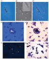
Interactions between uric acid crystals and HL-60 leukemia cells. Uric acid was generated enzymatically by adding 400 μM hypoxanthine and 0.5 U/mL XO to suspensions of HL-60 cells and incubating at 37 °C in 5% CO2 for 1.5 h. Then the HL-60 cells were examined using a wet mount preparation and cover slip (Panels A-C) or by concentration in the Cytospin centrifuge followed by staining (Panels D-G). Panel A, polarization microscopy showing a birefringent crystal 165 μm in length. Panel B, same field examined by phase contrast, showing two cells associated with the crystal. Panel B, Inset, higher power view showing blebbing of the cell surface of the two adherent HL-60 cells. Panel C, merged image of Panels A and B. Panels D and E show the same field examined using two different methods of illumination; polarization in Panel D and brightfield in Panel E, showing tight clustering of HL-60 cells around the uric acid crystal. Panels F and G again show a pair of images of the same microscope field, with Panel F showing at least 5 birefringent crystals. On brightfield examination, all of the crystals are covered by a tight layer of adhering cells. Note that only the largest crystal (center) is visible by brightfield exam alone.
Figure 3
Figure 3
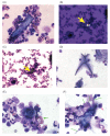
Interactions between uric acid crystals and rabbit peripheral blood leukocytes (PBLs). Uric acid was again generated by addition of 400 μM hypoxanthine +0.5 U/mL XO to suspensions of freshly isolated PBLs for 1.5 h, followed by Cytospin centrifugation and staining. In some conditions (Panels B and C), 600 U/mL catalase was added to break down hydrogen peroxide, while in others 2 U/mL uricase was added to remove uric acid by conversion to allantoin. Panel A, large cluster of PBLs surrounding a uric acid crystal 85 μm in length; original magnification, 400×. Panels B and C, a pair of images showing a uric acid crystal (Panel B, yellow arrow) completely surrounded by a large aggregate of PBLs (Panel C), where the yellow arrow again indicates the location of the crystal, which appears as a non-descript piece of debris on brightfield examination. Panel D, addition of uricase abolished the formation of crystals and also the aggregates of PBLs. Panel E, heterophil adhering to a uric acid crystal and showing release of its secretory granules (green arrow). Panel F, PBLs adhering to a uric acid crystals show elongation (green arrows), and a mononuclear cell shows signs of apoptosis (red arrow); original magnification of Panels E and F, 600 X.

References

    1. Harrison R. Milk xanthine oxidase: Properties and physiological roles. Inter Dairy J. 2006;16:546–54.
    1. Martin H, Hancock J, Salisbury V, et al. Role of xanthine oxidoreductase as an antimicrobial agent. Infect Immun. 2004;72:4933–9. - PMC - PubMed
    1. Hancock J, Salisbury V, Ovejero-Boglione M, et al. Antimicrobial properties of milk: Dependence on presence of xanthine oxidase and nitrite. Antimicrobial Agents Chemother. 2002;46:3308–10. - PMC - PubMed
    1. Kotloff KL, Nataro JP, Blackwelder WC, et al. Burden and aetiology of diarrhoeal disease in infants and young children in developing countries (the Global Enteric Multicenter Study, GEMS): A prospective, case-control study. Lancet. 2013;382:209–22. - PubMed
    1. Frank C, Werber D, Cramer JP, et al. Epidemic profile of shiga-toxin-producing Escherichia coli O104:H4 Outbreak in Germany. New Engl J Med. 2011;365:1771–80. - PubMed

Publication types

MeSH terms

"V体育安卓版" LinkOut - more resources