Skip to main page content
U.S. flag

An official website of the United States government

Dot gov

The . gov means it’s official. Federal government websites often end in . gov or . mil. Before sharing sensitive information, make sure you’re on a federal government site VSports app下载. .

Https

The site is secure. The https:// ensures that you are connecting to the official website and that any information you provide is encrypted and transmitted securely. V体育官网.

. 2013 Dec 12;39(6):1019-31.
doi: 10.1016/j.immuni.2013.10.019.

Cyclic GMP-AMP synthase is activated by double-stranded DNA-induced oligomerization

Affiliations

Cyclic GMP-AMP synthase is activated by double-stranded DNA-induced oligomerization

Xin Li et al. Immunity. .

Abstract

Cyclic GMP-AMP synthase (cGAS) is a cytosolic DNA sensor mediating innate antimicrobial immunity VSports手机版. It catalyzes the synthesis of a noncanonical cyclic dinucleotide, 2',5' cGAMP, that binds to STING and mediates the activation of TBK1 and IRF-3. Activated IRF-3 translocates to the nucleus and initiates the transcription of the IFN-β gene. The structure of mouse cGAS bound to an 18 bp dsDNA revealed that cGAS interacts with dsDNA through two binding sites, forming a 2:2 complex. Enzyme assays and IFN-β reporter assays of cGAS mutants demonstrated that interactions at both DNA binding sites are essential for cGAS activation. Mutagenesis and DNA binding studies showed that the two sites bind dsDNA cooperatively and that site B plays a critical role in DNA binding. The structure of mouse cGAS bound to dsDNA and 2',5' cGAMP provided insight into the catalytic mechanism of cGAS. These results demonstrated that cGAS is activated by dsDNA-induced oligomerization. .


"VSports最新版本" Figures

Figure 1
Figure 1
cGAS is activated by dsDNA and catalyzes the synthesis of 2′,5′ cGAMP, a high affinity ligand for STING. (A). cGAS activity assay by ion exchange chromatography. The reaction product was first purified by ultrafiltration and then analyzed using a MonoQ 5/50 GL column. Salmon sperm DNA at 0.2 mg/ml was used to stimulate mcGAS catalytic domain at 10 μM concentration. (B). The catalytic activity of cGAS is dependent on the length of dsDNA. Reaction products from mcGAS stimulated with different dsDNA were analyzed by ion exchange chromatography. The enzyme and DNA concentrations used in these assays are 10 and 50 μM, respectively (C). Binding study of mcGAS catalytic domain with a 20 bp dsDNA by isothermal titration calorimetry (ITC). (D). Binding study of 2′,5′ cGAMP with human STING ligand binding domain by ITC. (E). 2′,5′ cGAMP stimulates the secretion of IFN-β in human THP-1 cells. The concentration of IFN-β in the culture supernatant was analyzed by ELISA at 4 and 8 hours post stimulation. 3′,5′ cGAMP was used as a control. The concentration of 2′,5′ or 3′,5′ cGAMP used in these assays is 25 μg/ml. The error bars represent the standard deviations of signals from four independent measurements. See also Figure S1.
Figure 2
Figure 2
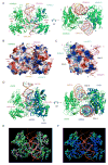
Structure of human cGAS catalytic domain. (A). Ribbon representation of the structure of hcGAS catalytic domain. The Zn2+ ion is shown by the red sphere. (B). Structure of the hcGAS dimer in the crystallographic asymmetric unit. (C). Comparison of ligand-free human (green, PDB 4LEV), mouse (blue, PDB 4K8V), and porcine (salmon, PDB 4JLX) cGAS dimer structures. (D). Superposition of ligand-free human, mouse, and porcine cGAS dimers rainbow-colored according to B-factors of the Cα atoms. Atoms in red have higher B-factors and atoms in blue have lower B-factors.
Figure 3
Figure 3
The structural basis of cGAS activation by dsDNA. (A). Crystal structure of mcGAS catalytic domain bound to an 18 bp dsDNA. Ribbon representations of the 2:2 mcGAS:dsDNA complex structure in two different orientations. (B). Electrostatic surface potential of the mcGAS dimer in the mcGAS:dsDNA complex. Positively charged surface is colored blue, and negatively charged surface red. The two dsDNA bound to the cGAS dimer are shown by ribbon representations. (C). Electrostatic surface potential at the DNA binding sites of a mcGAS monomer. The two dsDNA bound to mcGAS are shown by the cyan and purple stick models. (D). Comparison of the 2:2 mcGAS:dsDNA complex structure with the 1:1 OAS1:dsRNA complex structure (PDB, 4IG8). (E). Superposition of four cGAS:dsDNA complex structures. Structure of mcGAS bound an 18 bp dsDNA (this work, PDB 4LEY) is in green; mcGAS bound to a 16 bp dsDNA (PDB, 4K96) in yellow; mcGAS bound to a 16 bp dsDNA and pppG(2′,5′)pG (PDB, 4K98) in magenta; porcine cGAS bound to a 14 bp dsDNA (PDB, 4KB6) in cyan. The dsDNA bound to cGAS are shown by the orange ribbons. (F). Superposition of three mcGAS:dsDNA complex structures (PDB, 4LEY, 4K96, and 4K98). The proteins are rainbow-colored according to B-factors of the Cα atoms. Atoms in red have higher B-factors and atoms in blue have lower B-factors. See also Figures S2.
Figure 4
Figure 4
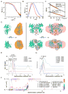
DNA binding by cGAS induces higher-order complex formation in solution (A). Small angle X-ray scattering (SAXS) data of mcGAS catalytic domain (red) and its complex with a 20 bp dsDNA (blue). (B). Normalized pair distance distribution function (PDDF) for mcGAS alone (red) and mcGAS:DNA complex (blue). (C). Guinier fits (red lines) of SAXS data for mcGAS alone (open cycles) and its complex with DNA (open squares). The SAXS data were normalized by the mass concentration of the samples. The fitted q ranges were limited to qmax*Rg < 1.3. Io is the forward scattering, i.e., scattering intensity at q=0. Cmass is the concentration of the samples in mg/ml. (D). Overlays of the crystal structures (cartoon) and SAXS molecular envelope (mesh) for mcGAS in isolation (left) and mcGAS:DNA complex (right). (E). Sedimentation velocity AUC analysis of wt and K382A mutant of mcGAS in the absence and presence of a 20 bp dsDNA, with experimentally determined mass estimates listed. The K382A dimer interface mutant formed a 1:1 complex with dsDNA but did not oligomerize. (F). Size-and-shape distribution analysis of the wt mcGAS:dsDNA sample reveals a complicated mixture of compact and elongated oligomeric species. Putative oligomeric species are identified based on experimentally determined molecular weights in Table S2. (G). Normalized sedimentation velocity AUC analysis of site A (K372E and R158E) and site B (R342E and K335E) mcGAS mutants in the presence of a 20 bp dsDNA. The wt mcGAS distribution is shown for comparison.
Figure 5
Figure 5
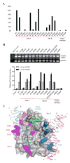
Mutations at the DNA binding sites and the dimer interface affect cGAS activity. (A). Enzyme activities of mcGAS mutants. The activities of wild-type and mutant mcGAS were analyzed by ion exchange chromatography. The activities of the mutants relative to the wild-type enzyme were calculated based on the peak areas of 2′,5′ cGAMP in the chromatograms. (B). IFN-β luciferase reporter assays in HEK 293T cells transfected with mouse STING and cGAS. Error bars represent the standard deviations of signals from three independent transfections. The upper panel shows the mRNA levels of mcGAS and its mutants in the transfected cells. The mRNA levels were determined after 20 cycles of RT-PCR amplification. GAPDH mRNA was amplified as an internal control. (C). Locations of the mutated residues on the surface of mcGAS and effects of these mutations on cGAS activity. The two dsDNAs bound to cGAS are shown by the cyan and purple stick models. Residues mutated at site A are colored magenta and those in site B are colored blue. Residues at the dimer interface are colored green. Mutations that caused more than 80% loss of enzyme activity (in Panel A) are underlined. See also Figures S3.
Figure 6
Figure 6
cGAS binds dsDNA cooperatively through two binding sites. (A). DNA binding study of wt mcGAS catalytic domain by electrophoretic mobility shift assay (EMSA). The concentration of the 45 bp interferon stimulatory DNA (ISD) used in this study is 1 μM and the protein concentrations are from 5 to 80 μM. (B to G). DNA binding studies of site A mutants of mcGAS. (H to K). DNA binding studies of site B mutants. (L and M). DNA binding studies of mutants at the dimer interface. (N). DNA binding study of 18 mcGAS mutants together on one gel. The DNA concentration used in this study is 1 μM and mcGAS is at 20 μM. See also Figures S4.
Figure 7
Figure 7
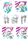
Structure of mcGAS bound to dsDNA and 2′,5′ cGAMP. (A). Ribbon representation of mcGAS bound to an 18 bp dsDNA and 2′,5′ cGAMP. The dinucleotide is shown by the yellow ball-and-stick model. Catalytic residues and residues that interact with 2′,5′ cGAMP are shown by the gray ball-and-stick models. For clarity, the two dsDNA bound to mcGAS are not shown. (B). Close-up view of 2′,5′ cGAMP bound to mcGAS. (C). Difference maps of 2′,5′ cGAMP bound to mcGAS in three different orientations. The σA weighted Fo-Fc map calculated without the ligand is contoured at 3.5σ. 2′,5′ cGAMP is shown by the yellow ball-and-stick model. (D). Comparison of structures of pppG(2′,5′)pG (blue sticks) and 2′,5′ cGAMP (yellow sticks) bound to mcGAS. The figure was generated by superposition of mcGAS bound to pppG(2′,5′)pG (PDB, 4K98) onto the complex structure of mcGAS bound to 2′,5′ cGAMP (this work, PDB 4LEZ). The structure of mcGAS determined in this work is shown by the cyan and purple ribbons. Residues at the active site of mcGAS are shown by the gray stick models. (E). Comparison of structures of 2′,5′ cGAMP bound to mcGAS in two different orientations. This figure was generated by superposition of a previously determined cGAS:DNA:2′,5′ cGAMP complex structure (PDB, 4K9B) onto the complex structure from this work (PDB, 4LEZ). See also Figure S5.

Comment in

References

    1. Ablasser A, Goldeck M, Cavlar T, Deimling T, Witte G, Rohl I, Hopfner KP, Ludwig J, Hornung V. cGAS produces a 2′-5′-linked cyclic dinucleotide second messenger that activates STING. Nature. 2013;498:380–384. - PMC - PubMed
    1. Adams PD, Afonine PV, Bunkoczi G, Chen VB, Davis IW, Echols N, Headd JJ, Hung LW, Kapral GJ, Grosse-Kunstleve RW, et al. PHENIX: a comprehensive Python-based system for macromolecular structure solution. Acta Crystallogr D Biol Crystallogr. 2010;66:213–221. - VSports - PMC - PubMed
    1. Barber GN. Innate immune DNA sensing pathways: STING, AIMII and the regulation of interferon production and inflammatory responses. Curr Opin Immunol. 2011a;23:10–20. - "V体育ios版" PMC - PubMed
    1. Barber GN. STING-dependent signaling. Nat Immunol. 2011b;12:929–930. - PubMed
    1. Burdette DL, Monroe KM, Sotelo-Troha K, Iwig JS, Eckert B, Hyodo M, Hayakawa Y, Vance RE. STING is a direct innate immune sensor of cyclic di-GMP. Nature. 2011;478:515–518. - PMC - PubMed

Publication types

Substances (V体育官网)

Associated data