V体育2025版 - Skip to main page content
U.S. flag

An official website of the United States government

Dot gov

The . gov means it’s official. Federal government websites often end in . gov or . mil. Before sharing sensitive information, make sure you’re on a federal government site VSports app下载. .

Https

The site is secure. The https:// ensures that you are connecting to the official website and that any information you provide is encrypted and transmitted securely. V体育官网.

. 2014 Jan 10;289(2):1142-50.
doi: 10.1074/jbc.M113.515080. Epub 2013 Nov 21.

3,4-methylenedioxy-β-nitrostyrene inhibits NLRP3 inflammasome activation by blocking assembly of the inflammasome

Affiliations

V体育ios版 - 3,4-methylenedioxy-β-nitrostyrene inhibits NLRP3 inflammasome activation by blocking assembly of the inflammasome

Yuan He et al. J Biol Chem. .

Abstract

The NLRP3 inflammasome is a critical component of the innate immune system VSports手机版. NLRP3 activation is induced by diverse stimuli associated with bacterial infection or tissue damage, but its inappropriate activation is involved in the pathogenesis of inherited and acquired inflammatory diseases. However, the mechanism by which NLRP3 is activated remains poorly understood. In this study, we explored the role of kinases in NLRP3 inflammasome activation by screening a kinase inhibitor library and identified 3,4-methylenedioxy-β-nitrostyrene (MNS) as an inhibitor for NLRP3 inflammasome activation. Notably, MNS did not affect the activation of the NLRC4 or AIM2 (absent in melanoma 2) inflammasome. Mechanistically, MNS specifically prevented NLRP3-mediated ASC speck formation and oligomerization without blocking potassium efflux induced by NLRP3 agonists. Surprisingly, Syk kinase, the reported target of MNS, did not mediate the inhibitory activity of MNS on NLRP3 inflammasome activation. We also found that the nitrovinyl group of MNS is essential for the inhibitory activity of MNS. Immunoprecipitation, mass spectrometry, and mutation studies suggest that both the nucleotide binding oligomerization domain and the leucine-rich repeat domain of NLRP3 were the intracellular targets of MNS. Administration of MNS also inhibited NLRP3 ATPase activity in vitro, suggesting that MNS blocks the NLRP3 inflammasome by directly targeting NLRP3 or NLRP3-associated complexes. These studies identified a novel chemical probe for studying the molecular mechanism of NLRP3 inflammasome activation which may advance the development of novel strategies to treat diseases associated with abnormal activation of NLRP3 inflammasome. .

Keywords: Caspase; Inflammation; Innate Immunity; Macrophages; NOD-like Receptor (NLR) V体育安卓版. .


"VSports在线直播" Figures
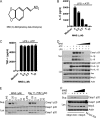
FIGURE 1.
FIGURE 1.
Identification of MNS as a potent inhibitor for ATP-induced NLRP3 inflammasome activation. A, chemical structure of MNS is shown. B and C, BMDMs were primed with LPS (100 ng/ml) for 4 h and then treated with indicated doses of MNS for 15 min before ATP stimulation, or left untreated. The culture supernatants were analyzed for IL-1β (B) or TNF-α (C) by ELISA. D, BMDMs were stimulated by ATP in the absence or presence of MNS (0.5, 1, 2.5, 5, 10 μm). Culture supernatants (Sup.) and cell extracts (Lys.) were immunoblotted for caspase-1, IL-1β, IL-18, Asc, or Nlrp3. Actin was a loading control. E, LPS-primed BMDMs were left unstimulated or stimulated with 5 mm ATP for 30 min in the presence of indicated concentrations of inhibitors. Culture supernatants and cell extracts were immunoblotted for caspase-1. F, immortalized Nlrp3 KO macrophages with reconstitution of Nlrp3 were stimulated with 5 mm ATP for 30 min in the absence or presence of different concentrations of MNS (1.25, 2.5, 5, 10 μm). After stimulation, culture supernatant and cell lysate were immunoblotted for caspase-1 and Nlrp3. Actin was a loading control. Data are representative of three independent experiments. Bar graphs shown are the mean ± S.D. (error bars) of triplicate wells.
FIGURE 2.
FIGURE 2.
MNS specifically inhibits the NLRP3 inflammasome. A and B, LPS-primed BMDMs were treated with indicated doses of MNS for 15 min and then stimulated by 10 μm nigericin for 1 h (A) or 500 μg/ml silica for 4 h (B). The culture supernatants were analyzed for IL-1β. C, BMDMs were primed with LPS and then stimulated with nigericin (1 h) or silica (4 h) in the absence or presence of 5 μm MNS. DMSO was used as a vehicle control. Culture supernatants (Sup.) and cell extracts (Lys.) were immunoblotted for caspase-1. Actin was a loading control. UT, untreated. D, primed BMDMs were infected with S. enterica sv. typhimurium (S. typhi) (m.o.i. = 10, 1 h) or stimulated by ATP (5 mm, 30 min) in the absence or presence of 5 μm MNS. Culture supernatants and cell extracts were immunoblotted for caspase-1. Actin was a loading control. E, primed BMDMs were transfected with 2 μg/ml poly(dA·dT) (4 h) or stimulated with 5 mm ATP (30 min) in the absence or presence of 5 μm MNS. Culture supernatants and cell extracts were immunoblotted for caspase-1. Actin was a loading control. F, cytotoxicity (lactate dehydrogenase (LDH release)) was measured from cells stimulated with ATP (5 mm, 30 min) or nigericin (10 μm, 1 h), infected with S. typhi (m.o.i. = 10, 1h) or transfected with poly(dA·dT) (2 μg/ml, 4 h) in the absence or presence of 5 μm MNS. Data are representative of three independent experiments. Bar graphs shown represent the mean ± S.D. (error bars) of triplicate wells. *, p < 0.05.
FIGURE 3.
FIGURE 3.
Syk kinase is dispensable for NLRP3 inflammasome activation. A, BMDMs from WT or Syk−/− mice were primed with 100 ng/ml LPS for 4 h and then stimulated with 5 mm ATP (30 min), 10 μm nigericin (1 h), or 500 μg/ml silica (4 h). B, culture supernatants (Sup.) and cell extracts (Lys.) were immunoblotted for caspase-1, IL-1β, IL-18, or Syk. Actin was a loading control. C–E, the culture supernatants were also analyzed for IL-1β (C), IL-18 (D), or TNF-α (E) by ELISA. Data are representative of three independent experiments. Bar graphs shown represent the mean ± S.D. (error bars) of triplicate wells. *, p < 0.05.
FIGURE 4.
FIGURE 4.
MNS inhibits NLRP3-mediated ASC speck formation without blocking potassium efflux. A, LPS-primed BMDMs were treated with DMSO or MNS (10 μm) for 15 min before ATP stimulation. The intracellular potassium was measured and expressed as the percentage of intracellular potassium in cells without ATP stimulation. B, LPS-primed BMDMs were stimulated with ATP (5 mm, 30 min) or nigericin (10 μm, 1 h), infected with S. enterica sv. typhimurium (S. typhi.) (m.o.i. = 10, 1 h), or transfected with poly(dA·dT) (2 μg/ml, 4h) in the absence or presence of 5 μm MNS. Cells were fixed, permeabilized, and stained for Asc (green). C, the percentage of cells containing ASC speck was quantified from three different view fields. D, LPS-primed BMDMs were untreated or pretreated with 5 μm MNS or 50 μm YVAD for 15 min. Cells were then stimulated with ATP (5 mm, 30 min) or nigericin (10 μm, 1 h), or infected with S. typhi. (m.o.i. = 10, 1 h). Macrophages were extracted with PBS buffer containing 0.5% Triton X-100, and the Triton-insoluble pellets were cross-linked with disuccinimidyl suberate. The cell lysates were immunoblotted for caspase-1. The cross-linked pellets were immunoblotted for Asc. Data are representative of three independent experiments. Bar graphs shown represent the mean ± S.D. (error bars) of triplicate wells. *, p < 0.05.
FIGURE 5.
FIGURE 5.
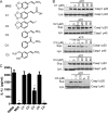
Structure-activity relationship of MNS analogues. A, chemical structures of MNS analogues. B, inhibitory activities of MNS analogues on NLRP3 inflammasome activation. LPS-primed BMDMs were pretreated with the indicated doses of chemical compounds for 15 min before ATP stimulation. The culture supernatants (Sup.) and cell lysates (Lys.) were collected and immunoblotted for caspase-1. C, LPS-primed BMDMs pretreated with DMSO or MNS derivatives (10 μm) for 15 min before ATP stimulation. IL-1β from culture supernatant was analyzed by ELISA. Data are representative of three independent experiments. Bar graphs shown represent the mean ± S.D. (error bars) of triplicate wells. *, p < 0.05.
FIGURE 6.
FIGURE 6.
MNS binds to NLRP3 and inhibits its ATPase activity. A, chemical structure of biotin-HMNS is shown. B, LPS-primed BMDMs were stimulated with 5 mm ATP for 30 min in the absence or presence of the indicated concentrations of biotin-HMNS. Culture supernatant (Sup.) and cell lysate (Lys.) were immunoblotted for caspase-1. C, NLRP3 was pulled down by biotin-HMNS from cell lysates. Lysates from LPS-primed BMDMs were incubated with biotin (1 μm) or biotin-HMNS (1 μm) at 4 °C for 1 h. Bound proteins were pulled down by streptavidin beads and fractioned on a SDS-polyacrylamide gel. The identity of bound proteins was determined by mass spectrometry. D, biotin-HMNS bound to NLRP3 in cells. BMDMs were pretreated (IP) with or without biotin-HMNS (10 μm) for 30 min and lysed in 0.5% Nonidet P-40 TBS buffer. The bound proteins in the lysate were pulled down by streptavidin beads. The cell lysates and bound proteins were immunoblotted for NLRP3 and NLRC4. GAPDH was used as a loading control. E, recombinant human NLRP3 was bound by biotin-HMNS. Recombinant human NLRP3 was incubated with biotin (1 μm) or biotin-HMNS (1 μm) in the presence or absence of free compound HMNS, 100 μm) at 4 °C for 1 h. The bound NLRP3 was pulled down by streptavidin beads and detected by Western blotting. F and G, HEK293T cells were transfected with FLAG-tagged full-length NLRP3 (FL), pyrin deletion mutant (ΔPyrin), LRR deletion mutant (ΔLRR), pyrin domain, NOD domain, or LRR domain for 16 h. Cells were treated with 10 μm biotin-HMNS for 30 min before lysis. The bound proteins were pulled down by streptavidin beads. The cell lysates and bound proteins were immunoblotted (IB) by using anti-FLAG antibody. Actin was a loading control. H, MNS inhibited NLRP3 ATPase activity. The release of NLRP3-hydrolyzed inorganic phosphate in the presence or absence of MNS was determined using a Pi ColorLock Gold phosphate detection system. The data were subtracted from the results of experiments without addition of NLRP3 and expressed as the percentage of control. I, lysates from LPS-primed BMDMs were incubated with biotin-HMNS (1 μm) in the presence of Bay 11-7082 (100 μm) or l-cysteine (100 μm) at 4 °C for 1 h. Bound proteins were pulled down by streptavidin beads and immunoblotted for NLRP3. The lysate blot was shown as an input control. Data are representative of two or three independent experiments.

References

    1. Dinarello C. A. (2009) Immunological and inflammatory functions of the interleukin-1 family. Annu. Rev. Immunol. 27, 519–550 - "VSports app下载" PubMed
    1. Martinon F., Mayor A., Tschopp J. (2009) The inflammasomes: guardians of the body. Annu. Rev. Immunol. 27, 229–265 - PubMed
    1. Franchi L., Muñoz-Planillo R., Núñez G. (2012) Sensing and reacting to microbes through the inflammasomes. Nat. Immunol. 13, 325–332 - "VSports最新版本" PMC - PubMed
    1. Schroder K., Tschopp J. (2010) The inflammasomes. Cell 140, 821–832 - PubMed
    1. Hornung V., Bauernfeind F., Halle A., Samstad E. O., Kono H., Rock K. L., Fitzgerald K. A., Latz E. (2008) Silica crystals and aluminum salts activate the NALP3 inflammasome through phagosomal destabilization. Nat. Immunol. 9, 847–856 - PMC - PubMed

Publication types

MeSH terms

Substances