Skip to main page content
U.S. flag

An official website of the United States government

Dot gov

The . gov means it’s official. Federal government websites often end in . gov or . mil. Before sharing sensitive information, make sure you’re on a federal government site VSports app下载. .

Https

The site is secure. The https:// ensures that you are connecting to the official website and that any information you provide is encrypted and transmitted securely V体育官网. .

. 2014 Jan;25(1):169-83.
doi: 10.1091/mbc.E13-09-0558. Epub 2013 Oct 30.

"V体育官网入口" NEAT1 long noncoding RNA regulates transcription via protein sequestration within subnuclear bodies

Affiliations

V体育ios版 - NEAT1 long noncoding RNA regulates transcription via protein sequestration within subnuclear bodies

V体育官网 - Tetsuro Hirose et al. Mol Biol Cell. 2014 Jan.

Abstract

Paraspeckles are subnuclear structures formed around nuclear paraspeckle assembly transcript 1 (NEAT1)/MENε/β long noncoding RNA (lncRNA). Here we show that paraspeckles become dramatically enlarged after proteasome inhibition. This enlargement is mainly caused by NEAT1 transcriptional up-regulation rather than accumulation of undegraded paraspeckle proteins. Of interest, however, using immuno-electron microscopy, we find that key paraspeckle proteins become effectively depleted from the nucleoplasm by 50% when paraspeckle assembly is enhanced, suggesting a sequestration mechanism. We also perform microarrays from NEAT1-knockdown cells and find that NEAT1 represses transcription of several genes, including the RNA-specific adenosine deaminase B2 (ADARB2) gene. In contrast, the NEAT1-binding paraspeckle protein splicing factor proline/glutamine-rich (SFPQ) is required for ADARB2 transcription. This leads us to hypothesize that ADARB2 expression is controlled by NEAT1-dependent sequestration of SFPQ. Accordingly, we find that ADARB2 expression is strongly reduced upon enhanced SFPQ sequestration by proteasome inhibition, with concomitant reduction in SFPQ binding to the ADARB2 promoter VSports手机版. Finally, NEAT1(-/-) fibroblasts are more sensitive to proteasome inhibition, which triggers cell death, suggesting that paraspeckles/NEAT1 attenuates the cell death pathway. These data further confirm that paraspeckles are stress-responsive nuclear bodies and provide a model in which induced NEAT1 controls target gene transcription by protein sequestration into paraspeckles. .


"VSports app下载" Figures
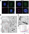
FIGURE 1:
FIGURE 1:
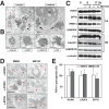
Proteasome inhibition results in paraspeckle elongation. (A) Paraspeckle enlargement upon proteasome inhibition. HeLa cells were treated with DMSO or 5 μM MG132 for 17 h. The paraspeckles were detected by RNA-FISH of NEAT1 lncRNAs (magenta) combined with immunofluorescence of PSPC1 (green). Scale bar, 10 μm. (B) Paraspeckle ultrastructure upon proteasome inhibition. HeLa cells treated with DMSO, 5 μM MG132, or 100 nM bortezomib for 17 h were analyzed by transmission EM after Epon embedding. Left, large fields; bar, 2 μm. Right, enlargement of dashed rectangles; bar, 0.5 μm. Red arrows, paraspeckle clusters; IG, interchromatin granule. (C) Size measurements of paraspeckle in control and MG132-treated HeLa cells (5 μM, 17 h). Short and long axes (Sx and Lx) were defined for 120 paraspeckles in each sample from Epon-embedded HeLa cells. Bar, 0.5 μm. (D) Dimensions were plotted by increasing Lx values, showing similar Sx values in both samples, whereas Lx values were augmented in the MG132-treated cells.
FIGURE 2:
FIGURE 2:
Proteasome inhibition does not result in accumulation of ubiquitinated proteins or reorganization of protein components within paraspeckles. (A) I-EM detection of ubiquitinated and SUMOylated proteins accumulating upon proteasome inhibition (thin sections of Lowicryl-embedded HeLa cells, MG132-treated 5 μM, 17 h). Left, dense aggregates (black arrow) of nuclear ubiquitinated proteins are formed in the vicinity of the nucleolus (No). In contrast, the expanded paraspeckles (red arrows) do not contain significant amount of ubiquitin. Right, SUMOylated proteins also accumulate in the ubiquitin-positive nuclear aggregates. Bars, 0.5 μm. (B) In some nuclear sections, MG132-induced protein aggregates form well-defined nuclear bodies, highly enriched at their periphery in ubiquitin- and SUMO-conjugated proteins and surrounded by a thin layer of PML protein. Bars, 0.5 μm. (C) The PSP levels were largely constant upon MG132 treatment. Seven essential PSPs (defined as those proteins whose knockdown in HeLa cells result in loss of paraspeckles) were detected in cells treated with DMSO or MG132 for 6 or 17 h by Western blotting. Category 1A proteins (1A) are required for both paraspeckle integrity and NEAT1_2 accumulation, whereas category 1B proteins (1B) do not affect NEAT1_2 levels but are required for paraspeckles. α-Tubulin is the control. (D) Abundance and distribution of NONO, CPSF6, and SFPQ in control and MG132-amplified paraspeckles was determined by I-EM. Red arrows indicate highly labeled paraspeckles. Bars, 0.5 μm. (E) Labeling densities (gold particles/μm2) of the three PSPs were quantified from the images shown in D, displaying a constant level of protein per unit surface of paraspeckle. At least 20 paraspeckles or 500 gold particles were counted for each sample. Bars, 0.5 μm.
FIGURE 3:
FIGURE 3:
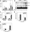
Proteasome inhibition activates NEAT1 lncRNA synthesis. (A, B) Proteasome inhibition results in elevation of NEAT1 lncRNA levels detected by qRT-PCR (A) and RNase protection assay (RPA; B). Quantitation of the RNase-protected bands is shown at the bottom. (C) Proteasome inhibition elevates the level of the newly synthesized nascent NEAT1 lncRNAs. Total and EU-labeled nascent RNAs prepared from the cells treated with DMSO or MG132 for 6 h were used for quantitation of NEAT1. For qRT-PCR, two primer sets corresponding to the NEAT1_1/1_2-overlapping region (NEAT1-#1) and the NEAT1_2-specific region (NEAT1_2-#1) were used. (D) Elevated binding of RNAPII phospho CTD serine 5 at the NEAT1 promoter in MG132-treated cells. Chromatin immunoprecipitation with αRNAPII phospho CTD serine 5 antibody was carried out in cells treated with DMSO or MG132 for 6 h. Levels of coimmunoprecipitated DNA fragments were quantified by qPCR with primer sets that span promoter regions of NEAT1 and glyceraldehyde-3-phosphate dehydrogenase (GAPDH) as control. **p < 0.01, *p < 0.05. (E) The NEAT1 promoter is activated by MG132. The graph shows qPCR of luciferase cDNA, relative to GAPDH, normalized to the control (untreated sample). RNA was extracted from MG132- and DMSO-treated HeLa cells transiently transfected 48 h before with plasmids encoding luciferase driven by the promoters as indicated. Error bars are SD as a result of two biological replicates.
FIGURE 4:
FIGURE 4:
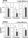
MG132 affects SFPQ and NONO nuclear distribution, resulting in depleted nucleoplasmic levels of the proteins. SFPQ or NONO density was determined by I-EM as gold particles/μm2 within paraspeckles, the surrounding nucleoplasm, and the cytoplasm of DMSO- and MG132-treated (5 μM, 17 h) HeLa cells. Although the proteins occur at different density values in the three locations (note the different values on the y axes), only the nucleoplasmic pool of SFPQ and NONO was specifically depleted with MG132, in parallel with paraspeckle enlargement under this condition. Densities were calculated on 120 and 140 μm2 of cytoplasm, 93 and 92 μm2 of nucleoplasm, and 33 and 51 paraspeckles in DMSO and MG132 cells, respectively, for SFPQ. Corresponding values for NONO are 76 and 75, 108 and 92, and 25 and 45.
FIGURE 5:
FIGURE 5:
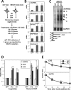
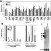
Intact paraspeckles act to suppress transcription of the ADARB2 gene. (A) Experimental strategy to identify the genes controlled by intact paraspeckles. Total RNAs were prepared from HeLa cells treated with either GFP ASO or NEAT1 ASO #12 for microarray analysis. The cells were harvested for RNA preparation 6, 12, and 24 h after ASO administration. The list of the target gene candidates is shown in Supplemental Table S1. (B) qRT-PCR validation identified five genes that were reproducibly up-regulated by two NEAT1 ASO (#12 and #17) treatments. (C) Northern blot analysis of ADARB2 mRNAs in NEAT1-eliminated cells (ΔNEAT1). PolyA+ RNAs prepared from HeLa cells treated with ASOs (green fluorescent protein [GFP] as a control, #12 and #17 for knockdown of NEAT1 lncRNA) were separated by electrophoresis on 1% agarose gels. The schematics of four putative ADARB2 mRNA isoforms (A–D) are shown. GAPDH mRNA is the loading control. (D) NEAT1 lncRNA elimination elevates the level of the nascent ADARB2 mRNA. Total RNA and pulse-labeled RNA with EU for 1 h (nascent RNA) were used as template for qRT-PCR to quantify ADARB2 mRNAs. (E) ADARB2 mRNA stability is unaltered upon NEAT1 lncRNA elimination. The mRNA decay curves (as measured by qRT-PCR) in HeLa cells treated with either GFP (solid lines) or #12 ASO (dashed lines) after actinomycin D treatment are shown. The two primer sets to detect the coding region of ADARB2 mRNA (CDS) and the 3′ UTR were used.
FIGURE 6:
FIGURE 6:
Two PSPs are required for transcription of the ADARB2 gene. (A) Identification of the PSPs required for ADARB2 gene expression. qRT-PCR to monitor ADARB2 mRNA level was carried out using RNA samples obtained from HeLa cells in which each of the PSPs was reduced by RNAi (Naganuma et al., 2012). Two primer sites (CDS and UTR) were used for ADARB2 qRT-PCR. The ADARB2 mRNA level in the cells treated with control siRNA was adjusted to 1.0. The control experiment to monitor GAPDH mRNAs is shown in Supplemental Figure S6. (B) Knockdown of SFPQ and HNRNPH1 down-regulates synthesis of nascent ADARB2 mRNA. Total RNA and EU pulse-labeled RNA (nascent RNA) from control (NC) and siRNA-treated cells were used for quantification of ADARB2 mRNAs by qRT-PCR. SFPQ and HNRNPH1 were knocked down with siRNAs #14 or #19 and H1#1, respectively. (C) Northern blot analysis of ADARB2 mRNAs in control (NC) and SFPQ-knockdown (#14 and #19) cells. ADARB2 isoforms are labeled as in Figure 5C.
FIGURE 7:
FIGURE 7:
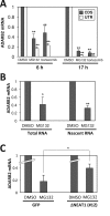
Proteasome inhibition reduces ADARB2 gene expression and requires intact paraspeckles for full suppression. (A, B) Proteasome inhibition down-regulates ADARB2 gene expression. (A) ADARB2 mRNA levels quantified by qRT-PCR from control and MG132- or bortezomib-treated HeLa cells for 6 or 17 h. (B) Proteasome inhibition (for 6 h) diminished the level of the newly synthesized nascent ADARB2 mRNA. The pulse labeling was carried out as in Figure 3C. (C) Full MG132-induced suppression of ADARB2 gene expression requires intact paraspeckles. The control (GFP) and NEAT1-eliminated (ΔNEAT1#12) cells were treated with 6 μM MG132 for 6 h. ADARB2 mRNA was monitored by qRT-PCR. **p < 0.01, *p < 0.05.
FIGURE 8:
FIGURE 8:
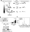
Proteasome inhibition induces SFPQ sequestration in the paraspeckles, which causes removal of SFPQ from the ADARB2 gene promoter. (A) Model of paraspeckle function. NEAT1 lncRNA expression dictates paraspeckle size and shape. NEAT1 sequesters the PSPs such as SFPQ (small black shapes) in paraspeckles. The paraspeckle unbound SFPQ is free to function as a transcriptional regulator of the several paraspeckle target genes such as ADARB2 through association with the promoters. (B) MG132 treatment results in increased association of NEAT1 RNA with SFPQ. RNA coimmunoprecipitations (RIP) with control immunoglobulin G (IgG) and anti-SFPQ (αSFPQ) were carried out from control (DMSO) and MG132-treated HeLa cells (MG132; top). The levels of coimmunoprecipitated NEAT1 lncRNAs were monitored by qRT-PCR with isoform-specific primer sets (NEAT1#1, NEAT1#2, NEAT1_2#1, and NEAT1_2#2) and normalized by immunoprecipitated SFPQ levels as shown at the bottom, in which immunoprecipitated SFPQ was quantified by Western blotting. α-Tubulin is the control. (C) Greatly reduced binding of SFPQ at the ADARB2 gene promoter in MG132-treated cells. Chromatin immunoprecipitation with αSFPQ antibody was carried out in cells treated with DMSO or MG132 for 12 h. The levels of coimmunoprecipitated DNA fragments were quantified by qPCR with the primer sets shown below, which span different segments of the ADARB2 gene locus. **p < 0.01, *p < 0.05.
FIGURE 9:
FIGURE 9:
Other paraspeckle-target genes are regulated with the same molecular mechanism as for the regulation of ADARB2 gene expression. (A) Proteasome inhibition down-regulates the paraspeckle-target genes. The mRNA levels were quantified as in Figure 7A. (B) The Influence of SFPQ RNAi on mRNA accumulation of the paraspeckle-target genes was monitored by qRT-PCR. RNAi was carried out as in Figure 5B. **p < 0.01.
FIGURE 10:
FIGURE 10:
NEAT1 lncRNA acts to attenuate MG132 induced apoptosis. (A) Induction of Neat1 expression by MG132 treatment in mouse embryonic fibroblast. The Neat1 levels were quantified with two primer sets (Neat1 and Neat1_2) in MEFs treated with DMSO (–) or MG132 (+) for 0, 3, and 6 h. (B) Neat1-knockout MEFs display greater sensitivity to MG132 in real-time growth assays. Neat1−/− and Neat1+/+ MEFs were analyzed in triplicate in a real-time growth assay over 40 h (Roche xCELLigence system), following the addition at time zero of 1 μM MG132 (blue for Neat1+/+, green for Neat1−/−) or DMSO (red for Neat1+/+, black for Neat1−/−) to the culture medium. Each 15-min time point is displayed; error bars are SD. (C) Neat1-knockout MEFs display a greater sensitivity to MG132 in the first 1–2 h of incubation. Experiments were performed as in B, with 0.25, 0.5, and 1 μM mG132 added to both Neat1+/+ and Neat1−/− MEFs. The 15-min time points in the first 4 h are plotted. (D) Incubation of Neat1+/+ and Neat1−/− MEFs with bortezomib shows a similar sensitivity of the Neat1−/− MEFs to proteasome inhibition relative to Neat1+/+ MEFs. Experiment was conducted as in B, with 7.5, 10, and 20 nM bortezomib added to cells and incubated for 40 h. Error bars are SD.

References

    1. Bond CS, Fox AH. Paraspeckles: nuclear bodies built on long noncoding RNA. J Cell Biol. 2009;186:637–644. - "V体育平台登录" PMC - PubMed
    1. Chen CX, Cho DS, Wang Q, Lai F, Carter KC, Nishikura K. A third member of the RNA-specific adenosine deaminase gene family, ADAR3, contains both single- and double-stranded RNA binding domains. RNA. 2000;6:755–767. - PMC - PubMed
    1. Chen JJ, Huang WC, Chen CC. Transcriptional regulation of cyclooxygenase-2 in response to proteasome inhibitors involves reactive oxygen species-mediated signaling pathway and recruitment of CCAAT/enhancer-binding protein delta and CREB-binding protein. Mol Biol Cell. 2005;16:5579–5591. - PMC - PubMed
    1. Chen LL, Carmichael GG. Altered nuclear retention of mRNAs containing inverted repeats in human embryonic stem cells: functional role of a nuclear noncoding RNA. Mol Cell. 2009;35:467–478. - PMC - PubMed
    1. Clemson CM, Hutchinson JN, Sara SA, Ensminger AW, Fox AH, Chess A, Lawrence JB. An architectural role for a nuclear noncoding RNA: NEAT1 RNA is essential for the structure of paraspeckles. Mol Cell. 2009;33:717–726. - PMC (V体育安卓版) - PubMed

Publication types

MeSH terms